• 검색 결과가 없습니다.

Interleukin-6 (-636 C/G) Gene Polymorphism in Korean Children With Kawasaki Disease

N/A
N/A
Protected

Academic year: 2022

Share "Interleukin-6 (-636 C/G) Gene Polymorphism in Korean Children With Kawasaki Disease"

Copied!
6
0
0

로드 중.... (전체 텍스트 보기)

전체 글

(1)

321

Open Access

Interleukin-6 (-636 C/G) Gene Polymorphism in Korean Children With Kawasaki Disease

Hye Mi Ahn, MD

1

, In Sook Park, MD

2

, Soo-Jong Hong, MD

2

, and Young Mi Hong, MD

1

1

Department of Pediatrics, School of Medicine, Ewha Womans University, Seoul,

2

Department of Pediatrics, College of Medicine, University of Ulsan, Seoul, Korea

ABSTRACT

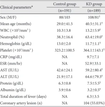
Background and Objectives: Kawasaki disease (KD) is a multi-systemic vasculitis with coronary artery involvement. Se- rum interleukin (IL)-6 levels during acute phase showed a significant correlation with the duration of fever in patients with KD who were not treated with intravenous immunoglobulin (IVIG), suggesting that the regulation of IL-6 expression in KD patients may differ from that in normal children. However, there are controversies surrounding the association between IL-6 (-636 C/G) gene polymorphism and development of KD. Subjects and Methods: One hundred and nine children with KD and 191 children with congenital heart disease were included in this study. Echocardiography was performed to examine cardiac involvement in patients with KD. Genotyping of the IL-6 (-636 C/G) gene polymorphism was performed using the single-base extension method, and serum IL-6 concentrations were estimated using the sandwich enzyme immu- noassay method. Results: Neutrophil, platelet count, liver function test, total protein and albumin concentrations were sig- nificantly different in the KD group and the serum IL-6 concentration was significantly higher in the KD group than the con- trol group. There was no difference between the patients with coronary arterial dilatation (CAD) and those without CAD in the IL-6 (-636 C/G) polymorphism. The serum albumin concentration was significantly lower in patients with KD who had the -636 C/G or GG genotype compared with the control group. The serum IL-6 concentration was significantly higher in pa- tients with KD who had the -636 C/G or GG genotype. Conclusion: There was no association between the IL-6 (-636 C/G) gene polymorphism and development of coronary arterial lesions in KD. Further multicenter studies are required to establish the relationship between the IL-6 (-636 C/G) gene polymorphism and development of KD. (Korean Circ J 2011;41:321-326) KEY WORDS: Mucocutaneous lymph node syndrome; Interleukins; Polymorphism, genetic.

Received: August 1, 2010

Revision Received: September 14, 2010 Accepted: October 24, 2010

Correspondence: Young Mi Hong, MD, Department of Pediatrics, School of Medicine, Ewha Womans University, 911-1, Mok-dong, Yangcheon- gu, Seoul 158-710, Korea

Tel: 82-2-2650-2841, Fax: 82-2-2653-3718 E-mail: [email protected]

• The authors have no financial conflicts of interest.

cc

This is an Open Access article distributed under the terms of the Cre- ative Commons Attribution Non-Commercial License (http://creativecom- mons.org/licenses/by-nc/3.0) which permits unrestricted non-commer- cial use, distribution, and reproduction in any medium, provided the origi- nal work is properly cited.

Introduction

Kawasaki disease (KD) is an acute, self-limited vasculitis that affects mainly infants and young children.

1)2)

Ten to fif- teen percent of patients with untreated KD develop coronary arterial lesions.

3-5)

Although it has been shown that coronary

complications can be significantly reduced by the use of high- dose intravenous immunoglobulin (IVIG) therapy combin- ed with oral acetylsalicylic acid, the etiology of KD still re- mains unknown.

6)

KD is known to involve an immune-mediated inflammato- ry process that leads to endothelial cell damage.

7)

The immu- nological changes in KD are evidenced by an increase in serum levels of cytokines, such as tumor necrosis factor (TNF)-α, in- terleukin (IL)-1, IL-2, IL-6, and interferon (IFN)-γ during the acute phase.

8)9)

The stimulus for increased serum levels of cytokines remains unknown.

Polymorphisms in genes encoding these crucial immuno-

modulatory molecules may result in an altered level of expres-

sion, and hence must be considered important candidate genes

for KD susceptibility and disease severity. The genetic suscep-

tibility to specific diseases may be identified by considering se-

veral genetic variants together.

(2)

IL-6 is a pro-inflammatory cytokine secreted by T cells and macrophages to stimulate an immune response leading to in- flammation.

10)

It is associated with atherosclerosis and myo- cardial infarction

11)

and is one of the most important media- tors of fever and of the acute phase response. Serum IL-6 dur- ing the acute phase showed a significant correlation with the duration of fever in patients who were not treated with IVIG.

This suggests that the regulation of IL-6 expression in KD pa- tients may differ from that in normal children, possibly due to gene polymorphisms.

12)

However, there are controversies ab- out the association between the IL-6 gene polymorphism and development of KD. Therefore, we studied the possible ge- netic influence on the expression of IL-6 in KD patients by ex- amining serum levels of IL-6 and the IL-6 (-636 C/G) gene po- lymorphism.

Subjects and Methods

The study group included 189 KD patients (108 males and 81 females) with a mean age of 40.5 months from the Depart- ment of Pediatrics at Ewha Womans University Hospital and the Asan Medical Center, Seoul, Korea. The subjects were all Koreans born in Seoul.

All of the KD patients were treated with IVIG (2 g/kg/day for 1 day), and oral aspirin (50 mg/kg/day). Echocardiography was performed by pediatric cardiologists to detect the presence of coronary artery lesions prior to IVIG administration. Coro- nary arteries were defined as abnormal if the internal lumen diameter was >3 mm in children younger than 5 years old and

>4 mm in children 5 years old or more; if the internal diame- ter of a segment measured ≥1.5 times that of an adjacent seg- ment; or if the coronary lumen was clearly irregular.

13)

The con- trol group consisted of 191 children (88 males and 103 fema- les) aged 29.0 months on average, with congenital heart disease which included ventricular septal defect (n=64), Tetralogy of Fallot (n=61), patent ductus arteriosus (n=33), atrial sep- tal defect (n=20), total anomalous pulmonary venous return (n=7), and interrupted aortic arch (n=6).

14)

None of the pa- tients had congestive heart failure.

Laboratory data were obtained from each child, including complete blood count (CBC), erythrocyte sedimentation rate (ESR), platelet count, alanine aminotransferase (ALT), aspar- tate aminotransferase (AST), serum total protein, albumin, and C-reactive protein (CRP). Genomic DNA samples were extracted from their peripheral bloods before IVIG therapy in the KD patient group and before operation for their con- genital cardiac abnormalities in the control group.

The study was carried out with the approval of the ethics committee of Ewha Womans University Hospital and the Asan Medical Center Institutional Review Board, and writ- ten informed consents were obtained from the parents of all subjects.

Genotyping of the interleukin-6 (-636 C/G) gene polymorphism

The genomic DNA was prepared from peripheral blood.

The IL-6 (-636 C/G) single nucleotide polymorphism (rs180 0796) was genotyped by using the single base extension me- thod. PCR was performed in reaction mixtures containing 1.25 pmol of each primer (sense, 5’-AGGATGGCCAGGC AGTTC-3’ and antisense 5’-CCAGTCATCTGAGTTCTT CTGTGTT-3’), 50 ng of genomic DNA, 250 mM dNTPs, and 0.15 U Taq DNA polymerase (Applied Biosystems, Fos- ter City, CA, USA) in the buffer provided by the manufac- turer. Amplification was performed in a GeneAmp PCR Sys- tem 9700 thermal cycler (Applied Biosystems).

To clean up the PCR products for the primer extension reaction, each reaction mixture was incubated with 1 U of sh- rimp alkaline phosphatase (SAP; Amersham Life Sciences, Cleveland, OH, USA) and 2 U of Exol (Amersham Life Sci- ences) for 1 hour at 37°C, and then at 72°C for 15 minutes for enzyme inactivation. Primer extension reactions were per- formed with a SNaP Shot ddNTP Primer Extension Kit (Ap- plied Biosystems), according to the manufacturer’s instruc- tions. The primer extension reaction products were cleaned up by incubation at 37°C for 1 hour in the presence of 1 U of SAP, followed by incubation at 72°C for 15 minutes. The ex- tension products were then mixed with the Genescan 120 Liz size standard solution (Applied Biosystems) and Hi-Di forma- mide (Applied Biosystems), incubated for 5 minutes at 95°C, placed on ice for 5 minutes, and then electrophoresed on an ABI Prism 3100 Genetic Analyzer (Applied Biosystems). The results were analyzed by using the ABI Prism GeneScan and Genotyper software packages (Applied Biosystems).

Measurement of serum IL-6

Serum samples were obtained from KD patients and con- trol subjects. Venous blood was withdrawn in the acute febrile stage before IVIG treatment in KD patients. Serum IL-6 lev- els were measured by using a sandwich enzyme immunoas- say-based Quantikine Human Interleukine-6 kit (R&D sys- tems Inc., Minneapolis, MN, USA), according to the manu- facturer’s instructions.

Statistical analysis

Statistical Package for the Social Sciences (SPSS) 12.0K for Windows (SPSS Inc., Chicago, IL, USA) was used for all sta- tistical analyses. Clinical characteristics, including CBC, ESR, platelet counts, ALT, AST, serum total protein, albumin, CRP, and total duration of fever were analyzed as a quantita- tive trait. Differences in clinical phenotypes between groups were analyzed using the Mann-Whitney U-test.

Unconditional logistic regression analysis adjusted for age and sex was used to calculate adjusted odds ratios (aOR), 95%

confidence intervals (CI). A p<0.05 was considered statistically

(3)

significant.

Results

Clinical characteristics of study subjects

Clinical characteristics are shown in Table 1. White blood cell (WBC) count (neutrophil), platelet count, ALT, AST, and total protein were significantly higher, and hemoglobin and albumin levels were significantly lower in the KD group com- pared with the control group (p<0.05). Coronary arterial le- sions in the KD group were noted in 104 patients (55.03%).

Allele frequencies of the interleukin-6 (-636 C/G) polymorphism in the Kawasaki disease group and the control group

The 189 study subjects and 191 patients in the control group were genotyped for the IL-6 (-636 C/G) polymorphism, and allele frequencies were examined. The genomic frequencies of the IL-6 (-636 C/G) polymorphism in the KD group were 57.1% (108/189) for CC, 37.6% (71/189) for CG, and 5.3%

(10/189) for GG. In the control group, these frequencies were 52.9% (101/191) for CC, 42.9% (82/191) for CG, and 4.2%

(8/191) for GG. The genotype frequencies of these samples were in accordance with Hardy-Weiberg equilibrium. The ge- nomic and allelic frequencies of this polymorphism did not significantly differ between the KD group and the control group (Table 2).

Association between the interleukin-6 (-636 C/G) polymorphism and clinical parameters in the Kawasaki disease group and the control group

Clinical parameters were compared between the KD group and the control group according to allele frequencies. WBC count (neutrophil), platelet count, AST, ALT, and total protein were significantly higher in the KD group compared with the control group. Interestingly, albumin concentrations were sig- nificantly lower in patients with KD with -636 C/G and those with GG genotypes, compared to those with the CC genotype (Table 3).

Allele frequencies of the interleukin-6 (-636 C/G) polymorphism in Kawasaki disease children with and without coronary arterial lesions

We determined whether the IL-6 polymorphism was asso- ciated with coronary arterial lesions in children with KD. In the KD group without coronary arterial lesions, allele frequ- encies were 54.2% (45/83) for CC, 39.8% (33/83) for CG, and 6.0% (5/83) for GG. In the KD group with coronary arterial lesions, allele frequencies were 59% (59/100) for CC, 37%

(37/100) for CG, and 4% (4/100) for GG. There was no dif- ference in the IL-6 (-636 C/G) polymorphism according to coronary arterial lesions (Table 4).

Serum interleukin-6 levels in the Kawasaki disease group and the control group

Serum IL-6 concentrations were 2.72±6.49 ng/mL in the control group and 51.0±13.85 ng/mL in the KD group. Serum IL-6 concentrations were markedly elevated in the KD group compared with the control group (Fig. 1).

Serum interleukin-6 levels in the Kawasaki disease group with CC and CG+GG genotypes

The serum IL-6 concentration was examined according to genotype. Serum IL-6 levels were 21.0±23.4 ng/mL in patients with KD who had the CC genotype and 69.3±30.9 ng/mL in Table 2. Allele frequencies of the IL-6 -636C/G polymorphism in children with Kawasaki disease and congenital heart disease Genotype

Control group

(n=191) KD group

(n=189)

No. (%) No. (%) aOR (95%CI) IL-6 -636C/G

CC 101 (52.9) 108 (57.1) 1.00

CG 082 (42.9) 071 (37.6) 0.81 (0.53-1.23)

GG 08 (4.2) 10 (5.3) 1.17 (0.44-3.08)

CG+GG 090 (47.1) 081 (42.9) 0.84 (0.56-1.26)

CC+CG 183 (95.8) 179 (94.7) 1.00

Frequency (C) 000 (74.3) 000 (75.9)

p>0.05. IL: interleukin, KD: Kawasaki disease, aOR: adjusted odds ratio

Table 1. Clinical characteristics of study subjects

Clinical parameters* Control group KD group

(n=191) (n=189)

Sex (M/F) 88/103 108/81

Mean age (months) 29.0±41.5 40.5±31.1

WBC (×10

3

/mm

3

) 10.3±3.80 013.2±5.9

00

Neutrophil (%) 38.3±16.4 63.4±19.0

Hemoglobin (g/dL) 13.0±2.00 11.7±1.1

0 Platelet (×10

3

/mm

3

) 323.2±100.5 364.1±143.1

CRP (mg/dL) NA 9.7±7.10

ESR (mm/hr) NA 52.9±33.10

AST (IU/L) 42.6±24.1 59.2±80.4

ALT (IU/L) 21.9±17.1 64.6±79.3

Protein (g/dL) 6.3±0.8 7.5±5.5

Albumin (g/dL) 3.9±0.6 3.2±0.5

Total duration of fever (days) NA 6.3±3.50 Coronary artery lesion (n) NA 104 (55.03%) The significance of clinical parameters according to groups was an- alyzed using the Mann-Whitney test. *Data are expressed as mean±

standard deviation,

p<0.05 significantly different from control

group. NA: not available, KD: Kawasaki disease, WBC: whole blood

cell, CRP: C-reactive protein, ESR: erythrocyte sedimentation rate,

AST: aspartate aminotransferase, ALT: alanine aminotransferase

(4)

patients with the -636 C/G and those with the GG genotype (Fig. 2).

Discussion

The etiology for KD remains unknown, but several studies have suggested the involvement of an immune-mediated pro-

cess. Cytokines such as IL-1, IL-6, IFN-γ, TNF-α are released inducing a series of inflammatory reactions leading to endo- thelial cell injury and inflammation of small and medium-siz- ed arteries, especially coronary arteries.

15-17)

Cytokines with po- lymorphic gene sequences are potential markers of disease susceptibility.

15)16)

In recent years, it has been well demonstrated that inflam- Table 3. Association between IL-6 -636 C/G alleles and clinical parameters in children with Kawasaki disease and children with congenital heart disease

Clinical parameters* Control group KD group

CC (n=101) CG+GG (n=90) CC (n=108) CG+GG (n=81)

Age (months) 27.6±36.3 028.9±45.0 42.6±31.5 38.3±30.8

WBC (×10

3

/mm

3

) 10.0±3.50 10.7±4.0 13.2±5.80 13.5±6.20

Neutrophil (%) 37.3±16.3 039.3±16.8 64.7±17.9 62.6±20.4

Hemoglobin (g/dL) 13.0±1.90 12.9±2.2 11.7±1.10 11.6±1.10

Platelet (×10

3

/mm

3

) 323.8±102.0 325.6±99.4 350.8±128.9 375.7±147.5

CRP (mg/dL) NA NA 10.5±7.70 8.9±6.2

ESR (mm/hr) NA NA 54.7±32.3 53.5±33.6

AST (IU/L) 38.8±16.0 046.9±30.0 59.0±85.4 061.6±76.4

ALT (IU/L) 19.2±10.7 025.0±21.8 63.2±80.4 067.4±80.2

Protein (g/dL) 6.4±0.8 06.2±0.8 8.1±7.6 6.8±0.8

Albumin (g/dL) 4.0±0.6 03.8±0.6 3.3±0.5 03.1±0.5

Total duration of fever (days) NA NA 6.3±3.1 6.4±4.0

*Data are expressed as mean±standard deviation,

p<0.05 compared between CC genotype and CG+GG genotype in children with control group. NA: not available, KD: Kawasaki disease, WBC: whole blood cell, CRP: C-reactive protein, ESR: erythrocyte sedimentation rate, AST:

aspartate aminotransferase, ALT: alanine aminotransferase

Table 4. Allele frequencies of the IL-6 -636 C/G polymorphism in Kawasaki disease children with and without early coronary arterial lesion Genotype IL-6 -636 C/G KD without coronary artery lesion (n=89) KD with coronary artery lesion (n=100)

aOR (95% CI)

No. (%) No. (%)

CC 48 (53.93) 59 (59.00) 1.00

CG 35 (39.32) 37 (37.00) 0.86 (0.47-1.58)

GG 6 (6.74) 4 (4.00) 0.61 (0.15-2.40)

CG+GG 41 (46.06) 41 (41.00) 0.82 (0.46-1.48)

CC+CG 83 (93.26) 96 (96.00) 1.00

Frequency (C) 0.74 0.78

p>0.05. IL-6: Interleukin-6, KD: Kawasaki disease, aOR: adjusted odds ratio

Fig. 1. Serum IL-6 levels in Kawasaki disease group and control gr- oup. Serum IL-6 level was markedly elevated in KD group com- pared with control group. *p<0.05 significantly different from control group. IL-6: interleukin-6, KD: Kawasaki disease.

70 60 50 40 30 20 10

0 Control KD

*

Se ru m IL -6 le ve l ( ng /m L)

Fig. 2. Serum IL-6 levels in KD group with CC and CG+GG geno- type. Regarding the IL-6 gene polymorphism, compared with the CC genotype, the serum IL-6 level was significantly higher in the CG+GG genotype in the KD group. *p<0.05 significantly different from CC group. IL-6: interleukin-6, KD: Kawasaki disease.

120 100 80 60 40 20

0 CC group CG+GG group

*

Se ru m IL -6 le ve l ( ng /m L)

(5)

matory molecules, as well as single nucleotide polymorphisms of genes encoding inflammatory mediators, may contribute to the development and progression of a large number of pa- thologic conditions including cardiovascular disease.

18-21)

One commonly used technique to identify genetic risk fac- tors for multifactorial diseases such as KD is the candidate gene approach, which directly examines the effects of genetic variants of a possibly contributing gene by an association study. To date, quite a number of genes have been examined, which are usually selected based on information regarding their function in inflammation, the immune response, and ot- her biological mechanisms.

18)

Single nucleotide polymorphisms (SNP) of pro-inflamma- tory and anti-inflammatory genes may strongly influence the plasma levels and biological activity of the corresponding pro- teins, with potentially important clinical implications.

22)23)

Burns et al.

24)

reported that the genetic variation in the IL-4 gene or regions linked to IL-4, plays an important role in KD pathogenesis and disease susceptibility. Jin et al.

25)

re- ported that the IL-10 (-627 A/C) gene polymorphism may be associated with coronary aneurysms and low albumin in Korean children with KD. IL-6 expression was noted in sev- eral diseases such as systemic lupus erythematosus,

26)

spora- dic Alzheimer’s disease,

27)

and systemic onset juvenile ch- ronic arthritis.

28)

The IL-6 gene -174G>C and -572G>C gene polymorphi- sms are associated with cerebral aneurysm.

28)

Whether this association is due to the development, progression, or rup- ture of such an aneurysm or represents survivor bias is un- clear. Sohn et al.

29)

examined the IL-6 gene polymorphism at +162 bp, +168 bp and -594 bp, and reported that the poly- morphism was not associated with KD. In KD, IL-6 has been reported to rise more than any other cytokine and correlates with coronary complications.

15)

There have been no reported studies about the association between the IL-6 (-636 C/G) polymorphism and KD. In this study, we investigated the association between the IL-6 (-636 C/G) polymorphism (rs1800796) and KD. The IL-6 (-636 C/G) polymorphism showed no difference in gene frequency between the KD group and the control group. There was no difference in the IL-6 (-636 C/G) polymorphism according to coronary arterial lesions either. Nonetheless, the serum IL-6 levels were significantly higher in the KD group than the control group and among the KD group, serum IL-6 levels were significantly higher in the CG+GG group compared to the CC group. Likewise, albumin concentrations were signific- antly lower in the KD group than the control group and among the KD group, the CG+GG group showed lower levels than the CC group. Therefore, our findings suggest that the IL-6 (-636 C/G) polymorphism may be associated with risk factors of KD, such as a low albumin concentration. In addi- tion, elevated serum IL-6 concentrations in the CG+GG ge-

notype suggests the possibility of a relationship between the IL-6 (-636 C/G) polymorphism and KD that needs to be an- alyzed further.

Our studies have some limitations. The studied population is relatively small in number. We enrolled patients with conge- nital heart disease as a control group instead of normal chil- dren. Also there is a possibility of linkage disequilibrium with an unknown risk single nucleotide polymorphism and the lack of function-related experiments. There have been no reported studies on the relationship of the transcription acti- vity of IL-6 in KD. SNPs on the IL-6 promoter could affect the transcription activity and furthermore the expression of IL- 6. Such SNPs could be a good candidate marker to study the transcriptional activity of IL-6.

In conclusion, we found that the IL-6 (-636 C/G) polymor- phism was not associated with the development of KD or coronary arterial lesions. This study suggests however, that the IL-6 (-636 C/G) polymorphism might prove useful as a ge- netic marker for the indicators of KD such as duration of fe- ver, platelet count, and albumin concentration. Given the li- mitations of our study, it is not possible to definitely rule out the direct correlation between the IL-6 (-636 C/G) polymor- phism and KD. Further large scale studies will be needed to establish the relationship between IL-6 gene polymorphisms and KD.

Acknowledgments

This study was sponsored by a grant from the Korean Ministry of He- alth and Welfare (01-PJ10-PG6-01GN15-0001).

REFERENCES

1) Yeo JS, Choi JW. Effectiveness of medium-dose intravenous immuno- globulin (1 g/kg) in the treatment of Kawasaki disease. Korean Circ J 2010;40:81-5.

2) Lee SJ, Ahn HM, Yoo JH, Hong YM. Carotid intima-media thickness and pulse wave velocity after recovery from Kawasaki disease. Korean Circ J 2009;39:264-9.

3) Wang CL, Wu YT, Liu CA, Kuo HC, Yang KD. Kawasaki disease in- fection, immunity and genetics. Pediatr Infect Dis J 2005;24:998-1004.

4) Fulton DR, Newberger JW. Long-term cardiac sequelae of Kawasaki disease. Curr Rheumatol Rep 2000;2:324-9.

5) Kato H, Sugimura T, Akagi T, et al. Long-term consequences of Kawa- saki disease: a 10- to 21-year follow-up study of 594 patients. Circul- ation 1996;94:1379-85.

6) Lloyd AJ, Walker C, Wilkinso M. Kawasaki disease: is it caused by an infectious agent? Br J Biomed Sci 2001;58:122-8.

7) Liang SJ, Cheong HI, Noh CI, Yun YS. Role of anti-endothelial cell antibody in the development of coronary arterial lesions in Kawasaki disease. Korean Circ J 2006;36:723-31.

8) Maury CP, Salo E, Pelkonen D. Circulating interleukin-1 beta in pa- tients with Kawasaki disease. N Engl J Med 1988;319:1670-1.

9) Ueno Y, Takano N, Kanegane H, et al. The acute phase nature of in- terluekin-6: studies in Kawasaki disease and other febrile illness. Clin Exp Immunol 1989;76:337-42.

10) Hirano T, Akira S, Taga T, Kishimoto T. Biological and clinical as- pects of interluekin-6. Immunol Today 1990;11:443-9.

11) Tanaka C, Mannami T, Kamide K, et al. Single nucleotide polymor-

phisms in the interleukin-6 gene associated with blood pressure and ath-

erosclerosis in a Japanese general population. Hypertens Res 2005;

(6)

28:35-41.

12) Kim MK, Kim DS. The interleukin-6 level in Kawasaki disease. J Korean Pediatr Soc 1992;35:515-26.

13) Research Committee on Kawasaki Disease. Report of Subcommittee on Standardization of Diagnostic Criteria and Reporting of Coronary Artery Lesions in Kawasaki Disease. Tokyo, Japan: Ministry of He- alth and Welfare;1984.

14) Hong YM, Jin HS, Park IS, Hong SJ. Association of the matrix me- talloproteinase-3 (-439C/G) gene polymorphism with Kawasaki dis- ease in Korean children. Heart Vessels 2008;23:341-7.

15) Hirao J, Hibi S, Andoh T, Ichimura T. High levels of circulating inter- leukin-4 and interleukin-10 in Kawasaki disease. Int Arch Allergy Im- munol 1997;112:152-6.

16) Cheung YF, Huang GY, Chen CB, et al. Inflammatory gene polymor- phisms and susceptibility to Kawasaki disease and its arterial sequel- ae. Pediatrics 2008;122:e608-14.

17) Leung DY, Geha RS, Newberger JW, et al. Two monokines, interleu- kin-1 and tumor necrosis factor, render cultured vascular endothelial cells susceptible to lysis by antibodies circulating during Kawasaki syndrome. J Exp Med 1986;164:1958-72.

18) Hata A, Onouchi Y. Susceptibility genes for Kawasaki disease: toward implementation of personalized medicine. J Hum Genet 2009;54:67-73.

19) Blake GJ, Ridker PM. Inflammatory bio-markers and cardiovascular risk prediction. J Intern Med 2002;252:283-94.

20) Zee RY, Cook NR, Cheng S, et al. Polymorphism in the P seletin and interleukin-4 genes as determinants of stroke: a population-based, pro- spective genetic analysis. Hum Mol Genet 2004;13:389-96.

21) Chapman CM, Beilby JP, Humphries SE, Palmer LJ, Thompson PL, Hung J. Association of an allelic variant of interleukin-6 with subclini- cal carotid atherosclerosis in an Australian community population. Eur

Heart J 2003;24:1494-9.

22) Jibiki T, Terai M, Shima M, et al. Monocyte chemoattractant protein 1 gene regulatory region polymorphism and serum levels of monocyte chemoattractant protein 1 in Japanese patients with Kawasaki disease.

Arthritis Rheum 2001;44:2211-2.

23) Fishman D, Faulds G, Jeffery R, et al. The effect of novel polymorph- ism in the interleukin-6 (IL-6) gene on IL-6 transcription and plasma IL-6 levels, and an association with systemic onset juvenile chronic ar- thritis. J Clin Invest 1998;102:1369-76.

24) Burns JC, Shimizu C, Shike H, et al. Family-based association analy- sis implications IL-4 in susceptibility to Kawasaki disease. Genes Im- mun 2005;6:438-44.

25) Jin HS, Kim HB, Kim BS, et al. The IL-10 (-627 A/C) gene polymorph- ism may be associated with coronary aneurysms and low albumin in Ko- rean children with Kawasaki disease. Pediatr Res 2007;61:584-7.

26) Linker-Israeli M, Wallace DJ, Prehn JL, Nand R, Li L, Klinenberg JR. A greater variability in the 3’flanking region of the IL-6 gene in pa- tients with systemic lupus erythematosus (SLE). Autoimmunity 1996;

23:199-209.

27) Papassotiropoulos A, Bagli M, Jessen F, et al. A genetic variation of the inflammatory cytokine interleukin-6 delays the initial onset and re- duces the risk for sporadic Alzheimer’s disease. Ann Neurol 1999;45:

666-8.

28) Morgan L, Cooper J, Montgomery H, Kitchen N, Humphries SE.

The interleukin-6 gene -174G>C and -572G>C promoter polymor- phisms are related to cerebral aneurysms. J Neurol Neurosurg Psychia- try 2006;77:915-7.

29) Sohn MH, Hur MW, Kim DS. Interleukin-6 gene gene polymorphism

is not associated with Kawasaki disease. Genes Immun 2001;2:357-62.

수치

Table 1.  Clinical characteristics of study subjects
Table 4. Allele frequencies of the IL-6 -636 C/G polymorphism in Kawasaki disease children with and without early coronary arterial lesion Genotype IL-6 -636 C/G KD without coronary artery lesion (n=89) KD with coronary artery lesion (n=100)

참조

관련 문서

Results: In this research, in the group with fibromyalgia patients group, systemic lupus erythematosus patients group and without systemic autoimmune

The change in active oxygen was decreased after exercise in the exercise group than in the control group, and there was a statistically significant

In the control group, lifestyle-related factors were significantly different only in blood glucose (p &lt;.01) and there was no significant difference in

Serum uric acid levels and risk for vascular disease in patients with metabolic syndrome... Prevalence if the metabolic syndrome in a Turkish

&lt;Table 5&gt; Difference of limb load asymmetry, postural sway and lumbar flexibility between control group and experimental group(In case

The AST levels in the male KRG administration group at 4 and 8 mo decreased sig- nificantly compared to the control group (p &lt; 0.05), and there was no significant

In conclusion, we found that MPV acted as a positive acute phase reactant in children with UTI, and MPV levels were significantly elevated in children with APN on DMSA

In the graft group, the weights of adipose tissue were significantly increased, compared to the contralateral control in both 3-and 7-day ( p &lt; 0.05) (Table 1, Fig..