www.ogscience.org 417
Case Report
Obstet Gynecol Sci 2018;61(3):417-420 https://doi.org/10.5468/ogs.2018.61.3.417 pISSN 2287-8572 · eISSN 2287-8580
Introduction
The rapid advancement of imaging technology during the prenatal period has enabled the diagnosis of fetuses with structural anomalies. In particular, prenatal ultrasound and magnetic resonance imaging (MRI) enable the detection of fe- tal neck masses and the extent of tracheoesophageal obstruc- tion [1]. Fetuses with anomalies that cause airway obstruction usually have poor outcomes because of the delayed time in securing an airway [2]. Hence, many interventions have been introduced to increase the time spent on airway establish- ment, and the first ex utero intrapartum treatment (EXIT) procedure was performed for a large neck mass[3]. Delivery of the fetal head and shoulders while maintaining the utero- placental circulation offers adequate time to secure the airway and prevent fetal hypoxia [4]. However, because of the rarity of fetal airway obstruction, only few cases of EXIT procedure have been reported in Korea. Here, we report our experiences of EXIT procedure for fetal airway obstruction, which were successfully performed without any complications.
Case report
1. Case 1
A 32-year-old nulligravida was referred to our institution
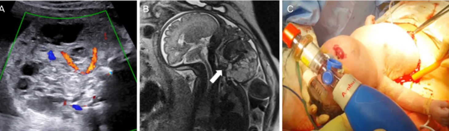
at 20.3 weeks of gestation with a diagnosis of a fetal neck mass. Ultrasonographic findings revealed a 5.3 cm-sized heterogeneous solid mass on the fetal neck, and color Dop- pler revelaed high vascularity. We diagnosed the mass as a cervical lymphangioma or teratoma. Increasing amniotic fluid index in ultrasonography suggested upper airway compres- sion by the mass. Serial ultrasound examination showed an increase in the tumor size to up to almost 8 cm at 30.3 weeks of gestation (Fig. 1A). To evaluate airway patency, MRI was performed at 31.0 weeks of gestation and revealed an 8.5 cm-sized mass arising from the mandible and anterior neck soft tissues and compressing the upper airway (Fig. 1B).
Delivery was performed via a cesarean section with the EXIT procedure at 38.3 weeks of gestation. A multidisciplinary
Ex utero intrapartum treatment procedure in two fetuses with airway obstruction
Joohee Lee
1, Mi-Young Lee
1, Yeni Kim
1, Jae-Yoon Shim
1, Hye-Sung Won
1, Euiseok Jeong
2, Byong Sop Lee
2, Ki-Soo Kim
2, Woo-Jong Choi
3, Yoon Se Lee
41Department of Obstetrics and Gynecology, 2Division of Neonatology, Department of Pediatrics, 3Departments of Anaesthesiology and Pain Medicine,
4Otolaryngology, University of Ulsan College of Medicine, Asan Medical Center, Seoul, Korea
The ex utero intrapartum treatment (EXIT) procedure was introduced to reduce fetal hypoxic damage while establishing an airway in fetuses with upper and lower airway obstruction. Delivery of the fetal head and shoulders while maintaining the uteroplacental circulation offers time to secure the fetal airway. Here, we report two cases of EXIT procedure for fetal airway obstruction, which were successfully managed with extensive preoperative planning by a professional multidisciplinary team.
Keywords: Fetal therapy; Laryngeal diseases; Lymphangioma; Prenatal diagnosis
Articles published in Obstet Gynecol Sci are open-access, distributed under the terms of the Creative Commons Attribution Non-Commercial License (http://creativecommons.
org/licenses/by-nc/3.0/) which permits unrestricted non-commercial use, distribution, and reproduction in any medium, provided the original work is properly cited.
Copyright © 2018 Korean Society of Obstetrics and Gynecology
Received: 2017.05.28. Revised: 2017.07.27. Accepted: 2017.08.07.
Corresponding author: Mi-Young Lee
Department of Obstetrics and Gynecology, University of Ulsan College of Medicine, Asan Medical Center, 88 Olympic-ro 43-gil, Songpa-gu, Seoul 05505, Korea
E-mail: [email protected] https://orcid.org/0000-0003-2691-4954
www.ogscience.org 418
Vol. 61, No. 3, 2018
team comprising obstetricians, neonatologists, anesthesiolo- gists, and otorhinolaryngologists was formed. The fetus was in cephalic presentation, and the placenta was located at the posterior body of the uterus. A Pfannenstiel skin incision and a lower uterine segment transverse hysterotomy were per- formed. The fetal head and upper body were delivered, while the lower body remained in the uterus (Fig. 1C). Compression of the fundus was avoided, and warm saline was continuously infused into the uterus to maintain the intrauterine volume and to prevent placental separation. Peripheral oxygen satura- tion (SpO2) and fetal heart rate were monitored. Initial fetal SpO2 was 23%, with a heart rate of 100 beats/min. During several intubation attempts, SpO2 varied from 48% to 56%, with a heart rate of 136 beats/min. Intubation was success- fully accomplished in 12 minutes. The baby was then deliv- ered, and the umbilical cord was clamped. The birth weight was 3,500 g, including the neck mass, and the Apgar score was 6 and 7 at 1 and 5 minutes, respectively.
After the delivery of the baby and the placenta, carbetocin was injected, and the uterine contraction was good. Postop- eratively, the estimated blood loss was 1.0 L, which was simi- lar to the volume loss during routine cesarean section. The he- moglobin level was 11.8 g/dL preoperatively and decreased to 9.3 g/dL on postoperative day 1. The mother was discharged on postoperative day 3 without any complications.
Postnatal MRI demonstrated a large cervicofacial lymphangi- oma with hemorrhage. Excision of the mass with tracheosto- my was performed at 62 days after birth. Complete resection was impossible, and histopathological examination confirmed a cavernous lymphangioma. After repeating several courses of ethanol sclerotherapy, the baby was discharged in toler-
able condition with tracheostomy at 4 weeks postoperatively.
Currently, at the age of 2 years, the baby remains in tracheos- tomy state but is doing well.
2. Case 2
A 31-year-old nulligravida was referred at 21.1 weeks of ges- tation with a suspected diagnosis of congenital high airway obstruction syndrome (CHAOS). Ultrasonography revealed symmetrically enlarged, hyperechoic, and homogeneous lungs with an inverted diaphragm, anteriorly displaced heart, and associated polyhydramnios, all of which suggested CHA- OS (Fig. 2A). Delivery was performed via a cesarean section with EXIT procedure at 37.0 weeks of gestation. The fetus was in cephalic position, and the placenta was located in the posterior body of the uterus. A similar operation to that in the case 1 was performed. Initial fetal SpO2 was 75%, with a heart rate 145 beats/min. Intubation was failed several times because of the invisible airway. The total amount of time spent on intubation attempts was approximately 5 minutes.
Tracheostomy was then performed within 3 minutes (Fig. 2B), and SpO2 recovered to 82%. After approximately 10 min- utes, the umbilical cord was clamped. The birth weight was 2,720 g, with an Apgar score of 6 and 8 at 1 and 5 minutes, respectively.
After delivery, the uterus showed poor contraction initially.
After injecting carbetocin with a continuous infusion of sul- prostone, uterine contraction was normalized; however, the expected blood loss was >2 L. The initial hemoglobin level was 13.2 g/dL, which decreased to 9.6 g/dL on the postop- erative day 1. The mother had no postoperative complications and was discharged on postoperative day 3.
Fig. 1. Ultrasonographic findings at 30.3 weeks of gestation showing a huge heterogeneous solid mass on the fetal neck with high vas- cularity (A). Magnetic resonance imaging at 31.0 weeks of gestation showing an 8.5 cm-sized neck mass with suspected airway obstruc- tion (arrow in B). After partially delivering the fetal head and shoulders, intubation was performed (C).
A B C
www.ogscience.org 419 Joohee Lee, et al. Ex utero intrapartum treatment procedure
Postnatal computerized tomography revealed a short seg- mental obliteration of the subglottis, with the visible lumen of the proximal trachea, suggesting laryngeal atresia. After con- servative treatment during 1 month, the baby was discharged.
At the age of 1 year, the baby remains in tracheostomy state but is doing well.
Discussion
To the best of our knowledge, to date, only five cases involv- ing the EXIT procedure have been reported in Korea [5-9]. Pre- vious reports mainly focused on the anesthetic management of the EXIT procedure to achieve adequate uterine relaxation, maintain uteroplacental circulation, and minimize fetal move- ment. Furthermore, management after the EXIT procedure has been discussed by pediatricians and otorhinolaryngolo- gists, with a focus on postnatal outcomes. On the basis of our successful experience with the EXIT procedure, we would like to mainly discuss the role of the obstetrician performing the EXIT procedure.
The EXIT procedure aims to manage fetuses with life- threatening diseases of the upper respiratory system, thereby increasing the survival rate of fetuses. Selecting appropriate candidates for the EXIT procedure is primarily important. Indi- cations for the EXIT procedure to secure the airway are as fol- lows: 1) masses that cause extrinsic airway compression such
as teratoma, lymphangioma, and epignathus; 2) severe mi- crognathia or agnathia; 3) CHAOS; and 4) removal of balloon occlusion in congenital diaphragmatic hernia [10]. The EXIT procedure can also be performed for resecting huge chest masses that cause airway obstruction, such as in congenital pulmonary airway malformation, bronchopulmonary seques- tration, and mediastinal or pericardial teratoma [11]. Other indications include severe congenital heart disease or severe congenital diaphragmatic hernia for applying extracorporeal membrane oxygenation and conjoined twins for separation [11]. Obstetricians should be aware of the abovementioned indications. Our second case of CHAOS safely managed with EXIT procedure was first reported in Korea, and our experi- ences may encourage the parents of fetuses with CHAOS to continue the pregnancy and to receive proper postnatal man- agement.
Once a diagnose is made, the obstetricians should obtain the data of mass size and its location as well as the charac- teristics of diseases causing airway obstruction. Although ultrasound is a standard tool for determining the need for the EXIT procedure, it has a limitation regarding the evaluation of airway obstruction. MRI is superior to ultrasound for assessing the extent of airway distortion [10]. MRI was not performed in our second case where the baby had only a pinpoint trachea.
If MRI was prenatally performed, the time spent on intubation could have been reduced.
The organization of a multidisciplinary team comprising obstetricians, neonatologists, anesthesiologists, and otorhino- Fig. 2. Ultrasonographic findings showing both lung hyperexpansion with diaphragm inversion, suggestive of congenital high airway ob- struction syndrome (A). Tracheostomy is in process while the baby is partially delivered (B).
A B
www.ogscience.org 420
Vol. 61, No. 3, 2018
laryngologists is essential for achieving successful outcomes of the EXIT procedure. Before performing the EXIT procedure, our multidisciplinary team gathered, discussed the need for tracheostomy, and simulated the condition during the EXIT procedure. The obstetricians should decide an appropriate time for delivery with considering both fetal and maternal conditions. The EXIT procedure should be performed before the onset of labor. Accordingly, 37–38 weeks of gestation is generally acceptable unless there are any other maternal or fetal indications for earlier delivery [4]. In addition, evaluation about the presence of polyhydramnios, fetal position, and placental location before the EXIT procedure is important.
Therefore, obstetricians require careful mapping of the pla- cental edges, which could alter the location of the uterine incision; they also need to determine the degree of exposure required, according to the fetal abnormalities [4].
During the EXIT procedure, obstetricians should focus on maximizing uterine relaxation time by not compressing the uterine fundus, delivering only the head and shoulders onto the surgical field, and infusing warm saline into the uterus to maintain the uterine volume and prevent placental sepa- ration and umbilical cord compression [4]. However, excess time spent on establishing the fetal airway during the pro- cedure can increase maternal complications, particulary postpartum hemorrhage. Although uterine atony resulted in an increased estimated blood loss in our second case, it was successfully managed with uterotonics. Because other complications such as uterine rupture or dehiscence at sub- sequent pregnancy may also occur, an inter-pregnancy inter- val of at least 24 months following EXIT procedure has been recommended [12].
The EXIT procedure can be a potent procedure for cases of life-threatening airway obstruction. Obstetricians play an im- portant role in managing the EXIT procedure for both fetuses and mothers. Minimizing fetal and maternal complications should be the goal for obstetricians performing the EXIT pro- cedure.
Conflict of interest
No potential conflict of interest relevant to this article was reported.
References
1. Hubbard AM, Crombleholme TM, Adzick NS. Prenatal MRI evaluation of giant neck masses in preparation for the fetal EXIT procedure. Am J Perinatol 1998;15:253-7.
2. Mychaliska GB, Bealer JF, Graf JL, Rosen MA, Adzick NS, Harrison MR. Operating on placental support: the ex utero intrapartum treatment procedure. J Pediatr Surg 1997;32:227-30.
3. Norris MC, Joseph J, Leighton BL. Anesthesia for perina- tal surgery. Am J Perinatol 1989;6:39-40.
4. Abraham RJ, Sau A, Maxwell D. A review of the EXIT (ex utero intrapartum treatment) procedure. J Obstet Gyn- aecol 2010;30:1-5.
5. Park JT, Chang HS, Kwon SY, Um DJ, Choi SJ. Anesthetic management during an ex utero intrapartum treatment (EXIT) procedure of the agnathic fetus: a case report.
Korean J Anesthesiol 2005;49:724-9.
6. Park HJ, Lee JH, Chung SM, Kim HS. A case of the EXIT (ex utero intrapartum treatment) procedure in congeni- tal fetal cervical immature teratoma. Korean J Otorhino- laryngol-Head Neck Surg 2008;51:681-5.
7. Lee H, Ryu JW, Kim DY, Lee GY. Anesthetic management of the ex utero intrapartum treatment (EXIT) procedure:
a case report. Korean J Anesthesiol 2010;59:S154-7.
8. Hwang I, Jung HR, Bae JG. A case of intrauterine lin- gual cyst with successful delivery assisting ex utero intrapartum treatment procedure. Korean J Perinatol 2014;25:22-6.
9. Park H, Jin HS, Kang SH, Park JS. A case of the ex utero intrapartum treatment procedure for fetal giant fourth branchial cleft cyst. Korean J Otorhinolaryngol-Head Neck Surg 2015;58:776-80.
10. Mota R, Ramalho C, Monteiro J, Correia-Pinto J, Ro- driques M, Guimarães H, et al. Evolving indications for the EXIT procedure: the usefulness of combining ultra- sound and fetal MRI. Fetal Diagn Ther 2007;22:107-11.
11. Dighe MK, Peterson SE, Dubinsky TJ, Perkins J, Cheng E.
EXIT procedure: technique and indications with prenatal imaging parameters for assessment of airway patency.
Radiographics 2011;31:511-26.
12. Zamora IJ, Ethun CG, Evans LM, Olutoye OO, Ivey RT, Haeri S, et al. Maternal morbidity and reproduc- tive outcomes related to fetal surgery. J Pediatr Surg 2013;48:951-5.