198
DOI 10.4070 / kcj.2009.39.5.198 Copyright ⓒ 2009 The Korean Society of Cardiology
Enhanced Cardiomyogenic Differentiation of P19 Embryonal Carcinoma Stem Cells
Jihyun Yang, Seok-Jin Ko, Beom-Suk Kim, Hyun-Seo Kim, Sangheon Park, Doran Hong, Soon-Woong Hong, Ji-Hyun Choi, MS, Chi-Yeon Park, MS, Seung-Cheol Choi, PhD, Sun-Jun Hong, MD and Do-Sun Lim, MD Department of Cardiology, College of Medicine, Korea University, Seoul, Korea
ABSTRACT
Background and Objectives: We investigated the effects of different concentrations of serum, 5-azacytidine, and culture time on the cardiomyogenic differentiation of P19 embryonal carcinoma stem cells in the course of devel- oping an efficient protocol for generating the cardiomyogenic lineage. Materials and Methods: P19 cells were plated at a density of 1×10⁶ cells on 10-cm bacterial dishes for 96 hours in the presence of 1% dimethyl sul- foxide to form embryoid bodies. The embryoid bodies were cultured in medium with 2% or 10% fetal bovine serum for an additional 10 or 15 consecutive days in the presence of 0, 1, or 3 μM 5-azacytidine. Results: Quan- titative real-time polymerase chain reaction (PCR) analysis showed that the messenger ribonucleic acid (mRNA) expression of cardiac muscle-specific genes, such as GATA4, α-actin, α-myosin heavy chain, and cardiac tropo- nin T, were significantly higher in the 15-day culture groups than in the 10-day culture groups. Furthermore, the cardiac muscle-specific genes were expressed more in the high-serum groups compared to the low-serum groups regardless of the culture time. Cardiomyogenic differentiation of the P19 cells was most effective in 1 μM 5-aza- cytidine regardless of the serum concentrations. In addition, the stimulation effects of 5-azacytidine on cardio- myogenic differentiation were more significant under low-serum culture conditions compared to high-serum culture conditions. Cardiomyogenic differentiation of P19 cells was further confirmed by immunostaining with cardiac mu- scle-specific antibodies. Conclusion: Taken together, these results demonstrated that cardiomyogenic differentiation of P19 cells was enhanced by a combination of different experimental factors. (Korean Circ J 2009;39:198-204) KEY WORDS: Cell differentiation; Embryonal carcinoma stem cells; Myocytes, cardiac; Serum.
Introduction
Ischemic heart disease is the leading cause of death worldwide. Recently, cell therapy for the treatment of heart failure has received considerable attention after new discoveries on the potential of adult stem cells showed an ability to differentiate into functional car- diomyocytes.1-4) However, the plasticity of bone marrow cells has been challenged by alternative mechanisms, such as cell fusion and paracrine action, but not transdiffer- entiation.5)6) Questions and controversies with regard to the mechanisms of myocardial regeneration still exist.
In this context, studies are needed to elucidate the mo- lecular mechanisms underlying cardiomyogenic differ-
entiation of adult stem cells, as well as embryonic stem cells, in order to make progress toward the clinical ap- plication of cell therapy in patients with cardiovascular diseases.
The P19 embryonal carcinoma stem cell line has been widely used as a model system for the study of molec- ular mechanisms underlying cardiomyogenic differen- tiation.7-9) These cells can be maintained in an undif- ferentiated state in a monolayer without a feeder-cell layer. Such conditions allow for easy introduction of ectopic genes and the performance of experiments re- quiring a large quantity of cells.9)10) Cardiomyogenic dif- ferentiation of P19 cells has generally been induced by embryoid body (EB) formation in the presence of 0.5- 1% dimethyl sulfoxide (DMSO) in bacterial dishes.7) We previously showed that 5-azacytidine could induce cardiac differentiation of P19 cells under confluent mo- nolayer culture conditions without prior EB formation and DMSO exposure.9) To date, however, the differenti- ation efficacy of P19 cells to develop into the cardio- myogenic lineage remains low. Therefore, in this study
Received: December 22, 2008 Accepted: February 11, 2009
Correspondence: Do-Sun Lim, MD,Department of Cardiology, College of Medicine, Korea University, Anam-dong 5-ga, Seongbuk-gu, Seoul 136-705, Korea
Tel: 82-2-920-5445, Fax: 82-2-927-1478 E-mail: dslmd@kumc.or.kr
we examined the effects of different culture conditions, including different concentrations of serum, 5-azacy- tidine, and culture time, on cardiomyogenic differenti- ation of P19 cells. The goal was to develop an efficient protocol for directing cardiomyogenic differentiation of P19 cells.
Materials and Methods
Culture and cardiac differentiation of P19 cells The P19 cells were obtained from the American Type Culture Collection (ATCC, Rockville, MD, USA). The cells were cultured in Dulbecco’s modified Eagle’s me- dium (DMEM; Gibco-BRL, Grand Island, NY, USA) supplemented with 10% fetal bovine serum (FBS, Gi- bco-BRL), 100 units of penicillin/mL, and 100 μg of streptomycin/mL. To induce cardiac differentiation, EB formation was induced by plating P19 cells on 10-cm bacterial dishes with 1×10⁶ cells in 10 mL of DMEM supplemented with 1% DMSO (Sigma, St. Louis, MO, USA), 10% FBS, 100 units of penicillin/mL, and 100 μg of streptomycin/mL for 96 hours. The formed EBs were transferred to 24-well plates, and cultured in DM- EM with 2% or 10% FBS for an additional 10 or 15 consecutive days in the presence of 0, 1, and 3 μM of 5-azacytidine (Sigma). The morphologic changes of the P19 cells were examined under an inverted microscope (Nikon, Tokyo, Japan) equipped with phase-contrast objectives and a digital camera.
Real-time polymerase chain reaction
Total ribonucleic acid (RNA) was extracted from P19 cells with Trizol reagent (Invitrogen, Carlsbad, CA, USA). Then, 0.5 μg of the total RNAs was treated with DNase (Promega, Madison, WI, USA) to remove the contaminated genomic deoxyribonucleic acid (DNA).
The first-strand complementary DNA (cDNA) was syn- thesized from 0.5 μg of DNase-treated total RNA us- ing 0.5 μg random hexamers, and 200 U Moloney mu- rine leukemia virus reverse transcriptase (Invitrogen) at 37℃ for 60 minutes in a volume of 20 μL. The first strand cDNA (1 μL) was used for polymerase chain reaction (PCR) amplification in a 25 μL reaction mix- ture. Real-time PCR was performed using an iQTM Cy- cler (Bio-Rad, Hercules, CA, USA); each reaction con- tained 25 μL of the iQTM SYBER Green Supermix (Bio-Rad), 3 μL of forward primer (5 μM), 3 μL of reverse primer (5 μM), 5 μL of a 1 : 20 dilution of a cDNA, and 14 μL of H2O. The PCR conditions in- cluded a denaturation step (95℃ for 3 minutes), am- plification and quantification repeated 45 times (94℃
for 15 seconds, 60℃ for 30 seconds, and 72℃ for 30 seconds), and melting curve analysis (55-95℃ with a heating rate of 0.05℃/second). The primers used for real-time PCR were as follows: GATA4 (CCTGCGGC
CTCTACATGA, AGGGTCTCACCAGCAGGA, 136 bp); alpha-cardiac muscle actin (α-actin; GGAGAAG AGCTATGAACTTCCTGA, GCCAGCAGATTCCA TACCA, 112 bp); alpha-cardiac myosin heavy chain (α-MHC; GGATTCTCTGAAAAGTTAACCAGAGT, GGCGTTCCTTCTCTGACTTTC, 108 bp); cardiac muscle troponin T (cTnT; GGCTCACTTCGAGAAC AGGA, TCATTGCGAATACGCTGCT, 108 bp); and GAPDH (TTCACCACCATGGAGAAGGC, GGCAT GGACTGTGGTCATGA, 237 bp). For the selection of primers for real-time PCR, we designed intron span- ning primers following the Roche Universal Probe Li- brary method (www.roche-applied-science.com) to avoid genomic DNA amplification. We then selected primers that generated a single band of PCR product with the expected size by performing a standard reverse trans- criptase-PCR (RT-PCR) (data not shown). The final primers were selected based on the linearity of the th- reshold cycle (Ct) values obtained in the serial dilutions of the template, with the negative controls containing no template.
The measurement of gene expression was assayed in triplicate. The relative gene expression levels were quan- tified based on the Ct, and normalized to the reference gene GAPDH.
Immunocytochemistry
For the immunostaining procedure, P19 cells were plated at a density of 1×10⁶ cells on 10-cm bacterial dishes for 96 hours in the presence of 1% DMSO. The formed EBs were transferred onto 0.1% gelatin-coated coverslips in 12-well dishes, and cultured in DMEM with 10% FBS for an additional 10 days in the pre- sence of 1 μM of 5-azacytidine. The formed EBs were then fixed during 20 minutes of incubation in PBS con- taining 4% paraformaldehyde, and then rinsed in PBS containing 0.1% Tween 20 (PBT) three times. The fixed cells were permeabilized for 30 minutes in PBS con- taining 0.5% Triton X-100, and then blocked with 10%
normal goat serum in PBT for 1 hour. The cells were then incubated with sarcomeric α-actinin, cardiac myo- sin heavy chain, cardiac myosin light chain, and cTnT (all from Sigma) at 4℃ overnight in 2% normal goat serum in PBT. After washing in PBT, the cells were then stained with secondary Alexa Fluor488-conjugated anti- rat IgG (Molecular Probes, Eugene, OR, USA) for 30 minutes at room temperature, and washed three times in PBT. For the control experiments, the cells were st- ained with secondary antibodies only. The nuclei were stained with 4’6’-diamidino-2-phenylindole (DAPI), and the cells were mounted with fluorescent mounting medium (Dako, Carpinteria, CA, USA).
The fluorescence images were obtained using the TE- FM Epi-Fluorescence system attached to an Olympus inverted microscope (Olympus, Tokyo, Japan).
Statistical analysis
The statistical analyses were performed using a t-test or the Student-Newman-Keuls’ multiple comparison test. Statistical significance was set a priori at a p<0.05.
All statistical values are expressed as the mean±stand- ard deviation (SD). All statistical analyses were performed using SigmaStat3.1 software (SPSS Inc., Chicago, IL, USA).
Results
Effects of culture time on cardiomyogenic differen- tiation of the P19 cells
We examined the effects of culture time on cardio- myogenic differentiation of the P19 cells by SYBR Green-based quantitative real-time PCR assays with primers for GATA4, α-actin, α-MHC, and cTnT as cardiac-specific markers. The P19 cells were maintained in a monolayer of DMEM+10% FBS before induction
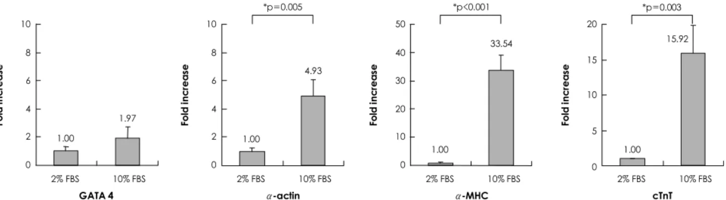
of cardiac differentiation (Fig. 1A). To induce cardiac differentiation, they were cultured in a suspension in bacterial dishes for 96 hours in DMEM+10% FBS containing 1% DMSO to form EBs (Fig. 1B); the EBs were further cultured for 10 or 15 consecutive days in DMEM+10% FBS. Expression of GATA4, α-actin, α-MHC, and cTnT mRNA cardiac-specific markers was -1.97, -4.93, -33.54, and -15.92-fold higher, res- pectively, in 20-day cultures of P19 cells than 15-day cultures of P19 cells (Fig. 2). This result shows that car- diomyogenic differentiation of P19 cells increases as the culture time is extended.
Effects of serum concentration on cardiomyoge- nic differentiation of the P19 cells
We also analyzed the effect of serum concentrations on cardiomyogenic differentiation of the P19 cells by quantitative real-time PCR assay. The P19 cells were cultured in suspension in bacterial dishes for 96 hours
Fold increase
10 8 6 4 2 0
1.00
1.97
2% FBS 10% FBS
Fold increase
10 8 6 4 2
0
2% FBS 10% FBS
Fold increase
50 40 30 20 10 0
1.00 2% FBS 10% FBS
Fold increase
20 15 10 5
0
2% FBS 10% FBS 1.00
4.93
15.92
*p=0.005
1.00
33.54
*p<0.001 *p=0.003
GATA 4 α-actin α-MHC cTnT
Fig. 2. Effects of culture period on cardiomyogenic differentiation of P19 cells. To form EBs, P19 cells were plated at a density of 1×10⁶ cells on 10-cm bacterial dishes for 96 hours in the presence of 1% DMSO. The formed EBs were transferred onto 24-well plates, and cultured in DMEM+10% FBS for an additional 10 or 15 consecutive days. Real-time PCR was carried out using total RNAs isolated from the P19 cells on days 10 and 15 of differentiation with primers for GATA4, α-actin, α-MHC, and cTnT. The relative gene expression le- vels were quantified based on the threshold cycle, and normalized to the reference gene GAPDH. α-actin: alpha-cardiac muscle actin, α-MHC: alpha-cardiac myosin heavy chain, cTnT: cardiac muscle troponin T, DMEM: Dulbecco’s modified Eagle’s medium, DMSO:
dimethyl sulfoxide, EB: embryoid body, FBS: fetal bovine serum, RNA: ribonucleic acid.
Fig. 1. DMSO-induced EB formation from P19 cells. P19 cells were plated at a density of 1×10⁶cells on 10-cm bacterial dishes for 96 hours in the presence of 1% DMSO to form EBs that contained cells differentiated into cardiomyogenic lineages. A: undifferentiated P19 cells were maintained in a monolayer. B: the formed EBs were examined under an inverted microscope at 96 hours for cardiac dif- ferentiation. Scale bars=100 μm. DMSO: dimethyl sulfoxide, EB: embryoid body.
A B
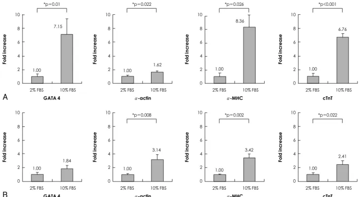
in DMEM+10% FBS containing 1% DMSO to form EBs; the EBs were further cultured for 10 or 15 conse- cutive days in DMEM containing 2% or 10% FBS. The expression of GATA4, α-actin, α-MHC, and cTnT mRNA in the EBs cultured in DMEM containing 10%
FBS were -7.15, -1.62, -8.35, and -6.75-fold higher, re- spectively, than the EBs cultured in DMEM containing 2% FBS for 10 consecutive days after EB formation (Fig. 3A). Furthermore, the expression of GATA4, α- actin, α-MHC, and cTnT mRNA in the EBs cultured in DMEM containing 10% FBS were also -1.84, -3.14, -3.42, and -2.41-fold higher, respectively, than the EBs cultured in DMEM containing 2% FBS for 15 con- secutive days after EB formation (Fig. 3B). This result demonstrates that cardiomyogenic differentiation of the P19 cells was enhanced under high-serum culture conditions (10% FBS) compared to low-serum culture conditions (2% FBS), regardless of the culture time.
Effects of 5-azacytidine on cardiomyogenic differ- entiation of the P19 cells under different serum concentrations
Several studies have reported that 5-azacytidine, a DNA hypomethylating agent, stimulates the cardiac dif- ferentiation of stem cells isolated from various tissues, such as mesenchymal stem cells,11) human embryonic
cells,12) cardiac stem cells,13) and P19 cells.9) Based on pre- vious reports, we investigated the effects of 5-azacytidine on cardiomyogenic differentiation of the P19 cells by qu- antitative real-time PCR assay. The P19 cells were cultured in suspension in bacterial dishes for 96 hours in DM- EM+10% FBS containing 1% DMSO to form EBs;
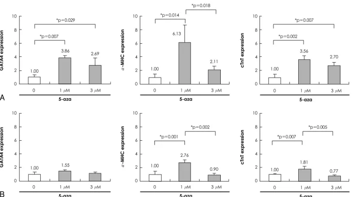
the EBs were further cultured for 10 consecutive days in DMEM containing 2% (Fig. 4A) or 10% FBS (Fig.
4B). The expression of cardiac-specific markers, such as GATA4, α-MHC, and cTnT mRNA was highest in the cells treated with 1 μM 5-azacytidine, regardless of the serum concentrations (Fig. 4). In addition, the cells treated with 3 μM 5-azacytidine showed a statistically significant increase in the expression of cardiac-specific markers compared to the control cells (Fig. 4). This re- sult demonstrates that cardiomyogenic differentiation of the P19 cells was the most effective with 1 μM of 5- azacytidine, regardless of the serum concentrations. In addition, stimulation by 5-azacytidine for cardiomyo- genic differentiation was more distinct under low-se- rum culture conditions (2% FBS) compared to high- serum culture conditions (10% FBS).
To further confirm the cardiomyogenic differentia- tion of the P19 cells at the protein level, we performed immunostaining with antibodies against cardiac-spe- cific markers, including sarcomeric α-actinin, cardiac
Fig. 3. Effects of serum concentration on cardiomyogenic differentiation of P19 cells. To form EBs, the P19 cells were plated at a density of 1×10⁶ cells on 10-cm bacterial dishes for 96 hours in the presence of 1% DMSO. The formed EBs were transferred into 24-well plates, and cultured in DMEM with 2% or 10% FBS for an additional 10 or 15 consecutive days. Real-time PCR was carried out using total RNAs isolated from the P19 cells cultured in DMEM with 2% or 10% FBS on day 10 (A) and day 15 (B) of differentiation with pri- mers for GATA4, α-actin, α-MHC, and cTnT. The relative gene expression levels were quantified based on the threshold cycle (Ct), and normalized to the reference gene GAPDH. α-actin: alpha-cardiac muscle actin, α-MHC: alpha-cardiac myosin heavy chain, cTnT: car- diac muscle troponin T, DMEM: Dulbecco’s modified Eagle’s medium, DMSO: dimethyl sulfoxide, EB: embryoid body, FBS: fetal bovine serum, PCR: polymerase chain reaction, RNA: ribonucleic acid.
Fold increase
10 8 6 4 2 0
1.00 7.15
2% FBS 10% FBS
Fold increase
10 8 6 4 2
0
2% FBS 10% FBS
Fold increase
10 8 6 4 2 0
1.00
2% FBS 10% FBS
Fold increase
10 8 6 4 2
0
2% FBS 10% FBS 1.00
1.62
6.76
*p=0.01
1.00 8.36
GATA 4 α-actin α-MHC cTnT
*p=0.026 *p<0.001
*p=0.022
Fold increase
10 8 6 4 2 0
1.00
1.84
2% FBS 10% FBS
Fold increase
10 8 6 4 2
0
2% FBS 10% FBS
Fold increase
10 8 6 4 2 0
1.00
2% FBS 10% FBS
Fold increase
10 8 6 4 2
0
2% FBS 10% FBS 1.00
2.41 1.00
3.42
GATA 4 α-actin α-MHC cTnT
*p=0.002 *p=0.022
*p=0.008
3.14
A
B
myosin heavy chain, cardiac myosin light chain, and cTnT after induction of cardiomyogenic differentiation of the P19 cells. The P19 cells were plated at a density of 1×10⁶ cells on 10-cm bacterial dishes for 96 hours in the presence of 1% DMSO. The formed EBs were trans- ferred onto 0.1% gelatin-coated coverslips in 12-well dishes, and cultured in DMEM with 10% FBS for an additional 10 consecutive days in the presence of 1 μM 5-azacytidine. A number of cells showing specific stain- ing patterns for cardiac-specific markers were observed in the cardiomyocytes differentiated from the P19 cells (Fig. 5).
Discussion
Cardiomyocyte differentiation of stem cells in vitro is known to be affected by various environmental factors, such as the incubation period, medium composition, serum components, growth factors, and chemicals. Th- erefore, in this study, we investigated the effects of different concentrations of serum and 5-azacytidine, and culture time on the cardiomyogenic differentia- tion of P19 cells. We showed that cardiomyogenic dif- ferentiation of the P19 cells was enhanced in parallel with the length of the culture period. This result was
in agreement with our previous findings that showed that the expression of cardiac-specific markers, such as GATA4, Nkx2.5, and cTnT after treatment with 1 μM 5-azacytidine in a P19 cell monolayer culture was up- regulated in a time-dependent manner.9)
In the present study, we compared the effects of se- rum concentrations on cardiac differentiation of the P19 cells. We showed that cardiomyogenic differen- tiation of the P19 cells was enhanced by high FBS (10%)- containing medium compared to low FBS (2%)-con- taining medium, regardless of the culture time period.
Investigation of the stimulatory or inhibitory effects of FBS concentration on the cardiac differentiation of stem cells has been previously reported. Medium containing 20% FBS has been shown to promote more rapid down- regulation of the pluripotency marker, Oct 4, and in- creased expression of endodermal and mesodermal genes at the time of EB formation of H1 human embryonic stem cells (ESCs), compared to medium with 20%
KnockOut serum replacement, in which spontaneous- ly-beating cardiomyocytes were only observed in the FBS-treated group.14) Moreover, bone morphogenetic protein 4 (BMP-4) was not shown to induce cardiomyo- cyte differentiation in mouse ESCs with serum-free models; at least a small amount of FBS in the hanging
Fig. 4. Effects of 5-azacytidine on cardiomyogenic differentiation of P19 cells under different serum concentrations. To form EBs, P19 cells were plated at a density of 1×10⁶ cells on 10-cm bacterial dishes for 96 hours in the presence of 1% DMSO. The formed EBs were transferred into 24-well plates, and cultured in DMEM with 2% or 10% FBS for an additional 10 consecutive days in the presence of 0, 1, and 3 μM 5-azacytidine. Real-time PCR was carried out using total RNAs isolated from the P19 cells cultured in DMEM with 2% FBS (A) or 10% FBS (B) in the presence of 0, 1 and 3 μM of 5-azacytidine on day 10 of differentiation with primers for GATA4, α-MHC, and cTnT. The relative gene expression levels were quantified based on the threshold cycle (Ct), and normalized to the reference gene GAPDH. α-actin: alpha-cardiac muscle actin, α-MHC: alpha-cardiac myosin heavy chain, cTnT: cardiac muscle troponin T, DMEM:
Dulbecco’s modified Eagle’s medium, DMSO: dimethyl sulfoxide, EB: embryoid body, FBS: fetal bovine serum, PCR: polymerase chain reaction, RNA: ribonucleic acid.
*p=0.018
GATA4 expression
10 8 6 4 2
0
0 1 μM 3 μM 5-aza
1.00
2.69
*p=0.002
A
3.86
*p=0.007
α-MHC expression
10 8 6 4 2
0
0 1 μM 3 μM 5-aza
1.00
2.11
*p=0.014
6.13
cTnT expression
10 8 6 4 2
0
0 1 μM 3 μM 5-aza
1.00
2.70 3.56
*p=0.007
*p=0.029
*p=0.002
GATA4 expression
10 8 6 4 2
0
0 1 μM 3 μM 5-aza
1.00
B
1.55 α-MHC expression 10
8 6 4 2
0
0 1 μM 3 μM 5-aza
1.00
0.90
*p=0.001
2.76
cTnT expression
10 8 6 4 2
0
0 1 μM 3 μM 5-aza
1.00 0.77
1.81
*p=0.005
*p=0.007
drop stage is necessary. These findings suggest that se- rum factors are not critical after the initial activation, but do enhance the differentiation of cardiomyocytes.15) By contrast, the number of beating areas in the co-cul- ture model with END-2 cells was increased 24-fold in the absence of fetal calf serum (FCS) compared to the cells in the presence of 20% FCS.16) These results sug- gest that pro- and anti-cardiogenic factors may be pre- sent in the serum. The stimulatory or inhibitory effects of the serum concentration on cardiac differentiation may be different in various stem cell lines and culture conditions.
Cardiomyogenic differentiation of P19 cells has gen- erally been induced with the combination of EB for- mation and DMSO in bacterial dishes.7) However, we previously reported that 5-azacytidine induced cardio- myogenic differentiation of P19 cells in confluent mo- nolayer cultures without prior EB formation and DM- SO exposure via, in part, activation of BMP signaling molecules.9) In this context, we investigated whether 5- azacytidine could enhance cardiomyogenic differenti- ation of P19 cells at the stage of preformed EB in the
presence of DMSO in bacterial dishes. Our results showed that 1 μM 5-azacytidine-treated cells had the most enhanced cardiomyogenic differentiation of P19 cells, both under low-serum culture conditions (2% FBS) and high-serum culture conditions (10% FBS), com- pared to the 5-azacytidine non-treated control group and the 3 μM 5-azacytidine-treated group. This ob- servation is consistent with the findings of our previous report9) that P19 cells differentiated into the cardio- myogenic cell lineage in response to 1 μM 5-azacytidine in a confluent monolayer culture. Various concentra- tions of 5-azacytidine with different treatment periods have been used to induce cardiomyogenic differentia- tion of mesenchymal stem cells derived from different species. Murine bone marrow stromal cells were treated with 3 μM 5-azacytidine for 24 hours, and sponta- neously-beating cells were observed after 2 weeks.11) Me- senchymal stem cells isolated from adult human bone marrow were reported to differentiate to a cardiomyo- genic lineage in vitro after treatment with 10 μM 5- azacytidine for 24 hours.17) Adult mesenchymal stem cells isolated from the fatty tissue of New Zealand White
Sarcomeric α-actinin Myosin Heavy Chain Myosin Light Chain Cardiac Troponin T
DAPI
Merge
DAPI DAPI DAPI
Merge Merge Merge
Fig. 5. Immunostaining of P19 cell-derived cardiomyocytes after 5-azacytidine treatment. To form EBs, the P19 cells were plated at a density of 1×10⁶ cells on 10-cm bacterial dishes for 96 hours in the presence of 1% DMSO. The formed EBs were transferred onto 0.1%
gelatin-coated coverslips in 12-well dishes, and cultured in DMEM with 10% FBS for an additional 10 consecutive days in the presence of 1 μM 5-azacytidine. The cells were stained with sarcomeric α-actinin, cardiac myosin heavy chain, cardiac myosin light chain, and cardiac troponin T as indicated. DAPI was used for staining the nuclei. Scale bars, 100 μm. DAPI: 4’6’-diamidino-2-phenylindole, DMEM: Dulbecco’s modified Eagle’s medium, DMSO: dimethyl sulfoxide, EB: embryoid body, FBS: fetal bovine serum.
rabbits were treated with 1, 3, 6, 9, and 12 μM 5-azacy- tidine and incubated for 12, 24, 48, and 72 hours.18) Taken together, our results and previous reports suggest that the optimal concentration and treatment time with 5-azacytidine for the induction of cardiomyogenic differentiation appears to depend on both the efficacy of cardiac differentiation and the cytotoxicity of the stem cells.
In conclusion, the results of this study demonstrated that cardiomyogenic differentiation of P19 cells could be enhanced by a combination of different experimen- tal factors, including the concentration of the serum and 5-azacytidine, as well as the culture time.
Acknowledgments
This research was supported by a grant (SC-4220) from the Stem Cell Research Center of the 21st Century Frontier Research Pro- gram, funded by the Ministry of Education, Science and Technology, Republic of Korea.
REFERENCES
1) Orlic D, Kajstura J, Chimenti S, et al. Bone marrow cells regen- erate infarcted myocardium. Nature 2001;410:701-5.
2) Kajstura J, Rota M, Whang B, et al. Bone marrow cells differ- entiate in cardiac cell lineages after infarction independently of cell fusion. Circ Res 2005;96:127-37.
3) Rota M, Kajstura J, Hosoda T, et al. Bone marrow cells adopt the cardiomyogenic fate in vivo. Proc Natl Acad Sci USA 2007;
104:17783-8.
4) Kim YS, Ahn Y, Hong MH, et al. Therapeutic potential of umbi- lical cord blood-derived mesenchymal stem cells in ischemic myocardium. Korean Circ J 2008;38:446-54.
5) Kinnaird T, Stabile E, Burnett MS, et al. Local delivery of mar- row-derived stromal cells augments collateral perfusion through paracrine mechanisms. Circulation 2004;109:1543-9.
6) Nygren JM, Jovinge S, Breitbach M, et al. Bone marrow-derived hematopoietic cells generate cardiomyocytes at a low frequency
through cell fusion, but not transdifferentiation. Nat Med 2004;
10:494-501.
7) McBurney MW, Jones-Villeneuve EM, Edwards MK, Anderson PJ. Control of muscle and neuronal differentiation in a cultured embryonal carcinoma cell line. Nature 1982;299:165-7.
8) van der Heyden MA, Defize LH. Twenty one years of P19 cells:
what an embryonal carcinoma cell line taught us about cardio- myocyte differentiation. Cardiovasc Res 2003;58:292-302.
9) Choi SC, Yoon J, Shim WJ, Ro YM, Lim DS. 5-azacytidine in- duces cardiac differentiation of P19 embryonic stem cells. Exp Mol Med 2004;36:515-23.
10) McBurney MW. P19 embryonal carcinoma cells. Int J Dev Biol 1993;37:135-40.
11) Makino S, Fukuda K, Miyoshi S, et al. Cardiomyocytes can be generated from marrow stromal cells in vitro. J Clin Invest 1999;
103:697-705.
12) Xu C, Police S, Rao N, Carpenter MK. Characterization and enrichment of cardiomyocytes derived from human embryonic stem cells. Circ Res 2002;91:501-8.
13) Oh H, Bradfute SB, Gallardo TD, et al. Cardiac progenitor cells from adult myocardium: homing, differentiation, and fusion after infarction. Proc Natl Acad Sci USA 2003;100:12313-8.
14) Bettiol E, Sartiani L, Chicha L, Krause KH, Cerbai E, Jaconi ME.
Fetal bovine serum enables cardiac differentiation of human em- bryonic stem cells. Differentiation 2007;75:669-81.
15) Taha MF, Valojerdi MR. Effect of bone morphogenetic protein-4 on cardiac differentiation from mouse embryonic stem cells in serum-free and low-serum media. Int J Cardiol 2008;127:78-87.
16) Passier R, Oostwaard DW, Snapper J, et al. Increased cardiomyo- cyte differentiation from human embryonic stem cells in serum- free cultures. Stem Cells 2005;23:772-80.
17) Antonitsis P, Ioannidou-Papagiannaki E, Kaidoglou A, Papakon- stantinou C. In vitro cardiomyogenic differentiation of adult hu- man bone marrow mesenchymal stem cells: the role of 5-azacy- tidine. Interact Cardiovasc Thorac Surg 2007;6:593-7.
18) Rangappa S, Fen C, Lee EH, Bongso A, Sim EK. Transforma- tion of adult mesenchymal stem cells isolated from the fatty tis- sue into cardiomyocytes. Ann Thorac Surg 2003;75:775-9.