713 Original Article
Korean Circulation J 2006;36:713-715
ISSN 1738-5520
ⓒ 2006, The Korean Society of Circulation CASE REPORT
Acrylic Cement Foreign Body and Thrombus in Right Atrium Causing Pulmonary Embolism after Percutaneous Vertebroplasty
Sang Eun Lee, MD1, Sung-A Chang, MD1, Min-Seok Kim, MD1, Song-Yi Kim, MD1, Jung-Kyu Han, MD1, Ho-Jun Jang, MD1, Yong-Jin Kim, MD1,
Dae-Won Sohn, MD1, Byung-Hee Oh, MD1 and Kyung-Hwan Kim, MD2
1Division of Cardiology, Department of Internal Medicine and 2Thoracic and Cardiovascular Surgery, Seoul National University College of Medicine, Seoul, Korea
ABSTRACT
A pulmonary embolism is a rare, but well described complication of percutaneous vertebroplasty; the majority of cases are caused by acrylic cement. Here, for the first time, we report a case of pulmonary embolism due to a th- rombus in the right atrium, which was caused by an acrylic cement foreign body in the right atrium and central veins 6 years after percutaneous vertebroplasty. This case suggests that an acrylic cement foreign body should be con- sidered as a potential source of thrombus formation in patients that develop a pulmonary embolism following per- cutaneous vertebroplasty. (Korean Circulation J 2006;36:713-715)
KEY WORDS:Embolism, pulmonary;Foreign bodies;Radiography, interventional;Embolism and thrombus.
Introduction
A pulmonary embolism caused by acrylic cement is a rare, but well described complication of percutaneous vertebroplasty. However, acrylic cement has never been reported as the source of thrombus formation leading to a pulmonary embolism. Here, we report a case of acrylic cement foreign body, with thrombus formation in the central veins and right atrium, causing a pulmonary em- bolism.
Case
A 55-year-old woman, with a history of percutaneous vertebroplasty for a compression fracture of the second lumbar vertebra(L2), preformed 6 years previously, pre- sented with mild dyspnea and general weakness of 2 months duration. She denied previous central venous ca- theterization or other instrumentation intervention of the central veins. Her initial chest radiograph revealed a long linear catheter-like foreign body and a fractured L2 vertebral body filled with bone cement(Fig. 1A). Her
transthoracic echocardiogram(Fig. 1B, C) and transe- sophageal echocardiogram(Fig. 1D) showed a catheter- like foreign body in the hepatic vein(HV), inferior vena cava(IVC) and right atrium(RA), with a large attached thrombus. CT angiography confirmed the presence of a 4×2 cm lobulated-mass attached to the RA wall, ob- literating the tricuspid valve(TV)(Fig. 2A), two sepa- rated catheter-like foreign bodies in the HV, IVC and RA(Fig. 2B, D), a pulmonary thromboembolus in the left lower pulmonary artery(Fig. 2E) and bone cement within the lumbar veins of the L2(Fig. 2F), suggesting the foreign bodies were caused by perivertebral cement leakage. Finally, a lung perfusion scan confirmed the pre- sence of a pulmonary embolism due to perfusion de- crease in the left lower lobe. The thrombus in the RA (Fig. 3A) and the foreign bodies in the central veins and RA(Fig. 3B) were removed during open-heart sur- gery, with a short period of total circulatory arrest under moderate hypothermia; the foreign bodies were identi- fied as acrylic bone cement(Fig. 3C).
Discussion
Since 1984, when percutaneous vertebroplasty was first described for the treatment of vertebral angiomata,1) its use for the treatment of pain associated with verte- bral body fractures has increased. Typically, approxima- tely 2 to 10 mL of a viscous mixture of polymethylme- thacrylate(PMMA) and opacifying agent, such as barium,
Received:June 23, 2006 Accepted:August 16, 2006
Correspondence:Kyung-Hwan Kim, MD, Department of Thoracic and Car- diovascular Surgery, Seoul National University College of Medicine, 28 Yon- gon-dong, Chongno-gu, Seoul 110-744, Korea
Tel: 82-2-2072-3971, Fax: 82-2-765-7117 E-mail: [email protected]
714·Korean Circulation J 2006;36:713-715
is injected into the affected vertebral body using 10 to 15 gauge needles. The procedure is usually visualized by fluoroscopy or CT. Moreover, although leakage of PMMA into the paraspinal veins is observed in up to 39% of patients that undergo vertebroplasty for osteo- porotic compression fractures,2) the incidence of pulmo-
nary embolisms in vertebroplasty patients due to acrylic cement has been reported to be within the range of 13) to 4.6%.4) On imaging studies, such emboli vary in shape from amorphous multidimensional masses to long cathe- ter-like shapes with radio-opaque properties.5)6) However, in the present case, the pulmonary embolus showed ra-
Fig. 1. Initial chest radiograph and echocardiogram. A: chest radiograph showing a long linear catheter-like foreign body (arrows) and fractured L2 vertebral body filled with bone cement (arrow heads). B: transthoracic echocardiogram subcostal view showing a catheter like foreign body (arrows) in the HV, IVC and RA, with a large thrombus in the RA (*). C: transthoracic echocardiogram parasternal short axis view showing an echogenic foreign body (arrows) and large thrombus in the RA (*). D: transesophageal echocardiogram showing a large thrombus (*) in the RA attached to an echogenic catheter-like foreign body (arrows). L2: second lumbar vertebra, HV: hepatic vein, IVC: inferior vena cava, RA: right atrium.
A B
C D
A B C
F E
D
Fig. 2. CT angiography. A: a 4×2 cm lobulated mass attached to the RA wall and obliterating the tricuspid valve (TV). Two separate catheter-like foreign bodies observed in the RA (B), IVC (C) and HV (D). E: a pulmonary thromboembolus in the left lower pulmonary artery. F: three di- mensional reconstruction of a CT angiograph, showing bone cement within the lumbar veins of the L2, suggestive of perivertebral cement leakage (arrows). RA: right atrium, IVC: inferior vena cava, HV: hepatic vein, L2: second lumbar vertebra, CT: computed tomography.
Sang Eun Lee, et al:Acrylic Cement Foreign Body and Pulmonary Thromboembolism after Percutaneous Vertebroplasty·715
dio-lucent properties. Also, a large thrombus in the RA was found to be the source of the embolus, with a ce- ment foreign body attached. Unlike other reported cases of pulmonary embolisms following percutaneous verte- broplasty, in this case the acrylic cement itself was not responsible, but a thrombus formed by the cement had caused the pulmonary embolism. Therefore, in cases of a pulmonary embolism following percutaneous vertebro- plasty, acrylic cement foreign bodies should be viewed, not simply as an embolus, but also as a potential source of a thrombus.
The choice between operative removal and expectant management of pulmonary arterial foreign bodies is con- troversial, with considerable disagreement concerning the optimal treatment strategy reported in the litera- ture. The majority of pulmonary embolisms due to ac- rylic cement are treated acutely with supportive therapy, either with or without anticoagulation, which results in satisfactory outcomes.7) Therefore, some authors have suggested that it is acceptable to just observe foreign bo- dies within the heart or pulmonary arteries, including acrylic pulmonary emboli.7-9) However, in the present case, aggressive intervention, i.e., open heart surgery, to remove the thrombus and foreign body were reasonable since the formed thrombus was large enough to caused recurrent embolic events. Moreover, the present case de- monstrates that asymptomatic cement foreign bodies can cause problems, even 6 years after the procedure; and thus, this report suggests it is reasonable to follow up foreign bodies much more cautiously or adopt inter- vention at an earlier stage. Although the patient in this report underwent open heart surgery due to a large th- rombus in the right atrium, percutaneous foreign body retrieval, using a snare device10) could be attempted in uncomplicated cases.
In the described case, a pulmonary embolism occurred 6 years after percutaneous vertebroplasty due to a th-
rombus in the RA caused by an acrylic cement foreign body. We suggest that an acrylic cement foreign body should be actively considered as a source of thrombus formation in patients with a pulmonary embolism follo- wing percutaneous vertebroplasty.
REFERENCES
1) Deramond H, Depriester C, Galibert P, le Gars D. Percutaneous vertebroplasty with polymethylmethacrylate: technique, indica- tions, and results. Radiol Clin North Am 1998;36:533-46.
2) Yeom JS, Kim WJ, Choy WS, Lee CK, Chang BS, Kang JW. Lea- kage of cement in percutaneous transpedicular vertebroplasty for painful osteoporotic compression fractures. J Bone Joint Surg Br 2003;85:83-9.
3) McGraw JK, Cardella J, Barr JD, et al. Society of Interventional Radiology quality improvement guidelines for percutaneous ver- tebroplasty. J Vasc Interv Radiol 2003;14:827-31.
4) Choe du H, Marom EM, Ahrar K, Truong MT, Madewell JE. Pul- monary embolism of polymethylmethacrylate during percutaneous vertebroplasty and kyphoplasty. AJR Am J Roentgenol 2004;183:
1097-102.
5) Tzzi P, Abdelmoumene Y, Corno AF, Gersbach PA, Hoogewoud HM, von Segesser LK. Management of pulmonary embolism du- ring acrylic vertebroplasty. Ann Thorac Surg 2002;74:1706-8.
6) Yoo KY, Jeong SW, Yoon W, Lee J. Acute respiratory distress syndrome associated with pulmonary cement embolism following percutaneous vertebroplasty with polymethylmethacrylate. Spine 2004;29:E294-7.
7) MacTaggart JN, Pipinos II, Johanning JM, Lynch TG. Acrylic cement pulmonary embolus masquerading as an embolized central venous catheter fragment. J Vasc Surg 2006;43:180-3.
8) Actis Dato GM, Arslanian A, di Marzio P, Filosso PL, Ruffini E.
Posttraumatic and iatrogenic foreign bodies in the heart: report of fourteen cases and review of the literature. J Thorac Cardiovasc Surg 2003;126:408-14.
9) Reynen K. 14-year follow-up of central embolization by a guide- wire. N Engl J Med 1993;329:970-1.
10) Cho SY, Park SJ, Cho CH, Chung NS, Shim WH, Lee WK. Non- surgical percutaneous retrieval of catheter emboli from the heart.
Korean Circ J 1987;17:131-7.
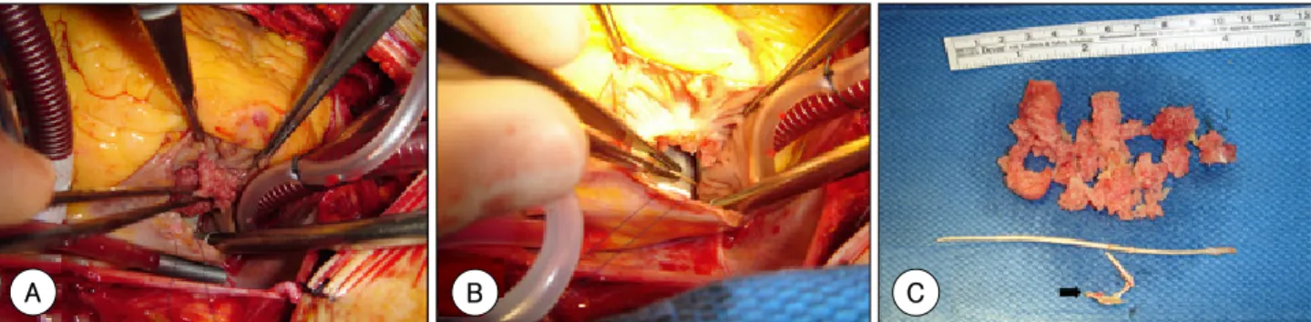
Fig. 3. Open heart surgery and gross specimens. The thrombus (A) and acrylic cement foreign bodies (B) removed by right atriotomy. C: a large thrombus and two catheter-like acrylic cement foreign bodies were finally removed. The smaller catheter-like foreign body was attached to the right atrium and constituted the nidus of the thrombus (arrow).
A B C