277
책임저자:장일성, 대전시 중구 대사동 640
301-721, 충남대학교병원 외과 Tel: 042-280-7179, Fax: 042-257-8024 E-mail: [email protected]
접수일: 2007년 3월 21일, 게재승인일:2007년 7월 4일
This work was supported by a grant from the National R&D Program for Cancer Control Ministry of Health & Welfare, Republic of Korea (No: 0720560).
유방암 환자에서 종양억제유전자 DNA 메틸화의 특성
충남대학교 의과대학 외과학교실, 의학연구소, 1지노믹트리, 2대전 지역암센터
이진선ㆍ오태정lㆍ김제룡ㆍ이증훈2ㆍ장일성
Promoter Methylation Profiles and Its Associa- tion with Clinicopathological Features in Breast Cancer
Jin Sun Lee, Tae Jung Oh1, Je Ryong Kim, Jeung Hoon Lee2 and Eil Sung Chang
Purpose: Aberrant DNA methylation of tumor suppressor genes has been accepted as a common feature and early event in human cancer. The aim of this study was to analyze the methylation profiles of 50 well established methyl- ation-associated genes in relation to various clinico-patho- logical features in breast cancer.
Methods: The methylation status of 50 genes were deter- mined in two breast cancer cell lines, MCF7 and MDA- MB231, using HpaII-MspI-PCR. 8 genes (APC, CALCA, CDH13, MTHFR, S100A2, H19, EDNRB and MUC2) were found to be methylated in at least 1 cell line. The methylation of all 8 genes was observed in tumor tissues, but with different methylation frequencies.
Results: The methylation frequencies of five genes in breast cancer were as follows: MTHFR (41.9%), APC (51.6%), EDNRB (77.4%), CALCA (80.6%), S100A2 (87.1%), CDH13 (93.5%), H19 (93.5%) and MUC2 (96.8%). The results in- dicate that a panel of these 8 genes would be useful in the detection of breast cancer. The prognostic significance of DNA methylation in this breast cancer series, the conven- tional markers of LN status (P=0.05), histologic grade (P=
0.007) and P53 gene status (P=0.049) showed significant prognostic value.
Conclusion: The methylation of APC, MTHFR, CALCA, CDH13, H19, MUC2, EDNRB and S00A2 would be useful in the detection of breast cancer. Detection of these abnor- malities may be useful in the risk assessment and early de-
tection of breast cancer. (J Korean Surg Soc 2007;73:277- 284)
Key Words: Methylation, Breast cancer, Tumor suppressor gene
중심단어: 메틸화, 유방암, 종양억제유전자
Department of Surgery, Research Institute for Medical Sciences, College of Medicine, Chungnam National Univer- sity, 1Research and Development Center of Genomictree,
2Daejeon Regional Cancer Center, Daejeon, Korea
서 론
유방암은 전 세계적으로 매년 40만 명의 사망률을 보여 전 세계 여성암 관련 사망의 가장 큰 원인 암 중의 하나이 다.(1) 다른 여러 종양과 마찬가지로 유방암도 종양유전자 (oncogene)의 활성화와 종양억제유전자(tumour suppressor gene)의 불활성화로 인해 발생하는 것으로 여겨지는데, 최 근 많은 연구에서 종양억제유전자의 메틸레이션 의존적 전 사억제(methylation induced transcriptional silencing)와 같은 후생유전학 기전(epigenetic mechanism)으로 이러한 유전자 의 불활성화가 발생한다는 것이 밝혀졌다. 프로모터 염기 서열 부위의 CpG dinucleotides-rich 구역은 중요한 성장조절 유전자의 전사억제(silencing)와 관련된 구역으로 여겨진 다.(2) 메틸화된 표적 유전자의 발견은 유방암의 분자생물 학적 발병기전을 밝히는 데 큰 도움이 될 것이며, 종양 특이 DNA 메틸화에 대한 연구는 유방암의 조기진단에 있어 임 상적 가치를 가질 수 있다. DNA 과메틸화 양식의 연구에서 가장 중요한 점은 치료에 대한 예후와 반응을 예측할 수 있는 특별한 생물학적 특성을 갖는 소집단을 찾아내는 것 이다. 유방암에서 유전자의 메틸화는 서로 다른 조직임상 적 특성을 가지는 표현형을 구분할 수 있을 것이라는 여러 연구들이 발표되고 있다.(8,9,17) 본 연구의 목표는 유방암 에서 유전자의 과메틸화와 주요 임상병리학적 인자의 특성 과의 관련성을 알아보는 것이다. 저자는 enzyme digestion (HpaII/MspI)-PCR assay를 이용하여 유방암조직의 종양억제 유전자 DNA 메틸화 특성을 연구하였다.
Table 1. Clinicopathologic features (n=31) Stage
I 7 (22.6%)
II 21 (67.8%)
III 3 (9.6%)
Median age (mean, years) 50.3 (28∼73) Median tumor size (mean, cm) 3.29 (0.4∼22) Histologic grade
Grade 1 4 (12.9%)
Grade 2 10 (32.3%)
Grade 3 17 (54.8%)
Mean no.of LN metastasis 1.29 (0∼14)
ER (positive, negative)% 14 (45.2%), 17 (54.8%) PR (positive, negative)% 8 (25.8%), 23 (74.2%) p53 (positive, negative)% 20 (64.5%), 11 (35.5%) c-erbB-2overexpression% 14 (45.2%)
ER = estrogen receptor; PR = progesterone receptor.
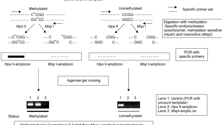
Fig. 1. Methylation assay by enzyme digestion (HpaII/MspI)-PCR.
방 법
1) 유방암 조직표본
2006년 2월부터 10월까지 충남대학교병원에서 유방암으 로 진단 후 유방전절제술을 받은 환자 31명을 대상으로 수
술 중 종양에서 얻은 조직을 이용하여 메틸화 상태를 평가 하였다. 대상환자의 평균나이는 50.3세였고, 병기는 2기가 21명(67.8%)으로 가장 많았으며, 조직학적 등급은 3등급이 17명(54.8%)으로 가장 많았다(Table 1).
2) Enzyme digestion (Hpa II/Msp I)-PCR
유방암에서 메틸화 특성을 알아 보기 위한 예비연구로 50 종의 다양한 암에서 메틸화가 보고된 유전자들의 유방암 세포주에서 메틸화 profile을 작성하였다. 2종의 유방암 세 포주(MCF7, UK, MDA-MB231)를 이용하여 메틸화 실험을 시 행하였다. 메틸화 실험은 Enzyme digestion (HpaII/MspI)- PCR 방법을 이용하였는데 이 방법은 HpaII 효소가 5’-CCGG-3’의 염기서열을 인지하여, 5’-CG-3’의 “C”가 메틸화되면 절단하 지 못하는 반면, MspI 효소는 HpaII의 isoschizomer로서 5’-CG-3’의 메틸화 여부에 상관없이 절단하는 특성을 이용 하는 것이다. MethPrimer를 이용하여 promoter염기서열 내 에 최소 1개 이상의 HpaII site를 포함하는 PCR target을 설 계하였다. Genomic DNA를 이들 두 가지 효소로 절단한 후, 각 절단물을 유전자 특이적인 PCR primer로 증폭한 후 전기 영동을 실시하면, 메틸화되어 있는 경우에는 HpaII-digested amplicon에서 PCR 산물이 만들어지지만, 메틸화되지 않았 을 경우에는 PCR 산물이 만들어지지 않는다. PCR에 대한 적합성을 확인하기 위하여 HpaII site가 없는 IFN2유전자에
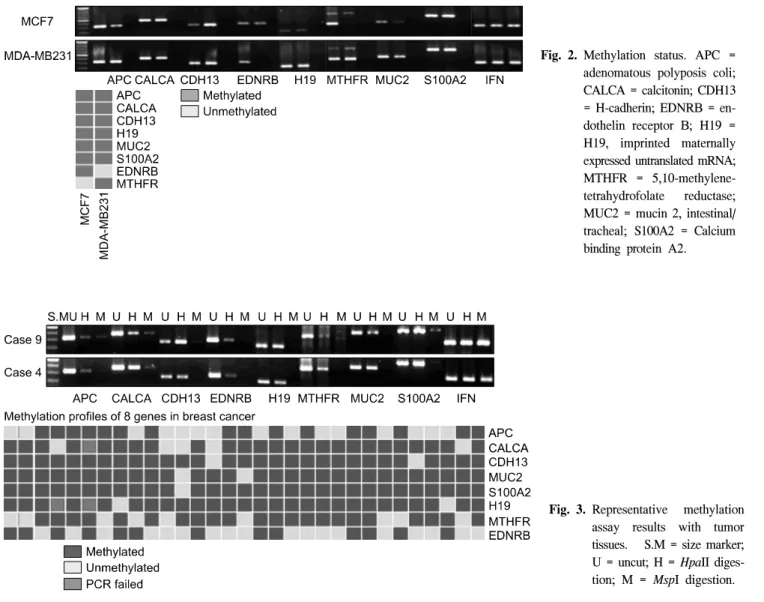
Fig. 3. Representative methylation assay results with tumor tissues. S.M = size marker;
U = uncut; H = HpaII diges- tion; M = MspI digestion.
Fig. 2. Methylation status. APC = adenomatous polyposis coli;
CALCA = calcitonin; CDH13
= H-cadherin; EDNRB = en- dothelin receptor B; H19 = H19, imprinted maternally expressed untranslated mRNA;
MTHFR = 5,10-methylene- tetrahydrofolate reductase;
MUC2 = mucin 2, intestinal/
tracheal; S100A2 = Calcium binding protein A2.
대한 PCR target을 설계하여 대조군으로 이용하였다. HpaII- digested amplicon의 band intensity가 MspI-digested amplicon 보다 2배 이상일 경우에 메틸화된 것으로 판단하였다.(3) (Fig. 1)
3) 통계적 분석
대상환자에서 얻은 유방암조직 31예를 대상으로 8개의 유전자(APC, CALCA, CDH13, H19, MUC2, S100A2, EDNRB, MTHFR)의 메틸화 상태를 조사하였다. 대상환자의 종양억 제유전자의 메틸화 상태와 유방암의 여러 병리조직학적 인 자들과의 관련성을 알아보기 위하여 χ2-test를 사용했다.
각각의 세포수가 5 미만일 경우에는 Fisher’s exact test를 이 용하였다. 모든 P값은 양측검정을 시행했다. 독립표본 t-test 를 이용하여 환자의 연령, 종양크기, 호르몬 수용체상태와 메틸화/비메틸화 그룹을 비교하였다. 모든 분석은 window 용 SPSS 12.0 통계프로그램을 이용하였다.
결 과
1) 유방암 세포주에서 메틸화 profile 분석결과 총 50개의 유전자 중 8개의 유전자가 2종의 유방암 세포주 (MCF7, MDA-MB231)에서 메틸화되어 있음을 확인하였다.
유방암 세포주에서 메틸화가 관찰된 8종의 유전자(APC, MTHFR, CALCA, CDH13, H19, MUC2, EDNRB, S100A2) 중 에서 EDNRB와 MTHFR은 2종의 유방암 세포주 중 1종의 세포주에서만 메틸화되어 있고, 나머지 6종은 두 세포주 모 두에서 메틸화되었다(Fig. 2).
2) 8종의 유전자를 이용한 유방암 조직의 메틸화 검증 유방전절제술을 받은 유방암 환자 31명을 대상으로 수술 중 종양에서 얻은 조직을 이용하여 유방암 세포주(MCF7, MDA-MB231)에서 메틸화가 확인된 8종의 유전자의 메틸 화 상태를 평가하였다(Fig. 3). 31예의 종양조직에서 관찰된
Table 2. Associations between gene promoter methylation and clinicopathological features of breast cancer (n=31)
Feature (n) APC CALCA CDH13 H19 MUC2 S100A2 EDNRB MTHFR
Methylatied 15 (51.6%) 25 (80.6%) 29 (93.5%) 29 (93.5%) 30 (96.8%) 27 (87.1%) 24 (77.4%) 13 (41.9%) Umethylated 16 (48.4%) 5 (16.1%) 2 (6.5%) 2 (6.5%) 1 (3.2%) 2 (6.5%) 7 (22.6%) 18 (58.1%)
PCR failed 0 1 (3.2%) 0 0 0 2 (6.5%) 0 0
Age 0.210 0.546 0.962 0.157 0.325 0.998 0.166 0.607
<50 yrs 10 12 15 14 15 14 14 6
>50 yrs 6 13 14 15 15 13 10 7
N stage 0.282 0.349 0.276 0.267 0.577 0.05 0.971 0.794
N0 10 19 21 21 21 19 17 9
N1 4 5 6 6 6 6 5 3
N2 1 1 1 1 2 2 1 0
N3 1 0 1 1 1 0 1 1
Histologic grade 0.200 0.527 0.887 0.107 0.263 0.979 0.469 0.925
Well/mod diff. 9 11 13 12 13 12 10 6
Poorly diff. 7 14 16 17 17 15 14 7
Histologic type 0.739 0.884 0.430 0.007 0.060 0.485 0.551 0.415
Non-ductal 4 6 7 5 6 6 6 2
Ductal 12 19 22 24 24 21 18 11
Tumor size 0.779 0.700 0.350 0.499 0.112 0.535 0.976 0.856
<2 cm 5 7 9 8 8 8 7 4
>2 cm 11 18 20 21 22 19 17 9
ER status 0.576 0.527 0.185 0.107 0.263 0.979 0.469 0.049
Negative 8 14 15 17 17 15 14 6
Positive 8 11 14 12 13 12 10 7
PR status 0.916 0.779 0.389 0.419 0.085 0.450 0.849 0.592
Negative 12 18 21 22 23 19 18 9
Positive 4 7 8 7 7 8 6 4
c-erbB-2 gene 0.576 0.276 0.887 0.185 0.356 0.979 0.889 0.524
Negative 8 13 16 15 16 15 13 8
Positive 8 12 13 14 14 12 11 5
TP53 gene status 0.208 0.369 0.278 0.049 0.170 0.283 0.173 0.641
Negative 4 8 11 9 10 11 7 4
Positive 12 17 18 20 20 16 17 9
APC = adenomatous polyposis coli; CALCA = calcitonin; CDH13 = H-cadherin; EDNRB = endothelin receptor B; H19 = H19, imprinted maternally expressed untranslated mRNA; MTHFR = 5,10-methylene-tetrahydrofolate reductase; MUC2 = mucin 2, intestinal/tracheal;
S100A2 = Calcium binding protein A2; ER = estrogen receptor; PR=progesterone receptor.
8개 종양억제 유전자 메틸화의 빈도는 MTHFR 13예(41.9%), APC 16예(51.6%), EDNRB 24예(77.4%), CALCA 25예 (80.6%), S100A2 27예(87.1%), CDH13 29예(93.5%), H19 29 예(93.5%), MUC2 30예(96.8%)였다. 8개 유전자 모두가 메 틸화된 경우는 2예(6.5%), 14예(45.2%)는 7개, 8예(25.8%)는 6개, 4예(12.9%)는 5개, 2예(6.5%)는 4개, 1예(3.2%)는 3개의 유전자에서 메틸화가 관찰되었다. 더 많은 수의 유전자가 메틸화된 경우(6∼8군데)와 적은 수의 메틸화가 발생한 경 우(0∼2군데)를 비교했을 때 질환의 경과는 큰 차이가 없었 다(Table 2).
3) 유전자 메틸화 상태와 여러 임상병리학적 인자들과 의 관련성
유방암에서 DNA 과메틸화의 예후적 중요성에 대해 평가 하기 위해 고식적인 임상병리적 인자인 액와림프절 전이 상태, 종양의 크기, 조직학적 등급, P53 발현, c-erbB-2 과발 현에 따라 그 특징을 비교연구하였다(Table 1). 그 결과 H19 는 조직학적 등급(P=0.007), P53 발현(P=0.049)과 상관관계 가 있었으며, S100A2 유전자는 N 병기와 상관성이 있었다 (P=0.05)(Table 3).
Table 3. Correlations between methylation of different genes in breast cancer
APC CALCA CDH13 H13 MUC2 S100A2 EDNRB MTHFR
APC 0.546 0.131 0.962 0.294 0.367 0.739 0.833
CALCA 0.399 0.399 0.068 0.001* 0.534 0.299
CDH13 0.701 0.79 0.854 0.430 0.214
H19 <0.001* 0.854 0.338 0.214
MUC2 0.926 0.583 0.388
S100A2 0.512 0.042*
EDNRB 0.415
APC = adenomatous polyposis coli; CALCA = calcitonin; CDH13 = H-cadherin; EDNRB = endothelin receptor B; H19 = H19, imprinted maternally expressed untranslated mRNA; MTHFR = 5,10-methylene-tetrahydrofolate reductase; MUC2 = mucin 2, intestinal/tracheal;
S100A2 = Calcium binding protein A2. χ2-test, *P<0.05
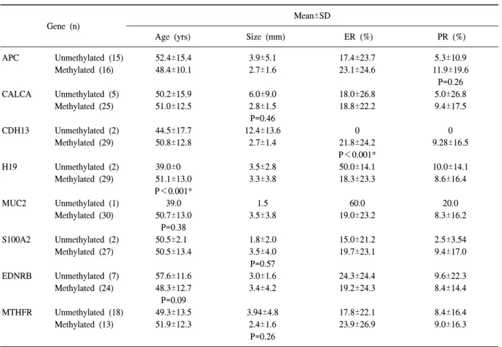
Table 4. Associations between gene promoter methylation and patient age, tumour size and hormone receptor content Gene (n)
Mean±SD
Age (yrs) Size (mm) ER (%) PR (%)
APC Unmethylated (15) 52.4±15.4 3.9±5.1 17.4±23.7 5.3±10.9
Methylated (16) 48.4±10.1 2.7±1.6 23.1±24.6 11.9±19.6
P=0.26
CALCA Unmethylated (5) 50.2±15.9 6.0±9.0 18.0±26.8 5.0±26.8
Methylated (25) 51.0±12.5 2.8±1.5 18.8±22.2 9.4±17.5
P=0.46
CDH13 Unmethylated (2) 44.5±17.7 12.4±13.6 0 0
Methylated (29) 50.8±12.8 2.7±1.4 21.8±24.2 9.28±16.5
P<0.001*
H19 Unmethylated (2) 39.0±0 3.5±2.8 50.0±14.1 10.0±14.1
Methylated (29) 51.1±13.0 3.3±3.8 18.3±23.3 8.6±16.4
P<0.001*
MUC2 Unmethylated (1) 39.0 1.5 60.0 20.0
Methylated (30) 50.7±13.0 3.5±3.8 19.0±23.2 8.3±16.2
P=0.38
S100A2 Unmethylated (2) 50.5±2.1 1.8±2.0 15.0±21.2 2.5±3.54
Methylated (27) 50.5±13.4 3.5±4.0 19.7±23.1 9.4±17.0
P=0.57
EDNRB Unmethylated (7) 57.6±11.6 3.0±1.6 24.3±24.4 9.6±22.3
Methylated (24) 48.3±12.7 3.4±4.2 19.2±24.3 8.4±14.4
P=0.09
MTHFR Unmethylated (18) 49.3±13.5 3.94±4.8 17.8±22.1 8.4±16.4
Methylated (13) 51.9±12.3 2.4±1.6 23.9±26.9 9.0±16.3
P=0.26
SD = Standard Deviation; ER = estrogen receptor; PR = progesterone receptor; APC = adenomatous polyposis coli; CALCA
= calcitonin; CDH13 = H-cadherin; EDNRB = endothelin receptor B; H19 = H19, imprinted maternally expressed untranslated mRNA; MTHFR = 5,10-methylene-tetrahydrofolate reductase; MUC2 = mucin 2, intestinal/tracheal; S100A2 = Calcium binding protein A2.
t-test, *P<0.05
메틸화된 유전자들 사이의 상관관계를 보면, CALCA와 S100A2 유전자(P=0.001), H19와 MUC2유전자(P<0.001)그 리고 S100A2와 MTHFR유전자 사이에는 메틸화 상태에 강 한 양의 상관관계가 있었다(Table 3).
Table 4는 각 유전자의 메틸화 유무에 따라 환자의 나이, 종양의 크기, 호르몬 수용체 상태를 비교한 표이다. H19 유 전자 메틸화는 높은 연령군(P<0.001)에서 더 많이 관찰되 었고, CHD13이 메틸화된 경우 ER 양성(P<0.001)을 보이는 경향이 뚜렷했다.
고 찰
DNA 과메틸화와 종양억제유전자의 전사억제 현상은 지 난 수십년간 종양에서 관심의 초점이 되고 있다. 종양 유전 자의 과메틸화에 대한 수많은 연구논문이 발표되었으며, 최근에는 유전체적 접근방법을 통해 유방암세포에서 메틸 화 특성을 밝히고 있다.(4-6) 종양세포에서 DNA의 비정상 적인 메틸화를 발견하는 것은 종양의 표지자로 CpG 과메틸 화를 사용하거나, 종양세포의 형질전환과 종양 진행의 분 자생물학적 기전을 설명하는 데 있어 중요한 임상적 의미 를 가진다. 기술의 발전으로 종양에서 DNA 메틸화를 평가 하고, 알려지지 않은 DNA서열을 가지는 유전자에서 서로 다르게 메틸화된 영역을 발견하고 확인할 수 있게 되었고, 서로 다른 조직학적 특성을 가지는 표현형을 구분할 수 있 는 여러 연구 결과들이 발표되고 있다. 예를 들면 최근 ar- ray-based method를 사용한 연구에서 덜 분화된 종양은 잘 분화된 종양에 비해 CpG islands의 과메틸화를 보이고,(7) PTEN pseudogene의 메틸화는 ERBB2의 과발현, 종양의 크 기, 높은 조직학적 등급과 관련이 있는 것으로 보고되었 다.(8) 침윤성 소엽암에 비해 침윤성 관암종에서 TWIST의 메틸화가 높지만,(9) DDND2의 메틸화는 종양 진행에 따라 다양하게 나타난다고 보고되었다.(10) 그밖에 호르몬과 비 호르몬 치료에 대한 유방암의 반응과 특이 유전자의 메틸 화 사이의 상관성에 대한 보고도 있다.(11) 현재까지 다양 한 연구에서 관련된 유전자가 발견되었지만, 유방암에서 특정 CpG island methylator의 표현형이 존재한다고 결론 내 리지는 못하고 있다.(12) 따라서 본 연구에서 유방암조직을 이용하여 특수한 병리학적 특성과 관련이 있을 것으로 보 이는 종양억제 유전자의 메틸화를 알아보았으며, 기존에 예후인자로서의 중요성을 보이는 여러 인자들과의 상호연 관성을 비교하여 임상적 유용성을 평가하였다. 우선 2개의 유방암 세포주에서 enzyme digestion (HpaII/MspI)-PCR assay 를 이용해 50개의 종양억제유전자 중 8개 유전자 (MUC2, H19, CDH13, S100A2, CALCA, EDNRB, APC, MTHFR)의 메틸화 상태를 확인하였고, 메틸화 빈도는 MUC2 (96.8%), H19 (93.5%), CDH13 (93.4%), S100A2 (87.1%) 그리고 CALCA (80.6%) 유전자에서 높게 나타났다.
MUC2는 mucine 단백을 암호화하는 유전자로 mucine은 장상피와 기도상피에서 주로 발현되는 주된 분비 당단백이 다.(13) 이는 유방, 대장, 전립선과 같은 다양한 기관에서 유 래한 모든 점액암종에서 일반적으로 발현된다. Mucin은 강 력한 예후인자로 작용한다고 보고되었으며,(14-16) 이의 발 현은 침윤성 유방암, 관내유방암종에서 공격적인 특성과 관련이 있다고 보고되었다.(17) 유방암에서 겔형성 MUC2 mucin이 높으면 악성 세포의 확산을 방지하는 장벽으로 작 용하여 점액암종이 덜 공격적인 성향을 보이지만,(18) 침윤 성관암종에서 MUC2가 과발현되면 공격적인 성향과 관련 이 된다고 보고되었다.(19,20)
H19 유전자는 모성 대립유전자에서 유래한 각인유전자 로 RNA molecule로 기능하며, 기질세포에 과표출되고, 상 피/기질 경계부에 위치하는 것을 선호하는 것으로 보아 상 피/기질 상호관계를 통해 H19 RNA 표출이 조절되는 것으 로 보인다. 나쁜 예후를 보이는 유방 선암종의 몇몇 예에서 H19는 상피세포에 과발현된다. 결국 어떠한 간엽세포 인자 가 상피세포에서 H19를 유도하는 것으로 보인다.(21) 본 연구에서는 MUC2유전자의 메틸화가 96.8%로 가장 높 았지만, 다른 병리 예후인자와의 상관성은 없었다. MUC2의 발현이 높을 경우 H19의 발현이 높게 나타났으며(P<0.001), H19의 메틸화는 침윤성 관암종에서 비관암종에 비해 높게 나타났으며(P=0.007), p53의 양성률이 높을수록(P=0.049), 연령이 많을수록 메틸화가 증가(P<0.001)하는 것으로 나타 났다.
CDH13유전자(H-cadherin)는 glycosylphosphatidylinositol-an- chored cell surface 단백질로 세포-세포 인식(cell-cell recog- nition)(22)에 관여하며 전립선, 폐, 대장 그리고 유방암에서 자주 메틸화된다. 조직이 양성인지 전암상태인지에 따라 그 빈도가 적어도 2배 이상 감소한다. 통계적으로 유의한 차이는 없었지만 양성 유방조직에서 종양조직에서보다 H-cadherin 메틸화의 빈도가 더 낮은 경향이 있다고 보고되 었다(P=0.21).(23,24)
H-cadherin (CDH13)은 유방암 세포주와 유방암 조직에서 감소되어있다. CDH13 cDNA의 도입은 종양세포 성장을 감 소시키며 결과적으로 정상적인 형태의 형태에서 침습성 형 태로 중요한 변화를 유도하게 된다는 것이 Matrigel out- growth assay를 통해 발견하였다.(25) CDH13의 메틸화는 유 방암과(18/55, 33%) 유방암세포주(7/20, 35%)에서 높게 발 생한다는 보고가 있었다.(26) 본 연구에서는 CDH13의 메틸 화가 29/31 (93.5%)로 이전의 여러 연구에 비해 높게 검출되 었는데, 실험 방법의 차이를 비교해 보아야 할 것으로 보인 다.
S100 단백은 calcium binding EF-hand protein으로 세포주 기진행과 분화에 있어 중요한 역할을 담당한다. S100A2는 신장, 폐, 유방상피에 많이 표출되며, 간, 심장근육, 근육에 서는 중등도로 표출되고, 부신, 장, 뇌에는 거의 존재하지
않으며,(27) 종양세포에서는 발현되지 않는다. 종양세포에 서 S100A2의 소실은 유방촬영에서 석회화로 관찰될 수 있 는데 이는 유방암의 조기 표식자로 이용될 수 있다. S100A2 유전자의 소실을 발견함으로써 석회화 이전에 유방암을 조 기에 발견할 수 있는 데 도움이 될 수 있을 것으로 보인다.
CALCA (calcitonin) 유전자 발현은 신경내분비세포에 국 한되어 나타나기 때문에 소세포폐암과 유방암의 일부처럼 신경내분비 요소를 가진 종양에서만 발현된다. Calcitonin 수용체는 vitamin D 수용체의 전사를 증가시키는데 이는 골 대사에서 중요한 역할을 하는 것으로 보인다. Calcitonin유 전자는 11p15염색체에 위치하며 종양억제 유전자로 알려져 있다. 이 부위의 소실은 산발적 유방암과 관련이 된다. Cal- citonin 유전자에는 CpG island가 있는데 유방관암종에서는 전사 시작 부위가 과메틸화되어 있지만, 양성 종양과 소엽 암종에서는 메틸화되어 있지 않았다.(28) 유방암에서 이 유 전자의 역할과 생물학적 중요성은 아직 확실히 밝혀지지는 않았다.
유전자의 메틸화와 연령과의 관계에 대한 이전의 다른 연구에서 CDH1, ER유전자의 메틸화는 연령이 적을수록 높 게 나타났으며, DDND, TWIST 유전자는 연령이 많을수록 메틸화가 높게 나타난다는 보고와,(29) HOXA5와 3OST3B 유전자가 높은 연령군에서 발생한다는 보고가 있었다.(30) 본 연구에서는 H19유전자의 메틸화가 높은 연령과 관련이 있는 것으로 나타났다(Table 4). 연령과 메틸화 사이의 관련 은 유전자의 서로 다른 기능에 의해 설명될 수 있을 것이다.
결 론
유방암환자 조직에서 8종의 종양억제유전자 MTHFR, APC, EDNRB, CALCA, S100A2 CDH13, H19, MUC2 메틸화 가 관찰되었으며, 특히 CDH, H19, MUC2의 메틸화는 90%
이상으로 유방암 조직에서 높게 나타났다. 각각의 유전자 와 임상병리학적 인자들과의 상관성을 살펴 본 결과 H19는 조직학적 등급(P=0.007), P53 발현(P=0.049)과 상관관계가 있었으며, S100A2 유전자는 N 병기와 상관성이 있었다 (P=0.05). DNA 메틸화에 대한 지속적인 연구를 통해 유방 암을 조기 발견하거나 비침윤성, 전암성 상태에서 조기에 발견하는 데 유용하게 사용될 수 있을 것이다.
REFERENCES
1. Parkin DM, Bray F, Ferlay J, Pisani P. Estimating the world cancer burden: Blobocan 2000. Int J Cancer 2001;94:153-6.
2. Herman JG, Baylin SB. Gene silencing in cancer in association with promoter hypermathylation, N Engl J Med 2003;349:
2042-54.
3. Tryndyak V, Kovalchuk O, Pogribny IP. Identification of dif- ferentially methylated sites within unmethylated DNA domains
in normal and cancer cells. Anal Biochem 2006;356:202-7.
4. Huang TH, Laux D, Hamlin BC, Tran P, Tran H, Lubahn DB.
Identification of DNA methylation markers for human breast carcinomas using the methylation-sensitive restriction finger- printing technique. Cancer Res 1997;57:1030-4.
5. Huang TH, Perry MR, LAux DE. Methylation profiling of CpG islands in human breast cancer cells. Hum Mol Genet 1999;8:459-70.
6. Chen CM, Chen HL, Hsiau TH, Hsiau AH, Shi H, Brock GJ, et al. Methylation target array for rapid analysis of CpG island hypermethylaiton in multiple tissue genomes. Am J Pathol 2003;163:37-45.
7. Yan PS, Perry MR, Laux DE, Asare AL, Caldwell CW, Huang TH, CpG island arrays: an application toward deciphering epi- genetic signatures of breast cancer. Clin Cancer Res 2000;6:
1432-8.
8. Garcia JM, Silva J, Pena C, Garcia V, Rodriguez R, Cruz MA, et al. Promoter methyaltion of the PTEN gene is a common molecular change in breast cancer. Genes Chromosomes Cancer 2004;41:117-24.
9. Fackler MJ, McVeigh M, Evron E, Garrett E, Mehrotra J ,Polyak K, et al. DNA methylation of RASSF1A,HIN-1,RAR- beta,Cyclin D2 and Twist in in situ and invasive lobular breast carcinoma, Int J Cancer 2003;107:970-5.
10. Lehmann U, Langer F, Feist H, Glockner S, Hasemeier B, Kreipe H. Quantitative assessment of promoter hypermethy- lation during breast cancer development. Am J Pathol 2002;
160:605-12.
11. Widschwendter M, Siegmund KD, Muller HM, Fiegl H, Marth C, Muller-Holzner E, et al. Association of breast cancer DNA methylation profiles with hormone receptor status and re- sponse to tamoxifen. Cancer Res 2004;64:3807-13.
12. Bae YK, Brown A, Garrett E, Bornman D, Fackler MJ, Sukumar S, et al. Hypermethylation in histologically distinct classes of breast cancer, Clin Cancer Res 2004;10:5998-6005.
13. Gum Jr JR, Hicks JW, Toribara NW, Siddiki B, Kim YS.
Molecular cloning of human intestinal mucin (MUC2) cDNA.
Identification of the amino terminus and overall sequence sim- ilarity to prepro-von Willebrand factor. J Biol Chem 1994;269:
2440-6.
14. Yamashita K, Yonezawa S, Tanaka S, Shirahama H, Sakoda K, Imai K, et al. Immunohistochemical study of mucin carbo- hydrates and core proteins in hepatolithiasis and cholangio- carcinoma. Int J Cancer 1993;55:82-91.
15. Zhang S, Zhang HS, Cordon-Cardo C, Ragupathi G, Living- ston PO. Selection of tumor antigens as targets for immune attack using immunohistochemistry: protein antigens. Clin Cancer Res 1998;4:2669-76.
16. Utsunomiya T, Yonezawa S, Sakamoto H, Kitamura H, Hokita S, Aiko T, et al. Expression of MUC1 and MUC2 mucins in gastric carcinomas: its relationship with the prognosis of the patients. Clin Cancer Res 1998;4:2605-14.
17. 1Diaz LK, Wiley EL, Morrow M. Expression of epithelial mu-
cins Muc1, Muc2, and Muc3 in ductal carcinoma in situ of the breast. Breast J 2001;7:40-45.
18. Matsukita S, Nomoto M, Kitajima S, Tanaka S, Goto M, Irimura T, et al. Expression of mucins (MUC1, MUC2, MUC5AC and MUC6) in mucinous carcinoma of the breast:
comparison with invasive ductal carcinoma. Histopathology 2003;42:26-36.
19. Walsh MD, McGuckin MA, Devine PL, Hohn BG, Wright RG. Expression of MUC2 epithelial mucin in breast carci- noma. J Clin Pathol 1993;46:922-5.
20. Xu Y, Kimura N, Yoshida R, Lin H, Yoshinaga K. Immuno- histochemical study of Muc1, Muc2 and human gastric mucin in breast carcinoma: relationship with prognostic factors.
Oncol Rep 2001;8:1177-82.
21. Adriaenssens E, Lottin S, Berteaux N, Hornez L. Cross-talk between mesenchyme and epithelium increases H19 gene ex- pression during scattering and morphogenesis of epithelial cells. Exp Cell Res 2002;275:215-29.
22. Koller E, Ranscht B. Differential targeting of T- and N-cadher- in in polarized epithelial cells. J Biol Chem 1996;271:30061-7.
23. Toyooka KO, Toyooka S, Virmani AK, Sathyanarayana UG, Euhus DM, Gilcrease M, et al. Loss of expression and aberrant methylation of the CDH13 (H-cadherin) gene in breast and lung carcinomas. Cancer Res 2001;61:4556-60.
24. Maruyama R, Toyooka S, Toyooka KO, Virmani AK, Zochba-
uer-Muller S, Farinas AJ, et al. Aberrant promoter methylation profile of prostate cancers and its relationship to clinicopatho- logical features. Clin Cancer Res 2002;8:514-9.
25. Lee SW. H-cadherin, a novel cadherin with growth inhibitory functions and diminished expression in human breast cancer.
Nature Me 1996;2:776-82.
26. Toyooka KO, Toyooka S, Virmani AK, Sathyanarayana UG, Euhus DM, Gilcrease M, et al. Loss of expression and aberrant methylation of the CDH13 (H-cadherin) gene in breast and lung carcinomas. Cancer Res 2001;61: 4556-60.
27. Glenney JR, Kindy MS, Zokas L. Isolation of a new member of the S 100 protein family: amino acid sequence, tissue, and subcellular distribution. J Cell Biol 1989;108:569-78.
28. Hakkarainen M, Wahlfors J, Myohanen S, Hiltunen MO, Eskelinen M, Johansson R, et al. Hypermethylation of calcito- nin gene regulatory sequences in human breast cancer as re- vealed by genomic sequencing. Int J Cancer 1996;69: 471-4.
29. Shao Ying Li, Minna Rong, Barry Iacopetta. DNA methylation in breast cancer and its association with clinicopathological features. Cancer Letters 2006;237:272-80.
30. Chen CM, Chen HL, Hsiaum TH, Shi H, Brock GJ, Wei SH, et al. Methylation target array for rapid analysis of CpG island hypermethylation in multiple tissue genomes. Am J Pathol 2003;163:37-45.