This is an Open Access article distributed under the terms of the Creative Commons Attribution Non-Commercial License (http: //creativecommons.org/licenses/by- nc/4.0/) which permits unrestricted non-commercial use, distribution, and reproduction in any medium, provided the original work is properly cited.
Copyright: © 2019 Korean Journal of Agricultural Science
https://doi.org/10.7744/kjoas.20190052
PLANT & FOREST
Anti-inflammatory activity of Ganoderma lucidum by inhibition of NF-κB p65
phosphorylation
Hyung Don Kim
1,†, Jeong-Yong Park
1,2,†, Hyung-Jun Noh
1, Seung Eun Lee
1, Jeong Hoon Lee
1, Kyung Hye Seo
1,*1
Department of Herbal Crop Research, National Institute of Horticultural and Herbal Science, Rural Development Administration, Eumseong 27709, Korea
2
Department of Food Science and Biotechnology, Chungbuk National University, Cheongju 28644, Korea
*Corresponding author: [email protected]
†
These authors equally contributed to this study as first author.
Abstract
Ganoderma lucidum, an oriental polypore fungus and medicinal mushroom, has a long history of use for promoting health and longevity in Korea, China, and other Asian countries.
This study was aimed at determining the anti-inflammatory activity and mechanism of action of Ganoderma lucidum in murine macrophage RAW 264.7 cells. Ganoderma lucidum was extracted with ethanol and freeze-dried. The anti-inflammatory effect (nitrite production) of Ganoderma lucidum extracts was tested using a nitric oxide (NO) colorimetric assay. Semi- quantitative reverse transcription polymerase chain reaction (RT-PCR) was performed to quantify the mRNA expression of cytokines including tumor necrosis factor-α (TNF-α), interleukin (IL)-1β, and IL-6. Western blotting was performed to measure the expression levels of inflammation-related proteins, such as inducible nitric oxide synthase (iNOS), cyclooxygenase-2 (COX-2), nuclear factor kappa B (NF-κB) p65, and phosphorylated NF-κB p65. The NO colorimetric assay showed that NO production increased with the treatment of lipopolysaccharide in (LPS)-activated RAW 264.7 macrophages and decreased with the co- treatment of Ganoderma lucidum extracts and LPS. Ganoderma lucidum extracts repressed the mRNA expressions of cytokines, which were increased after the LPS treatment. In addition, Ganoderma lucidum extracts inhibited the LPS-induced expression of iNOS and COX-2 and the LPS-induced phosphorylation of NF-κB p65. These results suggest that the Ganoderma lucidum extracts exert an anti-inflammatory activity by inhibiting NF-κB related proteins and cytokines.
Keywords: anti-inflammatory activity, Ganoderma lucidum, NF-κB p65 OPEN ACCESS
Accepted: August 12, 2019 Revised: August 05, 2019 Received: February 21, 2019 DOI:
Citation: Kim HD, Park JY, Noh HJ, Lee SE, Lee JH, Seo KH. 2019. Anti-inflammatory activity of Ganoderma lucidum by inhibition of NF-κB p65 phosphorylation.
Korean Journal of Agricultural Science.
https://doi.org/10.7744/kjoas.20190052
Introduction
영지버섯은 약리 효과가 뛰어나 아시아 지역에서 오래 전부터 약재로 사용되어 왔다. 특히 항암, 고혈압, 당뇨, 뇌 졸중 및 심장병 등 각종 성인병에 효과가 있는 것으로 알려져 있고(Cho et al., 2015), 항염증 활성 또한 밝혀져서 연구되 어 왔다(Song et al., 2004). 영지버섯의 주요 항염증 활성 물질에 대해서도 보고되었지만 활성 기전에 관한 연구는 미흡 한 실정이다(Dudhgaonkar et al., 2009). 염증 반응은 병원체의 감염, 화학적 또는 물리적 조직 손상 등에 대한 생체조직 의 방어 반응 중 하나이지만(Lee and Cho, 2015; Namkoong et al., 2015; Jang et al., 2016), 염증 반응이 장시간 지속될 경우 신 경퇴행질환과 같은 여러 만성질환 또는 피부 괴사, 암 등으로 발전할 수 있다(Simons et al., 1996; Guslandi, 1998; Wang et
al., 2007). 염증은 다양한 염증 매개 인자들이 대식세포에 인식되는 것으로부터 시작되는데, 대식세포는 단핵세포의 형태로 염증 반응뿐만 아니라 면역 반응에 중요한 역할을 한다(Jang et al., 2016). 그람 음성 박테리아 외벽에 존재하는 lipopolysaccharide (LPS)의 자극으로 대식세포의 염증 반응이 시작되면 전사인자 (transcription factor)인 nuclear factor kappa B (NF-κB)에 의해 tumor necrosis factor-α (TNF-α), interleukin (IL)-1β 및 IL-6와 같은 염증성 사이토카인의 분비가 촉진되 고 분비된 사이토카인이 다른 세포에 전달되면 그 세포에서는 다시 염증 반응이 활성화되면서 염증 반응은 증폭된다 (Kang et al., 2015; Son et al., 2018a; Son et al., 2018b). 스트레스, 흡연, 바이러스, 염증 반응, 종양 및 제 1형 당뇨병 등 여러 가 지 질병들은 NF-κB 신호 전달 경로와 관련되어 있으며 NF-κB 경로는 면역반응에 있어서 중요한 역할을 한다(Mamatha and Shanmuga, 2016). NF-κB는 자극을 받지 않는 상태에서는 inhibitory kappa B-α (IκB-α)와 결합하여 세포원형질에 있다 가 자극을 받으면 IκB-α가 프로테아좀 (proteasome)에 의해 분해가 되고 따라서 NF-κB는 세포질에서 핵 안으로 이동하 여 inducible nitric oxide synthase (iNOS)와 cyclooxygenase-2 (COX-2)와 같은 효소, 염증성 사이토카인 및 염증성 유전자의 전사인자로 활동하여 염증 발현을 조절하게 된다(Feng et al., 1999; Jang et al., 2016). 따라서 본 연구에서는 영지버섯 추출 물의 항염증 활성 기전을 밝히기 위해 대식세포주를 가지고 실험을 진행하였다. 대식세포주 RAW 264.7에서 영지버섯 의 NO 저해 활성을 분석하였으며, 염증 관련 인자의 발현 변화도 분석하였다. 특히 영지버섯이 염증 기전에 중요한 역 할을 하는 NF-κB의 서브유닛(subunit)인 p65 단백질 인산화에 미치는 영향을 확인하였다. 연구 결과는 영지버섯의 항염 증 활성을 설명하는 기초자료로 활용될 수 있을 것이라 기대된다.
Materials and Methods
버섯 추출물 제조
실험에 사용된 영지버섯은 영천영농조합에서 구입하였다. 버섯 추출물은 70% 에탄올을 사용하여 상온에서 3반복으 로 추출 및 여과한 후 감압 농축하여 용매를 제거한 후, 동결건조하고 dimethyl sulfoxide (DMSO; Sigma-Aldrich, St. Louis, USA)에 녹여 실험에 사용하였다.
세포주 배양
본 연구에 사용한 마우스 대식세포주(mouse leukaemic monocyte macrophage cell line) RAW 264.7은 한국 세포주 은 행(KCLB, Seoul, Korea)에서 구입하였다. 세포주는 10% Fetal Bovine Serum (FBS; HyClone, Logan, USA)과 100 units/mL
penicillin과 100 μg/mL streptomycin이 포함된 Dulbecco’s-modified Eagle’s medium (DMEM, high glucose; HyClone, Logan, USA) 배양액으로 배양하였으며, 37℃, 5% CO2 조건 (MCO-2OAIC, Sanyo, Moriguchi, Osaka, Japan)을 유지하였다.
세포 생존율 측정
배양된 세포는 96 well culture plate에 5000 cells/well을 분주 후, 1일 동안 37℃, 5% CO2 공기 조건하에서 24시간 배양하 였다. 1 μg/mL LPS (Sigma-Aldrich, St. Louis, USA) 를 세포에 4시간 처리 후, 100 μg/mL와 200 μg/mL의 최종농도가 되도록 버섯 추출물 시료 및 음성 대조군으로서 DMSO를 18시간 처리하였다. 그 후 세포에 CellTiter 96® AQueous One Solution Cell (Promega, WI, Madison, USA) 시약을 20 μL씩 각 well에 처리하고 3시간 방치한 후 microplate reader (BioTek Instruments, Inc., Winooski, USA)를 이용하여 490 nm에서 흡광도를 측정하였다.
Nitric oxide (NO) 저해 활성 측정
배양된 세포는 96 well culture plate에 5000 cells/well을 분주 후, 1일 동안 37℃, 5% CO2 공기 조건하에서 24시간 배양 하였다. 1 μg/mL LPS를 세포에 4시간 처리 후, 100 μg/mL와 200 μg/mL의 최종농도가 되도록 버섯 추출물 시료 및 음성 대조군으로서 DMSO를 18시간 처리하였다. 각각의 well에서 100 μL씩의 배지를 회수하여 griess reagent (Promega, WI, Madison, USA) 100 μL와 혼합하고 알루미늄 호일로 빛을 차단 후 15분간 반응시켰다. 반응 후 microplate reader를 이용하 여 540 nm에서 흡광도를 측정하였다.
Semi-quantitative reverse transcriptase polymerase chain reaction (RT-PCR)
마우스 대식세포 유전자들의 발현 정도를 조사하기 위해 1 μg/mL LPS를 4시간 전 처리하고 추출물 시료를 18시간 처 리하여 배양한 세포로부터 Trizol reagent를 이용하여 total RNA를 추출하였다. RNA 1 μg을 역전사 kit (Fermentas, Hanover,
USA)를 이용하여 cDNA를 제조한 다음, 동량의 cDNA를 PCR로 증폭시켰다. 유전자 증폭을 위한 primer sequences는 Table 1에 나타내었다.
단백질 발현 분석
Western blotting을 이용하여 마우스 대식세포의 염증 기작 관련 단백질의 발현 정도를 확인하였다. 1 μg/mL LPS를 4 시간 전 처리하고 추출물 시료를 18시간 처리하여 배양한 세포를 RIPA cell lysis buffer (Gibco, Grand Island, NY, USA)을 이 용하여 용해시키고, 13,000 rpm으로 4℃에서 20분간 원심분리 하여 단백질만 포함하고 있는 상층액을 얻었다. 정량한 단백질 20 μg/mL을 12% SDS-PAGE에 전기영동 시킨 후 nitrocellulose membranes (GE Healthcare, Chalfont St. Giles, UK)으 로 옮겨 1차 항체와 반응시킨 후 2차 항체인 horseradish peroxidase-conjugated anti-rabbit 또는 anti-mouse IgG를 반응시키고 enhanced chemiluminescence (ECL) detection reagents (West-Zol Plus, iNtRON, Seoul, Korea)를 사용하여 단백질의 발현 정도 를 확인하였다.
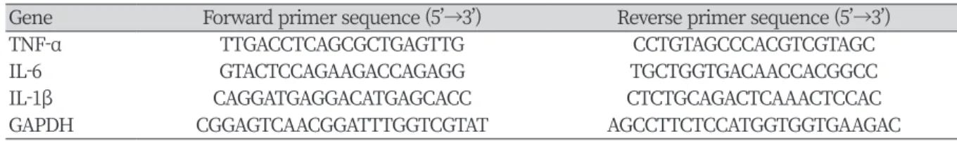
Table 1. Primer sequences for PCR amplification.
Gene Forward primer sequence (5’→3’) Reverse primer sequence (5’→3’)
TNF-α TTGACCTCAGCGCTGAGTTG CCTGTAGCCCACGTCGTAGC
IL-6 GTACTCCAGAAGACCAGAGG TGCTGGTGACAACCACGGCC
IL-1β CAGGATGAGGACATGAGCACC CTCTGCAGACTCAAACTCCAC
GAPDH CGGAGTCAACGGATTTGGTCGTAT AGCCTTCTCCATGGTGGTGAAGAC
TNF-α, tumor necrosis factor-α; IL, interleukin; GAPDH, glyceraldehyde 3-phosphate dehydrogenase.
통계처리
실험결과는 일원배치분산분석 (one-way ANOVA)을 실시하였고, 사후검정으로는 Duncan’s multiple range test를 적용하 였으며, p < 0.05 수준에서 유의성을 검정하였다.
Results and Discussion
마우스 대식세포주에서 영지버섯 추출물의 NO 저해 활성 측정
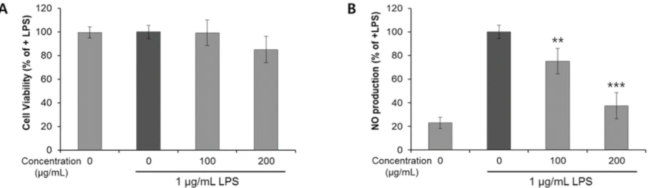
RAW 264.7 대식세포에서 영지버섯 추출물을 농도별로 처리하고 세포 생존율을 확인해 본 결과, 영지버섯 추출물 200 μg/mL까지 추출물 용매(vehicle)만을 처리한 대조군과 세포생존율에서 유의미한 차이를 보이지 않았다(Fig. 1A). 이 결과를 토대로 200 μg/mL을 추출물의 최대 처리 농도로 설정하였다. 그리고 영지버섯 추출물의 항염증 활성을 확인하 기 위해 nitric oxide (NO) assay를 실시하였다. NO는 높은 반응성을 가진 생체 생성분자로서, 염증 반응이 일어날 때 NOS (nitric oxide synthase)에 의해 L-arginine으로부터 생성되어 면역 반응에 중요한 역할을 하는 것으로 알려져 있다(Park and Jung, 2013; Lee, 2014). 실험 결과, 대식세포에서 무처리군에 비해 LPS 처리군에서 NO의 분비가 유의성 있게 증가하였고 영지버섯 추출물에 의해 생성된 NO가 농도 의존적으로 억제됨을 확인할 수 있었다(Fig. 1B). 이러한 결과는 이전에 보 고된 영지 물추출물 실험 결과와 같았다 (Song et al., 2004).
염증 관련 유전자 발현에 영지버섯 추출물이 미치는 영향 확인
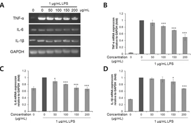
LPS에 의해 활성화되는 염증 반응에 영지버섯 추출물이 미치는 영향을 확인하기 위해, 염증성 사이토카인 유전자들 의 발현을 Semi-quantitative RT-PCR (reverse transcription polymerase chain reaction)로 확인하였다. RT-PCR은 RNA로 역전사 효소를 이용하여 cDNA를 만들고, 만들어진 cDNA를 중합효소연쇄반응으로 증폭하여 유전자 발현을 확인할 수 있는 방법이다. 사이토카인은 LPS에 의해 발현이 과하게 유도되며, 면역계의 신호전달물질로 염증 반응에 의해 분비된 사 이토카인은 iNOS와 COX-2를 자극하여 염증 반응을 더 증폭시키는 것으로 알려져 있다(Kim et al., 2019). 실험 결과, 대표 적인 염증 관련 사이토카인인 TNF-α, IL-6 그리고 IL-1β가 LPS에 의해 유의성 있게 발현이 증가하였고, 영지버섯 추출 물에 의해 농도 의존적으로 감소함을 확인할 수 있었다(Fig. 2). 이러한 결과는 영지버섯 추출물이 TNF-α, IL-6 및 IL-1β
Fig. 1. Effect of Ganoderma lucidumon extract on cell viability and NO production in lipopolysaccharide
(LPS)-induced RAW 264.7 cells. (A) Cell viability was experimented using a [3-(4,5-dimethylthiazol-2-yl)-5-(3- carboxymethoxyphenyl)-2-(4-sulfophenyl)-2H-tetrazolium (MTS) assay and (B) No production was measured by the Griess reagent. Values are the mean ± standard deviation (SD) of three independent experiments.***p < 0.001 and **p < 0.01 vs. LPS treated only.
의 mRNA 발현을 억제하여 항염증 활성을 나타내는 것으로 예상된다.
염증 관련 단백질 발현에 영지버섯 추출물이 미치는 영향 확인
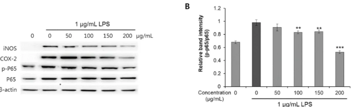
NF-κB 경로는 염증반응과 밀접한 관련이 있다. NF-κB의 서브유닛인 p65는 대부분의 세포에서 p65:p50 heterodimer 형 태로 존재하기 때문에 특히 중요하게 작용할 수 있다(Mamatha and Shanmuga, 2016). 본 연구에서는 영지버섯 추출물의 항염증 활성 기전을 확인하기 위해 추출물 처리에 의한 염증 관련 단백질과 NF-κB p65의 발현 변화를 분석하였다. 염 증 관련 단백질인 iNOS와 COX-2는 각각 염증 반응에 의해 생성되는 NO와 prostaglandin E2 (PGE2)을 생성시키는 효소들 이다. 실험 결과 LPS에 의해 iNOS와 COX-2의 발현이 유의성 있게 증가하였고, 영지버섯 추출물에 의해 농도 의존적으 로 감소하였다(Fig. 3A). 실험결과로 영지버섯 추출물에 의한 NO 분비 억제가 iNOS 발현 감소에 의한 것임을 확인할 수 있었다. 그리고 염증 반응에 있어서 중요한 전사인자 NF-κB의 서브유닛(subunit)인 p65의 발현 변화도 확인하였다. 염증 반응이 활성화되면 세포질에 존재하던 NF-κB p65 단백질이 인산화 되어 핵으로 이동하면서 COX-2나 iNOS와 같은 효 소들의 발현을 유도하여 NO의 생성을 촉진시키고 TNF-α, IL-1β 및 IL-6와 같은 염증 관련 유전자의 생성을 유도하는 것 으로 알려져 있다(Maguire et al., 2015). 실험 결과, LPS에 의해 p65의 발현이 유의성 있게 증가하였지만, 영지버섯 추출물 에 의해서는 발현량이 변하지 않았다(Fig. 3A). 하지만, p65 발현량 대비 인산화된 p65 (p-p65)의 발현량 비율을 확인해보 니, 영지버섯 추출물에 의해 농도 의존적으로 감소함을 확인할 수 있었다(Fig. 3B). 이러한 결과를 종합해볼 때, 영지버
Fig. 2. Effect of
Ganoderma lucidum extract on mRNA exprssion of cytokines in LPS-induced RAW 264.7 cells.(A) lipopolysaccharide (LPS)-induced RAW 264.7 cells were treated with various concentration of Ganoderma lucidum extract for 18 h. The mRNA expression level of tumor necrosis factor-α (TNF-α), interleukin (IL)-6, IL-1 β, and glyceraldehyde 3-phosphate dehydrogenase (GAPDH) were determined by RT-PCR (B - D). The band intensity of the genes was measured by the imageJ software, normalized to GAPDH and presented as the
섯 추출물은 p65의 인산화를 억제함으로써 NF-κB 활성화를 감소시킨다는 것을 확인할 수 있었다. 영지버섯 추출물에 의해 억제된 NF-κB로 인해 COX-2와 iNOS 단백질의 발현이 줄어들고 결과적으로 NO 분비가 억제된 것으로 예상된다.
또한 염증성 사이토카인 유전자들의 발현이 영지버섯 추출물에 의해 줄어들었던 것도 그에 따른 결과라고 볼 수 있다 (Fig. 2).
Conclusion
영지버섯의 항염증 활성에 대해서는 오래 전에 밝혀졌지만, 활성 기전에 관한 연구는 미흡한 실정이다. 따라서 본 연 구에서는 영지버섯의 항염증 활성 기전을 밝히기 위해 염증 관련 유전자와 단백질의 발현이 영지버섯 추출물에 의해 어떻게 변화하는지 확인하였다. 영지버섯은 염증 반응에 중요한 전사인자인 NF-κB의 p65 서브유닛 단백질의 인산화 를 억제해 NF-κB가 전사인자로 활동하는 것을 방해함으로써 하위 염증 관련 인자들의 발현을 감소시킨다는 것을 실험 을 통해 확인하였다. 이러한 연구결과는 영지버섯의 항염증 활성을 이해하는데 좋은 기초가 될 것이라 생각한다.
Acknowledgements
본 연구는 농촌진흥청 연구사업(세부과제번호: PJ01193203)의 지원에 의해 이루어진 결과로 이에 감사 드립니다.
Authors Information
Hyung Don Kim, https://orcid.org/0000-0003-0993-4347 Jeong-Yong Park, https://orcid.org/0000-0003-4964-4272
Hyung-Jun Noh, Department of Herbal Crop Research, RDA, Doctor Seung Eun Lee, https://orcid.org/0000-0003-1511-3262
Fig. 3. Effect of Ganoderma lucidum extract on expression of inflammation-related proteins in LPS-induced
RAW 264.7 cells. Lipopolysaccharide (LPS)-induced RAW 264.7 cells were treated with various concentration of Ganoderma lucidum extract for 18 h. (A) The protein expression level of inducible nitric oxide synthase (iNOS), cyclooxygenase-2 (COX-2), p65, p-p65, and β-actin were determined by western blot. (B) The relative band intensity results were quantified using the imageJ software. Values are the mean ± SD of three independent experiments. ***p < 0.001, **p < 0.01 vs. LPS treated only.Jeong Hoon Lee, https://orcid.org/0000-0001-6709-5508 Kyung Hye Seo, https://orcid.org/0000-0002-8155-8051
References
Cho JH, Park HS, Han JG, Lee KH, Jhune CS. 2015. Comparative analysis of ganoderic acid A, F, and H contents in the fruiting bodies of Ganoderma spp. Journal of Mushroom 13:319-325. [in Korean]
Dudhgaonkar S, Thyagarajan A, Sliva D. 2009. Suppression of the inflammatory response by triterpenes isolated from the mushroom Ganoderma lucidum. International Immunopharmacology 9:1272-1280.
Feng GJ, Goodridge HS, Harnett MM, Wei XQ, Nikolaev AV, Higson AP, Liew FY. 1999. Extracellular signal- related kinase (ERK) and p38 mitogen-activated protein (MAP) kinases differentially regulate the lipopolysaccharide-mediated induction of inducible nitric oxide synthase and IL-12 in macrophages:
Leishmania phosphoglycans subvert macrophage IL-12 production by targeting ERK MAP kinase. The Journal of Immunology 163:6403-6412.
Guslandi M. 1998. Nitric oxide and inflammatory bowel disease. European Journal of Clinical Investigation 28:904-907.
Jang JH, Jung HK, Ko JH, Sim MO, Woo KW, Kim TM, Lee KH, Ahn BK, Cho HW, Cho JH, Jung WS. 2016.
Anti-inflammatory effect of Sedum takesimense Nakai water extract in RAW 264.7 cells. Korean Journal of Medicinal Crop Science 24:228-236. [in Korean]
Kang BK, Kim KBWR, Kim MJ, Park SW, Park WM, Ahn NK, Choi YU, Bae NY, Park JH, Ahn DH. 2015. Anti- inflammatory effect of Sargassum coreanum ethanolic extract through suppression of NF-κB pathway in LPS induced RAW 264.7 cells in mice. Korean Journal of Microbiology and Biotechnology 43:112-119.
[in Korean]
Kim MS, Lim JS, Park TJ, Ko KW, Kim SY. 2019. Anti-inflammatory activity of agaricus blazei extract in lipopolysaccharide-stimulated RAW264.7 cells. Korean Society for Biotechnology and Bioengineering Journal 34:31-37. [in Korean]
Lee JH. 2014. Anti-oxidant and anti-inflammatory effects of Diospyros kaki Thumb leaves extracts. Korean Journal of Aesthetic and Cosmetology 12:719-724. [in Korean]
Lee SE, Cho SI. 2015. Anti-inflammatory effects of Salviae Miltiorrhizae Radix extract on RAW 264.7 cell via anti-oxidative activities. Korean Journal of Herbology 30:89-94. [in Korean]
Maguire O, O'Loughlin K, Minderman H. 2015. Simultaneous assessment of NF-κB/p65 phosphorylation and nuclear localization using imaging flow cytometry. Journal of Immunological Methods 423:3-11.
Mamatha S, Shanmuga RC. 2016. Function of nuclear factor kappa B (NF-κB) in human disease-A review.
South Indian Journal of Biological Sciences 2:368-387.
Namkoong KS, Jang SA, Sohn EH, Park JP, Sohn ES, Koo HJ, Yoon WJ, Kwon JE, Jeong YJ, Meng X, Han HS, Kang SC. 2015. Comparative study of Litsea japonica leaf and fruit extract on the anti-inflammatory effects. Korean Journal of Plant Resources 28:145-152. [in Korean]
Park JS, Jung SH. 2013. Effects of sandalwood essential oil on the iNOS expression and proinflammatory cytokine production. Yakhak Hoeji 57:70-75. [in Korean]
Simons RK, Junger WG, Loomis WH, Hoyt DB. 1996. Acyte lung injury in endotoxemic rats is associated with sustained circulating IL-6 levels and intrapulmonary CINC activity and neutrophil recruiment role of ciculating TNF-α and IL-1β? Shock 6:39-45.
Son JY, Park YW, Renchinkhand G, Paik SH, Nam MS. 2018a. Characterization of lactoferrin hydrolysates on inflammatory cytokine expression in Raw264.7 macrophages. Korean Journal of Agricultural Science 45:438-446
Son JY, Renchinkhand G, Bae HC, Paik SH, Lee JY, Nam MS. 2018b. Cytokine modulation in Raw 264.7 macrophages treated with ginseng fermented by Penibacillus MBT213. Korean Journal of Agricultural Science 45:769-777. [in Korean]
Song YS, Kim SH, Sa JH, Jin C, Lim CJ, Park EH. 2004. Anti-angiogenic and inhibitory activity on inducible nitric oxide production of the mushroom Ganoderma lucidum. Journal of Ethnopharmacology 90:17- Wang MT, Honn KV, Nie D. 2007. Cyclooxygenase, prostanoid, and tumor progression. Cancer and 20.
Metastasis Reviews 26:525-534.