NF- κB와 MAPKs 활성 저해를 통한 미야베 모자반(Sargassum miyabei Yendo) 에탄올 추출물의 항염증 활성
김민지1, 배난영1, 김꽃봉우리2, 박선희1, 장미란3, 임무혁4, 안동현1*
1부경대학교식품공학과/식품연구소
2부경대학교수산과학연구소
3식품의약품안전처건강기능식품정책과
4대구대학교식품공학과
Received: July 4, 2016 / Revised: September 1, 2016 / Accepted: October 24, 2016
서 론
염증반응은외부의물리적, 화학적자극, 세균에의한침 윤작용에대한면역체계반응의하나이며손상을수복, 재생
하려는기전이다[41]. 그러나만성염증반응은오히려인간
에게질병을일으키는원인이되기도한다. 대식세포는감염 체에대한식작용을나타내는대표적인백혈구로내독소인 lipopolysaccharide (LPS)와같은물질에의해활성이증가
되면 nitric oxide (NO)와 tumor necrosis factor-α (TNF- α), interleukin-6 (IL-6), IL-1β의 cytokine을생산하여초기 염증반응의중요한역할을한다[40]. 이들은조직상처나감 염부위에면역세포들을유인하게하며[5], helper T 세포, B 세포, 자연살해세포등의활성을증가시키는역할을한다
[8]. 하지만대식세포로부터염증반응에관여하는물질들이
지나치게분비하게되면과량생성된 NO는급성과민성염증 반응을유도하는무기자유라디칼물질로서작용하게되고
[24, 34], 염증인자들은염증성장질환을일으키며심지어관
절염, 퇴행성뇌질환, 암과같은만성질환을유발하기도한
다[2]. 따라서급성염증질환또는만성염증질환을치료하기
위한염증매개물질들을제어하고조절할수있는부작용 Anti-Inflammatory Activity of Ethanol Extract of Sargassum miyabei Yendo via Inhibition of NF-κB and MAPK Activation Min-Ji Kim1, Nan-Young Bae1, Koth-Bong-Woo-Ri Kim2, Sun-Hee Park1, Mi-Ran Jang3, Moo-Hyeog Im4, and Dong-Hyun Ahn1*
1Department of Food Science & Technology/Institute of Food Science, Pukyong National University, Busan 48513, Republic of Korea
2Institute of Fisheries Sciences, Pukyong National University, Busan 46041, Republic of Korea
3Health Functional Food Policy Division, Ministry of Food and Drug Safety, Chungcheongbuk-do 28519, Republic of Korea
4Department of Food Science and Biotechnology, Daegu University, Gyeongsan 38453, Republic of Korea
The aim of this study was to investigate the anti-inflammatory effect of Sargassum miyabei Yendo ethanol extract (SMYEE) using RAW 264.7 cells and croton oil-induced Balb/c mice. SMYEE inhibited the produc- tion of pro-inflammatory cytokines [interleukin (IL)-6, tumor necrosis factor (TNF)-α, and IL-1β] and nitric oxide in lipopolysaccharide (LPS)-induced inflammatory response. In addition, SMYEE suppressed the expression of inducible nitric oxide, cyclooxygenase-2, and nuclear factor-kappa B. Further, SMYEE inhib- ited the expression of mitogen-activated protein kinases (MAPKs), such as extra cellular signal-regulated kinase 1/2, p38, and c-Jun N-terminal kinase. In ear edema test, edema formation in the SMYEE treatment was lower than that in the positive control and was similar to that in the prednisolone treatment group.
Photomicrographs of mice ear tissue showed a reduction in dermal thickness and number of infiltrated mast cells. Therefore, our results indicate that SMYEE exerts an anti-inflammatory effect via inhibition of nuclear factor (NF)-κB and MAPK activation and can be used as a natural source of anti-inflammatory com- pounds.
Keywords: Anti-inflammatory activity, NF-κB, MAPKs, Sargassum miyabei Yendo
*Corresponding author
Tel: +82-51-629-5831, Fax: +82-51-629-5824 E-mail: [email protected]
© 2016, The Korean Society for Microbiology and Biotechnology
이적은천연항염증물질또는약제개발에관심이증가하 고있다.
대식세포에있어전염증성 cytokine 및단백질의유전자 발현은 nuclear factor kappa-B (NF-κB)에의해조절이된 다[29]. LPS와같은자극이발생하게되면 NF-κB가활성화 되게되면서다양한염증성매개체의유전자의발현을유도 하게 된다[6]. 또한 NF-κB의활성은 extracellular signal- regulated kinase (ERK), c-Jun-N-terminal kinase (JNK), p38 kinase를포함하는 mitogen-activated protein kinases (MAPKs)에의해조절되는것으로알려져있다[9, 25].
Sargassum 속은 Sargassaceae과에속하는갈조류로약
400종이알려져있으며[27], 전세계적으로분포하고있어식
품이나약재로서많은나라에서이용되고있다[15]. Sargassum
miyabei Yendo는주로남해안및제주도에서식하고있으
며, 생리활성에대한보고는다른 Sargassum 속에비해많 이알려져있지않다. 현재, 항돌연변이[23], α-glucosidase 저해활성[21], 항산화활성[32]에대해보고되었으며, 항염 증에대한연구는보고된적이없다. 이에본연구에서는 S.
miyabei Yendo를에탄올로추출하여 NF-κB와 MAPKs의 활성조절을통한 iNOS와 COX-2의활성억제와 NO와전 염증성 cytokine의분비억제효과를살펴보았으며, croton- oil로유도한귀부종동물모델에서귀부종형성및귀조직의 변화를관찰하여천연항염증소재로서이용가능성을알아 보았다.
재료 및 방법
에탄올 추출물 제조
부산백운포에서 2015년도에채취한미야베모자반은담 수로수회깨끗하게수세한후자연건조하였다. 그후동결 건조하여분말화하였으며진공포장상태로−20℃에서저장 하며실험에사용하였다. 10배(w/v)양의 95% ethanol을분 말에가하여교반기(H-0820, Dongwon Science Co., Korea) 를 이용해 24시간 추출하였다. 원심분리기(UNION 32R, Hanil Co., Korea)로 1,977 × g에서 10분간원심분리한후, 상층액을분리하여 37℃에서감압농축기(RE200, Yamoto Co.,
Japan)로농축하였다. 남은잔사는동일한방법을이용해
2회반복하여추출하여에탄올추출물을얻었다.
세포배양
한국세포주은행(KCLB 40071, Korea)에서 murine의대식 세포주 cell line인 RAW 264.7 세포를분양받아사용하였으 며, Dulbecco’s Modified Eagle Medium (DMEM) (GIBCO, USA)에 10% inactivated fetal bovine serum (FBS)와 1%
penicillin-streptomycin을첨가한 배지를 사용하여 37℃,
5% CO2조건에서배양하였다.
실험동물
귀부종실험을위해생후 8주령의수컷, ICR 마우스를오 리엔트바이오(Korea)에서구입하여사용하였으며, 마우스는 온도 20±2℃, 습도 50±10%, 12시간명암주기가유지되는 동물사육실에서 1주일간예비사육하여환경조건에적응시 켰다. 본동물실험은부경대학교동물실험윤리위원회로부 터동물실험승인을받아수행하였다(2015-04).
세포독성 측정
Park 등[33]의 MTT assay 방법을약간변형하여시료의 세포독성을평가하였다. RAW 264.7 세포를 1 × 10⁶ cells/ml 의농도로 96-well plate에분주해 20시간전배양한후미 야베모자반에탄올추출물(SMYEE)을농도별로(0.1, 1, 10, 50, 100 μg/ml) 첨가하여 22시간 본배양하였다. 5 mg/ml MTT (thiazol blue tetrazolium bromide, Sigma-Aldrich Chemical Co.) 용액을첨가하고 2시간재배양하였다. 배양 후 4℃, 879 × g에서 10분간원심분리하여상층액을걷어내 고 dimethyl sulfoxide 100 μl 분주하여생성된 formazan을 녹여내 microplate reader (Model 550, Bio-Rad, USA)를이용
하여 540 nm에서흡광도를측정하였다. 세포증식능은다
음식에의해계산하였다.
Proliferation index (% of control) = Sample 흡광도 / Control 흡광도× 100
Nitric oxide 분비량 측정
Griess 반응을이용하여배양액내의 nitrite 농도를측정 하였다[22]. RAW 264.7 cell을 2.5 × 10⁵ cells/ml로조절해 24-well plate에접종하고 5% CO2 incubator (MCO-15AC, Sanyo, Japan)에서 20시간전배양하였다. 그후 SMYEE 를 0.1, 1, 10, 50, 100 μg/ml 처리하고 1 μg/ml의 LPS로 자극하여 24시간 본 배양하였다. 배양 상층액은 4℃,
879 ×g에서 10분간원심분리하여정량실험에사용하였다.
Griess 시약(1% sulfanilamide + 0.1% naphthylendiamine dihydrochloride, 1:1)과배양상층액은 1:1로상온에서 10분 간반응시켜 microplate reader를이용해 540 nm에서흡광 도를 측정하였다. 세포 배양액 내 NO의 농도는 sodium nitrite (NaNO2)의농도별표준곡선과비교하여산출하였다.
Pro-inflammatory cytokines 분비량 측정
RAW 264.7 cell의세포배양액내의 TNF-α, IL-6 및 IL- 1β cytokine의 분비량을 ELISA kit (Mouse ELISA set, BD Bioscience, USA)를이용해측정하였다. RAW 264.7 cell
을 2.5 × 10⁵ cells/ml로조절하여 24-well plate에접종하고 18시간전배양하였다. 그후 0.1, 1, 10, 50, 100 μg/ml 농도 별 SMYEE와 1 μg/ml의 LPS를처리하고 12시간의본배양 을거쳐원심분리를통해세포배양상층액을얻었다. ELISA 는 microplate에 anti-mouse TNF-α, IL-6 및 IL-1β의 capture antibody를 coating buffer에희석후분주하여 4℃ 에서하룻밤동안 coating시켰다. 이후 0.05% Tween 20이 포함된 PBST로세척한후 10% FBS 용액으로 blocking 하 였고 PBST로세척한뒤각 microplate well에세포배양상 층액을분주하고실온에서 2시간반응시켰다. 반응후 PBST 로세척하고희석한 biotinylated anti-mouse TNF-α, IL-6 detection antibody와 streptavidin-horseradish peroxydase conjugate를분주하여실온에서 1시간반응시켰다. IL-1β의 경우, biotinylated anti-mouse IL-1β detection antibody를 첨가하고 1시간반응후, streptavidin horseradish peroxidase conjugate를첨가하여 30분반응시켰다. 반응후이를다시 PBST로세척하고 OPD 용액을첨가하여실온에서 30분동안 암반응시켰다. 2 N H2SO4로반응을종료시킨후 microplate reader를이용하여 490 nm에서흡광도를측정하였다.
Western blot에 의한 단백질 발현 분석
SMYEE가세포질내생성되는 iNOS, COX-2 및 NF-κB
p65의발현량에미치는영향을알아보기위하여 RAW 264.7
세포를배양하였다. 배양이끝난세포를수집하여 3회 PBS (phosphate buffered saline)로세척한 후, Sheeba와 Asha [37]의 방법에 따라 cytosol lysis buffer [50 mM HEPES (pH 7.4), 150 mM NaCl, 5 mM EDTA, 1% deoxycholate, 5 mM phenylmethylsulfonyl fluoride, 1 μg/ml aprotinin, 1% Triton X-100, 0.1% NP-40]를첨가하여 30분간 4℃에서 lysis 시킨후, 15,520 × g에서 20 분간원심분리하여세포 막성분등을제거하였다. NF-κB p65의경우 nucleus lysis buffer (10 mM HEPES, 100 mM NaCl, 1.5 mM MgCl2, 0.1 mM EDTA, 0.1 mM dithiothreitol)를첨가하여 30분간 4℃에서 lysis시킨후 15,520 × g에서 20분간원심분리하여 세포막 성분 등을 제거하였다. BCA protein assay kit (Pierce, USA)를사용하여단백질을정량하였으며 30 μl의 lysate를 Laemmli [19]의방법을사용하여 10% SDS-PAGE 로분리하였다. 분리된단백질은 Towbin 등[39]의방법을참 고하여 PVDF (polyvinylidene difluoride) membrane (Bio- Rad)에 1시간동안전사시켜 5% skim milk가포함된 TBSS (tris buffered saline; pH 7.5) 용액으로상온에서 2시간동 안 blocking 하였다. iNOS, COX-2 및 NF-κB의발현양을 검토하기위한항체로는 anti-mouse iNOS, COX-2 및 NF- κB (Santa Cruz Biotechnology, Inc., USA)를 사용하여
1:500으로희석하고상온에서 2시간반응시킨후 TBSS로
3회세정하였다. 2차항체로 HRP (horseradish peroxydase) 가결합된 anti-mouse IgG 및 anti-rabbit IgG를 1:2,000으 로희석하여상온에서 1시간반응시킨후, TBSS로 3회세
정하여 ECL 기질과 1-3분간반응후각각의단백질밴드는
Gene tool (GeneGnome5, Syngene, UK)을이용하여가시
화하였다. MAPKs의발현량을알아보기위하여 RAW 264.7
세포를 1 × 10⁶ cells/ml으로 18시간전배양하고 SMYEE를 처리하여 30분 동안 본배양한 후 p38, ERK, JNK 및 p- p38, p-ERK, p-JNK의 발현량을 검토하기 위하여 anti- mouse p-p38, p38, p-ERK, ERK, p-JNK 및 JNK (Cell Signaling Technology Inc., USA) 항체를 1:500으로희석하 여사용하였다. 이후의실험은 iNOS, COX-2 실험법과동일 하게진행하였다.
귀부종 측정 및 조직 관찰
SMYEE의항염증효과를 in vivo 상에서알아보기위하
여 Kim 등[14]와 Saraiva 등[36]의방법으로귀부종측정실 험을실시하였다. 생후 8주령의수컷, ICR 마우스(n = 5)에 SMYEE를 10, 50 및 250 mg/kg·body weight 농도로 200μl씩단회경구투여하고한시간후, 오른쪽귀에 2.5%
croton oil을 20 μl/ear 농도로도포하였다. 도포 5시간후귀 두께를측정하였고 croton oil의처리로귀두께가증가한것 을부종의형성으로간주하였다. 귀조직관찰은 ICR 마우 스의오른쪽귀에에탄올추출물을 100 mg/ml 농도로 20 μl 씩도포하고 15분뒤, 5% croton oil을 20 μl씩도포하였다. 6시간뒤, diethylether로마취사시키고, 귀조직을절제하 여 10% formaldehyde에 72시간고정하였다. 고정후파라
핀 블록을 만들어 박편을 제조하고 hematoxylin-eosin과
toluidine-blue 염색을하여조직을관찰하였다. 부종생성율 은다음과같은식에의해계산하였다.
Inhibition of edema (% of control) =
Sample의귀두께 / Control의귀두께 × 100
통계 처리
모든실험결과에대한유의차검정은 SAS software (ver.
9.3, SAS Institute, Inc., USA)에서평균값을분산분석한 후, Duncan's multiple range test 법에따라 p < 0.05 수준 에서검정하였다.
결과 및 고찰
RAW 264.7에 미치는 에탄올 추출물의 세포독성
SMYEE가 RAW 264.7 세포에미치는세포독성을알아보
기위하여농도에따라(0.1, 1, 10, 50, 100 μg/ml) 추출물을
처리하고세포생존의변화여부를 MTT assay를이용해측 정하였다. 그결과, 0.1−100 μg/ml 농도까지 PBS 처리군과 비교시유의적인차이가없음을확인하였다(Fig. 1). 이를 통해 SMYEE는 0.1−100 μg/ml에서세포생존율에영향을 미치지않아독성이없는것으로사료되어이농도에서염 증억제활성을확인하기위한실험을진행하였다. 이는큰 잎모자반에탄올추출물의세포독성실험결과, 농도의존적 으로세포독성이나타나지않은결과와유사하다[13].
Nitric Oxide 생성 억제 효과
NO는인체내생리적이나병적인반응에서중요한물질 로서 L-arginine을기질로하여 NOS에의해 L-citrulline으 로합성되는무기유리체로적절한수준에서는혈소판억제, 면역조절, 신경전달, 혈관확장등의역할을한다. 하지만 NO 가과도하게생성되면염증반응을심화시키고 shock에의 한혈관확장, 기관지염, 다발성경화증과같은병적반응및 자가면역질환을일으킨다[11, 18]. 본연구에서는 SMYEE의 NO 생성억제효과를알아보기위하여 LPS로염증반응을유 발한 RAW 264.7 cell에서의 NO 생성량을 griess 시약을이 용해측정하였다. 세포의생존율에영향을미치지않는농도 (0.1, 1, 10, 50, 100 μg/ml)에서 LPS로염증반응을유발한 RAW 264.7 cell의 NO 생성량을확인한결과, LPS 만으로 염증을유발하였을때 NO 생성량이 16.26±0.15 μM이었으 며 SMYEE를 100 μg/ml로처리하였을때 9.89±0.15 μM 로 44%의 NO 억제효과를나타내었다(Fig. 2). 본연구결과 는큰잎모자반추출물이농도의존적으로 NO 생성량의감 소를보인결과[13]와참모자반에탄올추출물의 NO 저해 능을확인한연구결과[10]와유사함을확인하였다. 따라서
SMYEE는 LPS로유도된대식세포에서증가된 NO의생성
억제하여염증반응조절제로서의효과가있는것으로확인 되었다.
Pro-inflammatory cytokines 생성 억제 효과
LPS로처리한 RAW 264.7 세포에서, SMYEE가 cytokine 의분비에미치는영향을확인하기위해농도별로처리하여 IL-6, TNF-α및 IL-1β의분비량변화를 ELISA kit를이용 하여 정량하였다. 그 결과(Fig. 3), LPS 의해 증가한모든
cytokine의분비량을농도의존적으로억제하는것을확인하
였다. 먼저 IL-6의 경우(Fig. 3A), LPS에 의해 2194.93± 35.79 pg/ml까지증가한분비량을 SMYEE를 100 μg/ml로 처리했을시 577.07±15.34 pg/ml로억제하여 74%의뛰어 난억제효과를나타내었다. TNF-α의경우, 농도의존적으로 분비량이억제되었으며, LPS를단독으로처리하여 2008.59± 7.73 pg/ml의분비량을나타낸것과비교하여 100 μg/ml 처 리구에서는분비량이 1289.62±10.62 pg/ml로 36% 이상으 로억제되었다(Fig. 3B). LPS에의해증가된 IL-1β의분비량 은 100 μg/ml로처리하였을때 72% 이상의뛰어난억제효 과를보임을확인하였다(Fig. 3C). 이는참모자반추출물을 처리한후 pro-inflammatory cytokines의분비량을농도의 존적으로억제한효과[10]과유사하였다. LPS는대식세포의
활성화에관여하는대표적인 mitogen이자대식세포의표면
수용체인 toll-like receptor4 (TLR4)에 결합하는 ligand로
표면수용체복합체의활성화를통해 MyD88, TIRAP/Mal,
TRIF와같은 TIR 영역을포함하는어댑터단백질과결합하
여반응하게된다[25]. 이러한 LPS에의해생성되는 cytokine Fig. 1. Effects of Sargassum miyabei Yendo ethanol extract on
proliferation in RAW 264.7 cells. Proliferation index (% of control) = (absorbance of sample / absorbance of control) × 100.
Means with letter (a) above the bars are not significantly different (p > 0.05).
Fig. 2. Effect of Sargassum miyabei Yendo ethanol extract on nitric oxide production in RAW 264.7 cells. Cells were incu- bated in the presence of LPS (1 μg/ml) alone or in combination with SMYEE (0.1, 1, 10, 50, and 100 μg/ml) for 24 h. The culture media of the treated cells were used to measure NO levels.
Means with different letters (a-e) above the bars are significantly different (p < 0.05).
의 분비는 PGE2와 NO의 발현을 야기하고 과도한 pro- inflammatory cytokine의분비량증가는패혈증및심한조
직손상, 전신성염증반응증후군을야기한다[28]. 그중 IL- 6는 T cell, monocyte, macrophage, synovial fibroblast 등의 면역세포에서 발현되는 염증성 cytokine으로 B cell을 plasma cell로분화시켜항체생산을촉진한다[4]. TNF-α는 염증반응과정중초기주요내인성매개자로서대식세포외 에도 NK cell이나 CD4 + lymphocyte에서도생성되어염증 부위로의백혈구를증가시키는다른염증성 cytokine과내 피세포부착분자의발현을촉진시켜패혈증이나암, 염증 성장질환등의자가면역질환의요인이된다[41]. 또한 IL- 1β는염증반응시생성되는촉진성사이토카인으로 caspase- 1에의해활성형으로전환되는데낮은농도에서는세포성장 이나항상성유지에필수적이지만염증반응및면역적자극 에의해대량생산될경우 T cell을활성화시키고 B cell을성 숙시켜증상을악화시킨다[20]. 따라서본연구결과 SMYEE 가이러한염증성사이토카인을조절하는물질로서염증반 응으로유도된다양한질병을조절할수있는가능성이있 음을알수있다.
에탄올 추출물의 iNOS, COX-2, NF-κB 발현 억제 효과 SMYEE의처리가 iNOS와 COX-2의발현조절과관련이
있는지를조사하기위하여 western blot 분석을수행하였
다. 그결과(Fig. 4), LPS 처리구에서는 iNOS와 COX-2 단백 질발현이현저히증가하였으나 SMYEE를농도별(0.1, 1, 10, 50, 100 μg/ml)로처리한그룹에서는농도의존적으로감
소한경향이보였다. 특히 iNOS의발현량이억제되는현상
은 SMYEE 처리가 NO 생성에대한억제효과를보이는것
과일치하였다. 전사인자인 NF-κB는 LPS에의해유도되는 염증성매개물질인 iNOS와 COX-2의발현을촉진시킨다. 핵 분획의 NF-κB p65 단백질의발현은 LPS 처리군에서는대 조구에비해유의성있게증가하였으나 SMYEE 처리구에서 는농도의존적인감소경향을나타내었다(Fig. 4). 만성적인 염증성질환은 NF-κB의비정상적인활성과관련이있다. 따 라서대부분의천연물또는기존의항염증제는핵전사인자 인 NF-κB 활성을억제하여항염증효과를나타낸다고알려 져있으며[30], NF-κB 활성을억제시킴으로염증성질병과 암의예방및치료에이용하고자하는연구가활발히진행 중이다[1]. 최근연구에따르면, Kim 등[16]과 Bae 등[3]은 각각갈조류인잘피에탄올추출물, 참도박에탄올추출물을 LPS로유도된대식세포에처리하였을때, NF-κB의활성억 제를통해 iNOS 및 COX-2의발현량이감소함을밝혔으며,
해조류유래추출물인 SMYEE를이용한본연구결과와유
사함을보였다.
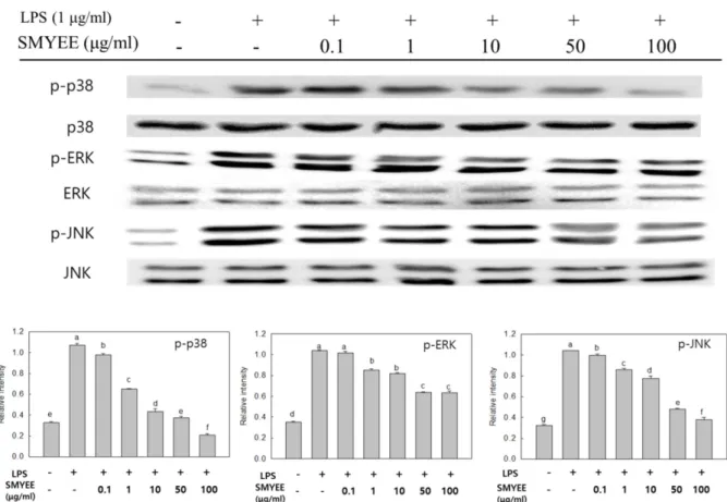
에탄올 추출물의 MAPKs 발현 억제 효과
대식세포의활성화에는여러가지세포내신호전달계가 Fig. 3. Effect of Sargassum miyabei Yendo ethanol extract on
production of IL-6 (A), TNF-α (B), and IL-1β (C) in RAW 264.7 cells. Cells were incubated in the presence of LPS (1 μg/ml) alone or with various concentrations of SMYEE (0.1, 1, 10, 50, and 100 μg/ml). The levels of pro-inflammatory cytokine in the cell cul- ture media were measured by ELISA. Means with different letters (a-g) above the bars are significantly different (p < 0.05).
관여하며이들신호를제어함으로써대식세포가활성화되 어발생되는염증성물질을조절하는연구가여러선행연 구에서보여지고있다[7]. 이들중 MAPKs (ERK, JNK, p- 38)의활성은인산화에의해서나타나며대식세포의활성에 관여하고특히 NF-κB를활성화시켜 iNOS, COX-2 및전염 증성매개물질의발현에중요한역할을한다고알려져있다
[9]. 따라서 SMYEE 처리에 따른 대식세포 내 인산화된
MAPKs 발현량에미치는영향을 western blot으로분석한 결과(Fig. 5), LPS로유도된 RAW 264.7 세포에서 MAPKs 에속한단백질이모두활성화되었음을알수있었다. 반면, SMYEE를처리한결과, 인산화된 ERK, JNK, p-38의발현
이 LPS 처리군에비해농도의존적인감소를보였으며, 특히
인산화된 p-38 및 JNK의발현량이 50 및 100 μg/ml에서
LPS를처리하지않은대조군과비슷한수준까지발현량이
감소함을보였다. 본연구결과는잘피에탄올추출물의항
염증활성효과[16] 및꽃지누아리에탄올추출물의항염증
활성효과[17]에서추출물농도의존적으로 MAPKs의인산 화농도가감소함을보인것과유사하다. 이상의결과를종
합해볼때 SMYEE를처리함으로써 LPS로유도된염증성사
이토카인, iNOS 및 COX-2와같은염증성인자들의활성화
에관련하여 MAPKs 신호전달경로를효과적으로억제함에
따라항염증작용을보인것으로사료된다. 귀부종 억제 효과 및 조직 관찰
염증은인체의물리적, 화학적손상에대한보호작용으로 발열이나부종, 통증등을동반하며세포질내과립을다량 가진비만세포는활성화되어 protease나 histamine 등과같 은혈관확장물질들을분비한다. 혈류량증가로인해혈액 내의 neutrophils 등이혈관밖의조직으로부과되어부종을 유발하며염증부위의 prostaglandin 증가를통해통증이발
생한다[12]. SMYEE가미치는부종의완화효과및조직내
의 mast cell 침윤 억제효과를 확인하기 위해 10, 50 및 Fig. 4. Effect of Sargassum miyabei Yendo ethanol extract on LPS-induced iNOS, COX-2, and NF-κB p65 expression in RAW 246.7 cells. The levels of iNOS and COX-2 in cytosolic protein and NF-κB p65 in nuclear protein were determined by a western blot analysis.
RAW 264.7 cells were treated with the indicated concentrations of SMYEE (0.1, 1, 10, 50, and 100 μg/ml) and LPS (1 μg/ml) for 18 h or 30 min and the proteins were detected using specific antibodies. Means with different letters (a-f) above the bars are significantly dif- ferent (p < 0.05).
250 mg/kg 농도로경구투여한후 croton oil로염증을유발 하고귀두께를측정하였다. 마우스모델에서부종실험을진 행한결과(Fig. 6), croton oil 만을처리하여부종이발생한
대조군과비교시 SMYEE 처리에의해모든농도에서유의
적으로귀두께가감소하였다. Croton oil은피부에도포하 면즉각적으로강한급성염증반응을유도하는데[38] croton oil로유발한부종을 추출물 250 mg/kg 농도에서 positive control인 prednisolone이 10 mg/kg 농도로처리하였을때와
유사하게약 27% 이상억제하는효과를확인하였다. 이는
조직관찰에서추출물을 100 mg/ml 농도를처리하였을시, 진피의두께가현저히얇아진것을확인하였으며(Fig. 7A), 조직내 mast cell 관찰결과에서도 SMYEE를처리함으써 mast cell의침윤을 positive control과유사한수준으로억
제하였다(Fig. 7B). 염증과통증의완화를위해사용되는비
스테로이드계소염진통제는여러부작용이보고[35]된바 있어현재천연물로부터 항염증및진통제로서의가능성을 찾기위한연구가활발히진행되고있는데앞의연구결과
로미루어볼때, SMYEE는기존의항염증제와유사한정
도의부종억제효과를나타냄에따라뛰어난항염증효과를 가져이를이용한치료제개발에도가능성이있을 것으로 사료된다.
Fig. 5. Effect of Sargassum miyabei Yendo ethanol extract on MAPKs expression in RAW 246.7 cells. The levels of p-p38, p38, p- ERK, ERK, p-JNK, and JNK in the cytosolic protein were determined by western blot analysis. RAW 264.7 cells were treated with the indi- cated concentrations of SMYEE (0.1, 1, 10, 50, and 100 μg/ml) and LPS (1 μg/ml) for 30 min, and the proteins were detected using spe- cific antibodies. Means with different letters (a-g) above the bars are significantly different (p < 0.05).
Fig. 6. Inhibition of Sargassum miyabei Yendo ethanol extract against croton oil-induced mouse ear edema. Means with dif- ferent letters (a-e) above the bars are significantly different (p <
0.05).
요 약
본연구에서는미야베에탄올추출물의항염증활성을확 인하기위해 LPS로활성화된 RAW 264.7 세포와 croton-oil 로유도된귀부종동물모델을이용하였다. 그결과, SMYEE 50 및 100 μg/ml 농도처리시, LPS로유도된염증반응에서 NF-κB 활성억제와더불어 MAPKs의인산화를효과적으로
억제함을보였다. LPS에의해증가된 NO와전염증성사이
토카인의분비량도농도의존적으로감소하였다. 또한 SMYEE
는 croton oil로부종을유발한마우스모델에서귀부종억제
효과를나타내었고, 조직의진피두께및 mast cell의수가 현저히감소하였음을확인하였다. 이를통해 SMYEE는염 증반응의전사인자인 NF-κB 및 MAPKs의활성을조절함 으로써 iNOS와 COX-2의발현및전염증성매개인자인 NO, IL-6, TNF-α및 IL-1β의분비를억제하여항염증활성을가 지는것을확인하였다. 현재까지미야베모자반내의항염증 효능물질에관한연구는보고되지않고있으며향후실험을 통해미야베모자반에탄올추출물로부터항염증효과를가 지는유효성분을밝히기위한분리연구를진행할예정이다.
Acknowledgments
This research was supported by Basic Science Research Program through the National Research Foundation of Korea (NRF) funded by the Ministry of Education (2012R1A6A1028677).
References
1. Aggarwal BB. 2004. Nuclear factor-κB: The enemy within. Can-
cer Cell 6: 203-208.
2. Atreya R, Neurath MF. 2005. Involvement of IL-6 in the patho- genesis of inflammatory bowel disease and colon cancer. Clin.
Rev. Allergy Immunol. 28: 187-196.
3. Bae NY, Kim MJ, Kim KBWR, Ahn NK, Choi YU, Park JH, et al.
2015. Anti-inflammatory effect of ethanol extract from Grate- loupia elliptica Holmes on lipopolysaccharide-induced inflam- matory responses in RAW 264.7 cells and mice ears. J. Korean Soc. Food Sci. Nutr. 44: 1128-1136.
4. Bhattacharyya A, Pathak S, Datta S, Chattopadhyay S, Basu J, Kundu M. 2002. Mitogen-activated protein kinases and NF- kappaB regulate H. pylori-mediated IL-8 release from macro- phages. Biochem. J. 368: 121-129.
5. Bosca LM, Zeini M, Traves PG, Hortelano S. 2005. Nitric oxide and cell viability in inflammatory cells: A role for NO in macro- phage function and fate. Toxicology 208: 249-258.
6. Chen XL, Kunsch C. 2004. Induction of cytoprotective genes through Nrf2/antioxidant response element pathway a new therapeutic approach for the treatment of inflammatory dis- eases. Curr. Pharm. Des. 10: 879-891.
7. Guha M, Mackman M. 2001. LPS induction of gene expression in human monocytes. Cell. Signal. 13: 85-94.
8. Hibbs JB Jr, Taintor RR, Vavrin L, Rachlin EM. 1988. Nitric oxide:
A cytotoxic activated macrophage effector molecule. Biochem.
Biophys. Res. Commun. 157: 87-94.
9. Jang BC, Paik JH, Kim SP, Shin DH, Song DK, Park JG, et al. 2005.
Catalase induced expression of inflammatory mediators via activation of NF-kappaB, PI3K/AKT, p70S6K, and JNKs in BV2 microglia. Cell. Signal. 17: 625-633.
10. Jeong DH, Kim KBWR, Kim MJ, Kang BK, Bark SW, Pak WM, et al.
2014. Anti-inflammatory effect of ethanol extract from Sargas- sum fulvellum on lipopolysaccharide induced inflammatory responses in RAW 264.7 cells and mice ears. J. Korean Soc. Food Fig. 7. Photomicrograph of transverse sections of mice ears sensitized with topical application of 5% croton oil (v/v) in acetone (a-c) or vehicle acetone (d, non-inflamed), stained with hematoxylin-eosin (A) and toluidine-blue (B) examined under light microscopy (magnification: 200×). Treatments: vehicle 2% Tween 80 (a), prednisolone 0.08 mg/ear (b) and SMYEE 20 μl/ear (c). The numbers 1 and 2 indicate dermis and epidermis, respectively and the arrows indicate mast cell infiltration.
Sci. Nutr. 43: 1158-1165.
11. Jeong HR, Sung MS, Kim YH, Ham HM, Choi YM, Lee JS. 2012.
Anti-inflammatory activity of Salvia plebeia R. Br. leaf through heme oxygenase-1 induction in LPS-stimulated RAW 264.7 macrophages. J. Korean Soc. Food Sci. Nutr. 41: 888-894.
12. Ju MS, Jeong HU, Kim HG, Park GH, Youn YS, Kim YO, et al. 2010.
Anti-nociceptive and anti-inflammatory effects of Geranii herba. Korea J. Herbol. 25: 97-101.
13. Kang BK, Kim KBWR, Kim MJ, Bark SW, Pak WM, Ahn NK, et al.
2015. Anti-inflammatory effect of Sargassum coreanum ethan- olic extract through suppression of NF-κB pathway in LPS induced RAW 264.7 cells in mice. Microbiol. Biotechnol. Lett. 43:
112-119.
14. Kim DW, Chi YS, Son KH, Chang HW, Kim JS, Kang SS, et al.
2002. Effects of sophoraflavanone G, a prenylated flavonoid from Sophora flavescens, on cyclooxygenase-2 and in vivo inflammatory response. Arch. Pharm. Res. 25: 329-335.
15. Kim KN, Kim J, Yoon WJ, Yang HM, Heo SY, Ko JY, et al. 2013.
Inhibitory effect of Sargassum patens on inflammation and melanogenesis. Int. J. Pharmacol. 9: 524-532.
16. Kim MJ, Bae NY, Kim KBWR, Park JH, Park SH, Cho YJ, et al. 2015.
Anti-inflammatory effect of Zostera marina ethanolic extract on LPS-induced RAW 264.7 cells and mouse model. Korean Soc.
Biotechnol. Bioeng. J. 30: 182-190.
17. Kim MJ, Bae NY, Kim KBWR, Park JH, Park SH, Choi JS, et al. 2016.
Anti-inflammatory effect of Grateloupia imbricata Holmes eth- anol extract on LPS-induced RAW 264.7 cells. J. Korean Soc.
Food Sci. Nutr. 45: 181-187.
18. Kim YS, Lee SJ, Hwang JW, Kim EH, Park PJ, Jeong JH. 2012.
Anti-inflammatory effects of extracts from Ligustrum ovalifo- lium H. Leaves on RAW 264.7 macrophages. J. Korean Soc. Food Sci. Nutr. 41: 1205-1210.
19. Laemmli UK. 1970. Cleavage of structural proteins during the assembly of the head of bacteriophage T4. Nature 227: 680- 685.
20. Lebovic DI, Bentzien F, Chao VA, Garrett EN, Meng YG, Taylor RN. 2000. Induction of an angiogenic phenotype in endome- triotic stromal cell cultures by interleukin-1beta. Mol. Hum.
Reprod. 6: 269-275.
21. Lee EH, Han J, Ahn HR, Kim MC, Kim CY, Pan CH, et al. 2009.
Inhibitory effects of the compounds isolated from Sargassum yezoense on α-glucosidase and oxidative stress. Korean J. Phar- macogn. 40: 150-154.
22. Lee ST, Jeong YR, Ha MH, Kim SH, Byun MW, Jo SK. 2000. Induc- tion of nitric oxide and TNF-α by herbal plant extracts in mouse macrophages. J. Korean Soc. Food Sci. Nutr. 29: 342-348.
23. Lei C, Hui T, Ting F, Jianbo X. 2016. Agrimonolide from Agrimo- nia pilosa suppresses inflammatory responses through down- regulation of COX-2/iNOS and inactivation of NF-κB in lipo- polysaccharide-stimulated macrophages. Phytomedicine 23:
846-855.
24. Libby P. 2006. Inflammation and cardiovascular disease mech- anisms. Am. J. Clin. Nutr. 83: 456S-460S.
25. Majdalawieh A, Ro HS. 2010. Regulation of IκBα function and NF-κB signaling: AEBP1 is a novel proinflammatory mediator in macrophages. Mediators Inflamm. 2010: 1-27.
26. Marks-Konczalik J, Chu SC, Moss J. 1998. Cytokine-mediated transcriptional induction of the human inducible nitric oxide synthase gene requires both activator protein 1 and nuclear factor kappaB-binding sites. J. Biol. Chem. 273: 22201-22208.
27. Mattio L, Payri CE. 2011. 190 years of Sargassum taxonomy, fac- ing the advent of DNA phylogenies. Bot. Rev. 77: 31-70.
28. Nathan C, Xie QW. 1994. Nitric oxide synthases : roles, tolls, and controls. Cell 78: 915-918.
29. Pahan K, Sheikh FG, Liu X, Hilger S, McKinney M, Petro TM.
2001. Induction of nitric-oxide synthase and activation of NF- kappaB by interleukin-12 p40 in microgial cells. J. Biol. Chem.
276: 7899-7905.
30. Pande V, Ramos MJ. 2005. NF-κB in human disease : current inhibitors and prospects for de novo structure based design of inhibitors. Curr. Med. Chem. 12, 357-374.
31. Park YB, Kim IS, Yoo SJ, Ahn JK, Lee TG, Park DC, et al. 1998. Elu- cidation of anti-tumor initiator and promoter derived from seaweed-2: investigation of seaweed extracts suppressing mutagenic activity of PhIP and MeIQx. J. Korean Fish. Soc. 31:
581-586.
32. Park YB. 2005. Determination of nitrite-scavenging activity of seaweed. J. Korean Soc. Food Sci. Nutr. 34: 1293-1296.
33. Park YM, Won JH, Yun KJ, Ryu JH, Han YN, Choi SK, et al. 2006.
Preventive effect of Ginkgo biloba extract (GBB) on the lipo- polysaccharide-induced expressions of inducible nitric oxide synthase and cyclooxygenase-2 via suppression nuclear fac- tor-κB in RAW 264.7 cells. Biol. Pharm. Bull. 29: 985-990.
34. Salerno L, Sorrenti V, Di Giacomo C, Romeo G, Siracusa MA.
2002. Progress in the development of selective nitric oxide synthase (NOS) inhibitors. Curr. Pharm. Des. 8: 177-200.
35. Sánchez-Borges M. 2010. NSAID hypersensitivity (respiratory, cutaneous, and generalized anaphylactic symptoms). Med.
Clin. North Am. 94: 853-864.
36. Saraiva RA, Araruna MK, Oliveira RC, Menezes KD, Leite GO, Kerntopf MR, et al. 2011. Topical anti-inflammatory effect of Caryocar coriaceum Wittm. (Caryocaraceae) fruit pulp fixed oil on mice ear edema induced by different irritant agents. J. Eth- nopharmacol. 136: 504-510.
37. Sheeba MS, Asha VV. 2009. Cardiospermum halicacabum etha- nol extract inhibits LPS induced COX-2, TNF-alpha and iNOS expression, which is mediated by NF-kappaB regulation, in RAW264.7 cells. J. Ethnopharmacol. 124: 39-44.
38. Towbin H, Pignat W, Wiesenberg I. 1995. Time-dependent cytokine production in the croton oil-induced mouse ear oedema and inhibition by prednisolone. Inflamm. Res. 44:
S160-161.
39. Towbin HT, Staehelin T, Gordon J. 1979. Electrophoretic trans- fer of proteins from polyacrylamide gels to nitrocellulose sheets: procedure and some applications. Proc. Natl. Acad. Sci.
USA 76: 4350-4354.
40. Xie QW, Whisnant R, Nathan C. 1993. Promoter of the mouse gene encoding calcium-independent nitric oxide synthase confers inducibility by interferon gamma and bacterial lipo- polysaccharide. J. Exp. Med. 177: 1779-1784.
41. Xin C, Jingshan M, Hao W, Fang Z, Jie H, Peng G, et al. 2015. The anti-inflammatory activities of Ainsliaea fragrans Champ.
extract and its components in lipopolysaccharide-stimulated RAW 264.7 macrophages through inhibition of NF-κB path- way. J. Ethnopharmacol. 170: 72-80.
42. Zamora R, Vodovtz Y, Billiar TR. 2000. Inducible nitric oxide syn- thase and inflammatory diseases. Mol. Med. 6: 347-373.