Kinase와 Heat-Shock Protein 70의 결핵균에 대한 방어면역효능 분석
1대한결핵협회 결핵연구원, 2연세대학교 의과대학 미생물학교실
이승헌1, 이은계2, 김수연2, 조상래2, 박영길1, 배길한1
Protective Efficacy of Recombinant Proteins Adenylate Kinase, Nucleoside Diphosphate Kinase, and Heat-Shock Protein 70 against Mycobacterium tuberculosis Infection in Mice
Seung-Heon Lee1, Eun-Gae Lee2, Su-Yeon Kim2, Sang-Nae Cho2, Young-Kil Park1, Gill-Han Bai1
1Department of Molecular Biology, Korean Institute of Tuberculosis, Seoul, Korea,
2Department of Microbiology, Yonsei University College of Medicine, Seoul, Korea
Background : Priming and boosting vaccination strategy has been widely explored for new vaccine development against tuberculosis. As an effort to identify other vaccine candidates, this study was initiated to evaluate protective efficacy of adenylate kinase (AK), nucleoside diphosphate kinase (NdK), and heat shock protein 70 (Hsp70) of Mycobacterium tuberculosis.
Method : M. tuberculosis genes encoding AK, NdK, and Hsp70 proteins were amplified by PCR and cloned into E.
coli expression vector, pQE30. Recombinant AK, NdK, and Hsp70 was purified through Ni-NTA resin. To evaluate immune responses, we performed enzyme-linked immunosorbent assay (ELISA) for IgG isotype and IFN-γ after mice were immunized subcutaneously with recombinant proteins delivered in dimethyl dioctadecylammonium bromide (DDA). Immunized- and control groups were challenged by aerosol with M. tuberculosis. The spleens and lungs of mice were removed aseptically and cultured for CFU of M. tuberculosis.
Result : Vaccination with recombinant proteins AK, NdK, and Hsp70 delivered in DDA elicited significant level of antibody and IFN-γ responses to corresponding antigens but no protective immunity comparable to that achieved with Mycobacterium bovis BCG.
Conclusion : Recombinant proteins AK, NdK, and Hsp70 do not effectively control growth of M. tuberculosis in mice when immunized with DDA as an adjuvant. (Tuberc Respir Dis 2005; 58:142-151)
Key words : Adenylate kinase, Nucleoside diphosphate kinase, Heat shock protein70, Mycobacterium tuberculosis, Protective efficacy
본 연구는 보건복지부 보건의료기술연구개발사업의 지원에 의하여 이루어진 것임(03-PJ1-PG1-21400-0001).
Address for correspondence : Gill-Han Bai Ph.D.
Korean Institute of Tuberculosis
14 Woomyundong, Sochogu, Seoul, 137-140, Korea Phone : 02-577-5766 Fax : 02-573-1914 E-mail : [email protected]
Received : Dec. 20. 2004 Accepted : Jan. 18. 2005
서 론
세계적으로 한 해 190 만명 이상의 사망자가 발생 하는 결핵은 여전히 주요 보건문제의 하나로 남아있 으며, HIV/AIDS의 확산과 다제내성 (multidrug-res
istant) 결핵의 출현은 결핵문제를 더욱 심각하게 하 고 있다1. 현재 전세계적으로 신생아들에게 접종되고
있는 결핵예방백신은 우형 결핵균의 독성을 약하게 만든 BCG (Bacille Calmette-Guérin)이지만 결핵에 대한 예방 효과는 0~80%로 보고되고 있어 보다 개선된 효 능의 백신이 필요하다2. 근래에 결핵균 (Mycobacterium tuberculosis)에 대한 유전자의 염기 서열이 분석됨으 로써 결핵균에 대한 T세포 항원을 이용한 새로운 백 신 개발이 여러 차례 시도되고 있다3.
이 새로운 T 세포 항원 중에 adenylate kinase (AK) 는 ATP와 AMP사이에서 가역적 고에너지 인산화 작 용을 촉매하여 ADP를 생성하는 효소(Mg . ATP + AMP ↔ Mg . 2ADP)로서 에너지 물질대사와 nucleotide 합성에 관여한다. Markaryan 등은 ATP가 존재한 상 태에서 Pseudomonas aeruginosa의 AK가 대식세포 (macrophage)의 사멸을 유도하는 독성인자임을 증명 하였고4, Lehmann 등은 결핵균의 AK가 짧은 단편으
로 구성된 세균성 AK의 새로운 아과(subfamily)에 포 함된다고 보고하였다5.
ATP를 기질로 이용하는 효소로서 nucleoside di
phosphate kinase (NdK)는 nucleoside (NTP) 혹은 deoxynucleoside triphosphate (dNTP)들의 γ-인산기 (phosphate)를 nucleoside (NDP) 혹은 deoxynucleoside diphosphate (dNDP) 들에 전이함으로써 NTP와 dNTP 들을 생성하는 중요한 효소로서 성장, 신호전달계와 병원성에 중요한 역할을 한다6. 결핵균의 NdK는 X-ray 구조상 짧은 polypeptide chain으로 구성된 2.6Å의 매 우 안전한 육량체로서7, ATP에 의한 마우스 대식세포 의 사멸을 유도한다는 보고가 있다8.
항원 70kDa는 M. leprae와 M. bovis BCG의 배양 추출물에서 처음 동정되었으며, 결핵균의 항원 71kDa 과 교차 반응이 일어나는 것으로 보고되었다9,10. 이들 항원들은 사람의 T 세포 면역원으로 알려져 있으며, 이에 대한 근거로 BCG의 70kDa 단백질이 BCG를 면 역주사한 사람과 tuberculoid leprosy환자에서 T 세포 반응을 일으킨다는 보고가 있다11,12. 또한 이들 단백질 들은 염기서열분석을 통하여 70kDa Heat shock protein (Hsp70) 종류의 단백질로서 인식되어 오고 있으며, 2D-전기영동을 이용하여 동정된 결핵균의 단백질 중 에서 Hsp70은 진단 및 백신 개발에 이용할 결핵균 항 원 후보물질로서 제안되어 왔다13.
본 실험에서는 독성이 약하고 세포성 면역반응(cell- mediated immunity)을 유도하는 dimethyl dioctade
cylammonium bromide (DDA) 면역보강제를 이용하 여 결핵균 AK, NdK와 Hsp70가 백신 후보물질로서 가능한지를 알아보고자 하였다.
대상 및 방법
1. 마우스
특이적 병원성이 없는 6-7주된 C57BL/6 암컷 마우 스를 구입하였으며, 연세대학교 임상의학센터 BL-3 biohazard 동물실에 유치하였다.
2. 균 주
Mycobacterium tuberculosis H37Rv와 Mycobacterium
bovis BCG Pasteur 1173P2는 Saution배지를 이용하 여 37℃에서 정체상태로 배양하였다.
3. 재조합 단백질들의 클로닝 및 정제
ak (rv0733), ndk (rv2445c)와 hsp70 (rv0350) 유전 자를 포함한 M. tuberculosis H37Rv 게놈을 주형으로 클로닝에 이용할 제한효소 염기서열이 삽입된 primer 들을 이용하여 중합효소 연쇄반응하였다. 중합효소 연쇄반응 산물을 pDrive T-vector (QIAGEN, Hilden, German)에 클로닝한 후, 염기서열분석법을 통하여 중 합효소 연쇄반응 산물(pDrive:ak, ndk, hsp70)을 확인 하였다. 확인된 이들 클론들에서 중합효소 연쇄반응 산물을 각각의 제한효소를 이용하여 분리하고, 동일한 제한효소로 절단된 E.coli 발현벡터 pQE30 (QIAGEN, Hilden, German)에 삽입하였다. 이 재조합 플라스미 드를 E.coli M15에 형질전환하였고, 재조합 단백질들 을 QIAGEN 실험방법을 이용하여 Ni-NTA resin을 이용한 친화성 크로마토그래피(affinity chromatography) 법으로 분리하였다.
4. 면역법
실험방법은 Andersen14의 방법을 기초로 변형하여 실시하였다. 생리식염수 200 μl에 DDA (250 μg/dose;
Sigma, St. Louis, MO, USA)와 재조합 단백질 (40 μ g/dose)들을 혼합하여 2주 간격으로 세 번에 걸쳐 마 우스 C57BL/6의 등에 피하주사하였다.
양성대조군으로는 BCG Pasteur 1173P2 균주(1 x 10⁶ CFU)를 동일한 위치에 한번 주사하였다.
5. 체액성 면역반응 (humoral immune response)
마지막 면역주사 후, 4, 8, 12주째에 마우스로부터 혈청을 분리하여 효소면역측정법(enzyme-linked im
munosorbent assay, ELISA)을 이용하여 항체 생성을 통한 체액성 면역반응을 알아보았다.
0.05 M bicarbonate 완충액(pH 9.6)에 각각의 재조 합 단백질 혹은 결핵균 CFP (culture filtrate protein;
2 μg/mL)를 첨가하여 4 ℃에서 16시간 방치함으로써 96-well microtiter 판(Costar, Cambridge, MA)에 코 팅(coating)하였다. 0.05 % Tween 20이 포함된 PBS (PBST)용액을 이용하여 microtiter 판을 세척한 후 10 % fatal bovine serum이 포함된 PBS 용액 (10 % FBS-PBS)을 첨가하고 37 ℃에서 1시간 방치하였다.
다시 PBST 용액으로 세척한 microtiter 판에 10 % FBS-PBS 용액에 1/100 희석한 혈청을 첨가하고 37
℃에서 2시간 혹은 4 ℃에서 16시간 방치한 뒤 다시 PBST 용액으로 세척하였다. 여기에 biotinated anti- mouse IgG (Gibco BRL, Rockville, MD, USA)를 첨 가하여 37 ℃에서 1시간 반응시키고, PBST 용액으로 세척한 후 anti-biotin-horseradish peroxidase (HRP;
Sigma, St. Louis, MO, USA)를 첨가하고 37 ℃에서 1시간 반응시켰다. PBST 용액으로 microtiter 판을 여러 차례 세척하고 ortho-phenylene-diamine (0.4 mg/mL)과 30 % H2O2 (0.4 μl/mL)가 혼합된 0.05 M citrate butter (pH 5.0)를 첨가하여 37 ℃에서 15분간 암실에서 반응시킨 뒤 2.5 N H2SO4를 첨가하여 반응 을 종결시켰다. 반응결과는 490 nm optical density에 서 ELISA 판독기를 이용하여 측정하였다.
위와 동일한 방법으로 biotinated anti-mouse IgG1 과 IgG2a (Serotec, Raleigh, NC, USA)를 이용하여 항체 isotype 생성능도 실시하였다.
위 실험들은 마지막 면역주사 후, 4, 8, 12주째에 각 그룹 당 3마리 마우스에서 분리한 혈청을 이용하여 2 회 실시하였다.
6. 비장세포 분리 및 IFN-γ 생성능 측정
마지막 면역주사 후, 8, 12주째에 마우스를 사멸하 고 비장을 무균적으로 분리하였다. 3마리 마우스에서 분리한 세포를 모으고, 50 μM 2-mercaptoethanol, 1
% penicillin-streptomycin, 1 mM glutamate와 10
%(v/v) fetal calf serum이 포함된 RPMI 1640배지에 well당 2 x 10⁶량의 비장세포와 재조합 단백질(10 μ g/mL) 혹은 결핵균 CFP (10 μg/mL)을 첨가하고 37
℃, CO2에서 배양하였다. 배양 6일후 배양 상층액을 채취하여 mouse IFN-γ OptEIATM set (Pharmingen,
San Diego, CA, USA)를 이용하여 IFN-γ 생성능을 측정하였다.
위 실험들은 마지막 면역주사 후, 8, 12주째에 각 그 룹 당 3마리 마우스에서 분리한 비장세포를 이용하여 2회 실시하였다.
7. 결핵균의 공기감염(aerosol challenge) 및 방어 면역효능(protective efficacy)
마지막 면역주사 후, 12주째에 M. tuberculosis H37Rv 15 x 106 cell/5 mL을 각 그룹 당 마우스 10마 리씩 공기 감염시켰다. 이는 마우스의 폐 당 140 결핵 균 수를 감염시킬 수 있는 양이었다. 공기 감염 후 6 주, 18주째에 각 그룹 당 5마리 마우스의 폐와 비장을 무균적으로 분리하여 동질 분쇄하였다. 결핵균 수를 확인하기 위하여 동질 분쇄한 조직액을 희석하고 7H11 배지(Difco, Sparks, MD, USA)에 분주하여 37 ℃에 서 3주간 배양하였다.
각 그룹간의 결핵균 수의 차이는 Student's t-test 에 의해 신뢰수준 95% 범위에서의 유의성(P<0.05)을 통계학적으로 분석하였다.
결 과 1. 항체 생성능
마지막 면역주사 후, 4, 8, 12주째에 마우스로부터 혈청을 분리하고 효소면역측정법을 이용하여 항체 생 성을 통한 체액성 면역반응을 알아보았다. 재조합 단 백질을 면역주사 한 마우스에서 각 단백질에 대한 IgG 생성능은 높은 항체 생성능을 나타내면서 조금씩 감소하였다. 특히 BCG를 면역주사 한 마우스에서는 4주째의 반응에서 AK와 NdK에 대한 항체 생성능이 높게 나타났으나 8주째에 급격히 감소하였다. IgG1과 IgG2a에 대한 생성능에서는 IgG2a에 대한 생성능에 비하여 IgG1 항체 생성능이 매우 높게 나타났다(Fig.
1-3).
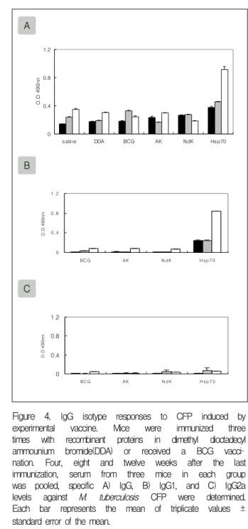
결핵균 CFP에 대한 항체 반응에서는 Hsp70을 면 역주사 한 마우스에서만 IgG와 IgG1 생성능이 높게
A
0 0 .5 1 1 .5 2 2 .5
sa lin e D D A BC G H sp7 0
O.D 490nm
B
0 0 .5 1 1 .5 2 2 .5
BC G H sp7 0
O.D 490nm
C
0 0 .5 1 1 .5 2 2 .5
BC G H sp7 0
O.D 490nm
Figure 1. IgG isotype responses to Hsp70 induced by experimental vaccine. Mice were immunized three times with recombinant Hsp70 in dimethyl dioctadecyl ammounium bromide(DDA) or received a BCG vacci
nation. Four, eight and twelve weeks after the last immunization, serum from three mice in each group was pooled, specific A) IgG, B) IgG1, and C) IgG2a levels against Hsp70 were determined. Each bar repr
esents the mean of triplicate values ± standard error of the mean.
■:4wk, ▨:8wk, and □:12wk after the last immunization
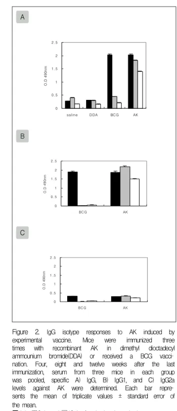
A
0 0.5 1 1.5 2 2.5
salin e DDA BCG AK
O.D 490nm
B
0 0 .5 1 1 .5 2 2 .5
BC G AK
O.D 490nm
C
0 0 .5 1 1 .5 2 2 .5
BC G AK
O.D 490nm
Figure 2. IgG isotype responses to AK induced by experimental vaccine. Mice were immunized three times with recombinant AK in dimethyl dioctadecyl ammounium bromide(DDA) or received a BCG vacci
nation. Four, eight and twelve weeks after the last immunization, serum from three mice in each group was pooled, specific A) IgG, B) IgG1, and C) IgG2a levels against AK were determined. Each bar repre
sents the mean of triplicate values ± standard error of the mean.
■:4wk, ▨:8wk, and □:12wk after the last immunization 나타났다(Fig.4).
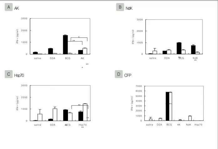
2. IFN-γ 생성능
AK에 대한 IFN-γ 생성능에서는, 마지막 면역주사
후 8주째에 BCG를 면역주사 한 마우스에서 음성대조 군인 생리식염수와 DDA를 면역주사 한 마우스와 비 교하여 통계적으로 IFN-γ의 생성이 증가되었고(P<
0.01), 12주째에서는 증가된 양상을 나타내지 못하였 다. 재조합 단백질 AK를 면역주사 한 마우스에서는
A
0 0.5 1 1.5 2 2.5
saline DDA BCG N dK
O.D 490nm
B
0 0 .5 1 1 .5 2 2 .5
BC G N dK
O.D 490nm
C
0 0 .5 1 1 .5 2 2 .5
BC G N dK
O.D 490nm
Figure 3. IgG isotype responses to NdK induced by experimental vaccine. Mice were immunized three times with recombinant NdK in dimethyl dioctadecyl ammounium bromide(DDA) or received a BCG vaccin
ation. Four, eight and twelve weeks after the last immunization, serum from three mice in each group was pooled, specific A) IgG, B) IgG1, and C) IgG2a levels against NdK were determined. Each bar repre
sents the mean of triplicate values ± standard error of the mean.
■:4wk, ▨:8wk, and □:12wk after the last immunization
A
0 0.4 0.8 1.2
saline DDA BCG AK NdK Hsp70
O.D 490nm
B
0 0 .4 0 .8 1 .2
BC G AK N d K H sp 7 0
O.D 490nm
C
0 0.4 0.8 1.2
BC G AK N d K H sp 7 0
O.D 490nm
Figure 4. IgG isotype responses to CFP induced by experimental vaccine. Mice were immunized three times with recombinant proteins in dimethyl dioctadecyl ammounium bromide(DDA) or received a BCG vacci
nation. Four, eight and twelve weeks after the last immunization, serum from three mice in each group was pooled, specific A) IgG, B) IgG1, and C) IgG2a levels against M. tuberculosis CFP were determined.
Each bar represents the mean of triplicate values ± standard error of the mean.
■:4wk, ▨:8wk, and □:12wk after the last immunization 생리식염수를 면역주사 한 마우스와 비교하여 면역주
사 후 8주째(P<0.05)와 12주째(P<0.01)에서 IFN-γ의 생성이 증가되었고, DDA를 면역주사 한 마우스와 비 교에서는 12주째에 증가되었다(P<0.05). BCG와 재조 합 단백질 AK를 면역주사 한 마우스간의 비교에서는
면역주사 후 8주째에는 BCG를 면역주사 한 마우스에 서 IFN-γ의 생성이 증가되었고(P<0.01), 12주째에는 재조합 단백질 AK를 면역주사 한 마우스에서 IFN-γ 의 생성이 증가되었다(P<0.05).
NdK에 대한 IFN-γ 생성능에서는, 마지막 면역주 사 후 8주째에 BCG를 면역주사 한 마우스에서 생리 식염수와 DDA를 면역주사 한 마우스와 비교하여 통 계적으로 IFN-γ의 생성이 증가되었고(P<0.01), 12주
A AK B NdK
0
1 0 0 0 2 0 0 0 3 0 0 0
sa lin e DDA BC G AK
IFN-r (pg/ml)
0
1 0 00 2 0 00 3 0 00
sa lin e D D A BC G N dK
IFN-r (pg/ml)
C Hsp70 D CFP
0
1 0 0 0 2 0 0 0 3 0 0 0
sa lin e D D A BC G H sp7 0
IFN-r (pg/ml)
0
1 0 00 2 0 00 3 0 00 4 0 00 5 0 00 6 0 00 7 0 00
sa lin e DDA BC G AK N dK Hsp7 0
IFN-r (pg/ml)
Figure 5. IFN-γ production by splenocyte of immunized mice.
Spleen cells prepared from mice eight and twelve weeks after last immunization. Cells were pooled from three mice per group and restimulated in vitro with recombinant proteins or M. tuberculosis CFP. Each point represents the mean of triplicate values ± standard error of the mean. *, P<0.05; **, P<0.01 compared to controls or BCG-immunized mice.
■:8wk, and □:12wk after the last immunization
째에서는 증가된 양상을 나타내지 못하였다. 재조합 단백질 NdK를 면역주사 한 마우스에서는 면역주사 후 8주째에 생리식염수(P<0.01)와 DDA(P<0.05)를 면역주사 한 마우스와 비교하여 IFN-γ의 생성이 증가 되었으며 BCG와 재조합 단백질 NdK를 면역주사 한 마우스간의 비교에서는 증가된 양상을 나타내지 못하 였다.
Hsp70에 대한 IFN-γ 생성능에서는, 마지막 면역주 사 후 8주째에 BCG를 면역주사 한 마우스에서 생리 식염수와 DDA를 면역주사 한 마우스와 비교하여 통 계적으로 IFN-γ의 생성이 증가되었고(P<0.01), 12주 째에서는 증가된 양상을 나타내지 못하였다. 재조합 단백질 Hsp70을 면역주사 한 마우스에서는 면역주사 후 8주째와 12주째에 생리식염수와 DDA를 면역주사
한 마우스와 비교하여 IFN-γ의 생성이 증가되었고 (P<0.01), BCG와 재조합 단백질 Hsp70을 면역주사 한 마우스간의 비교에서는 면역주사 후 12주째에 재 조합 단백질 Hsp70을 면역주사 한 마우스에서 IFN-γ 의 생성이 증가되었다(P<0.01).
결핵균 CFP(culture filtrate protein)에 대한 IFN-γ 생성능에서는, BCG를 면역주사 한 마우스에만 면역 주사 후 8주째(P<0.01)와 12주째(P<0.01)에서 IFN-γ 의 생성이 증가되었다(Fig.5).
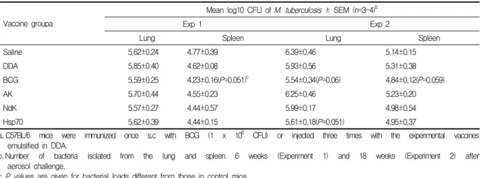
3. 결핵균에 대한 방어면역효능
결핵균에 대한 방어면역효능에서는 생리식염수를 면역주사 한 마우스의 결핵균 수와 비교한 결과, 신뢰
**
**
** *
*
** **
** **
**
** **
**
Table 1. Protective efficacy of recombinant proteins emulsified in DDA in a mouse model of tuberculosis.
Vaccine groupa
Mean log10 CFU of M. tuberculosis ± SEM (n=3-4)b
Exp 1 Exp 2
Lung Spleen Lung Spleen
Saline 5.62±0.24 4.77±0.39 6.39±0.46 5.14±0.15
DDA 5.85±0.40 4.62±0.08 5.93±0.56 5.31±0.38
BCG 5.59±0.25 4.23±0.16(P=0.051)c 5.54±0.34(P=0.06) 4.84±0.12(P=0.059)
AK 5.70±0.44 4.55±0.23 6.25±0.46 5.23±0.20
NdK 5.57±0.27 4.44±0.57 5.99±0.17 4.98±0.54
Hsp70 5.62±0.39 4.44±0.15 5.61±0.18(P=0.051) 4.95±0.37
a. C57BL/6 mice were immunized once s.c with BCG (1 x 10⁶ CFU) or injected three times with the experimental vaccines emulsified in DDA.
b. Number of bacteria isolated from the lung and spleen 6 weeks (Experiment 1) and 18 weeks (Experiment 2) after aerosol challenge.
c. P values are given for bacterial loads different from those in control mice.
수준 95% 범위(P<0.05)에서 유의성이 없어서 결핵에 대한 재조합 단백질들의 백신 후보 물질로서의 효능 성을 나타내지 못하였다. 그러나 생리식염수를 면역 주사 한 마우스와 비교하여 BCG를 면역주사 한 마우 스에서 공기 감염 후, 6주째 비장에서 통계학적 유의 성 경향을 보였고(P=0.051), 재조합 단백질 Hsp70을 면역주사 한 마우스에서도 공기 감염 후 18주째 폐에 서 유의성 경향을 나타내었다(P=0.051)(Table 1).
고 찰
다수의 실험들에 의해 결핵균의 CF (culture filtrate), 세포벽, 및 세포질에 포함된 단백질들이 높은 면역원 을 지니고 있어 백신에 이용할 후보 물질로서 판명되 고 있다15-17. 이들은 ESAT-6, 85 complex, 38kDa 등의 단백질로서 여러 유형의 백신 연구에 이용되고 있다.
새로운 백신 후보물질의 탐색을 목적으로, 본 실험 은 마우스에 대한 공기 감염 모델을 이용하여 결핵균 에 내재된 AK, NdK와 Hsp70의 결핵균에 대한 방어 면역효능을 측정하였다.
각 단백질에 대한 IgG 항체 생성능은 BCG를 면역 주사 한 마우스와 비교하여 재조합 단백질을 면역주 사 한 마우스에서 높게 나타났으나, 결핵균 CFP에 대 한 IgG 항체 생성능에서는 Hsp70을 면역주사 한 마우 스에서만 높은 생성능을 나타내었다. 이는 CFP내에 분포하는 AK와 NdK의 양이 매우 적기 때문인 것으
로 예측되었다.
또한 CD4+ T 세포의 분화 방향을 알아보기 위하여 IgG isotype중 IgG1과 IgG2a에 대한 생성능을 확인한 결과, IgG1에 대해서 높은 생성능을 나타내어 Th2 세 포로 분화되는 것으로 판단되었다. 그러나, 시간이 경 과함에 따라 BCG를 주사한 마우스는 IgG2a/IgG1 비 율을 비추어 볼 때 Th1/Th2 세포의 혼합된 양상으로 분화하는 것으로 나타내었다.
Lindbland 등은 C57BL/6J 마우스 모델로 결핵균 CFP와 여러 면역보강제를 이용한 백신 실험에서 DDA를 주사한 마우스에서 강한 IgG2a 반응이 나타 나 Th1 세포를 자극한다고 보고하였으나18, 이전의 보 고에서는 항원 농도가 높음에 따라 IgG1의 생성이 두드 러지게 나타나 Th2 세포를 자극한다고 보고하였다19.
Martin은 C57BL/6 마우스 모델을 이용한 백신 실 험에서 IgG2a 생성능을 조사하는 점에 대해서 문제 제기를 하였는데 Igh1-b allele를 포함한 마우스(C57BL /6, C57BL/10, SJL등)들은 IgG2a의 유전자가 제거되 어 있고 상품화된 anti-IgG2a 혈청과 반응을 하지 않 는 IgG2c isotype을 발현하기 때문에 IgG2a 생성능을 측정하는 것은 불가능하다고 보고하였다20. 이러한 내 용을 기초로, 본 실험에서도 IgG2b 항체 생성능을 측 정하였으나 오히려 IgG2a 항체 생성능에 비해 낮게 나타내었다.
IFN-γ 생성능의 결과에서는 재조합 단백질들을 면 역주사 한 마우스에서 각 항원에 대한 IFN-γ 생성이
대조군과 비교하여 증가되었으나 결핵균 CFP에 대해 서는 IFN-γ 생성이 낮게 나타나 이들 재조합 단백질과 DDA를 이용한 면역반응은 세포성 면역반응이 아닌 체 액성 면역반응으로 일어나는 것으로 확인하였다.
면역 효과를 증가시키기 위하여 면역보강제(adjuvant) 로 세포성 면역반응을 유도하고 낮은 독성을 지닌 DDA (dimethyl dioctadecylammonium bromide)를 선택하였다. 이 면역보강제는 결핵균의 단백질 백신 연구에 이용되고 있으며14,18,19,21
, 여러 면역보강제와 결핵균 항원을 이용한 백신 실험에서 인체 내에 주로 이용되는 Alum에 비해 결핵균에 대한 방어면역효능 이 높은 것으로 나타났다. 그러나 본 실험에서는 결핵 균에 대한 방어면역효능을 나타내지 못하였다. 이러 한 결과는 백신 후보물질로서의 단일 항원의 면역원 능과 그에 따른 면역보강제의 선택에 따른 것으로 판 단된다. 면역보강제의 선정을 목적으로 한 실험들에 서는 결핵균 CFP와 DDA를 이용하여 면역주사 한 마우 스에서 결핵균에 대한 방어면역효능을 나타내었다18,19. 그러나 동일한 면역보강제와 단일 항원을 이용한 백 신 실험에서는 단백질의 종류에 따라 상이한 결과를 나타내었는데, 항원 85B를 이용한 실험에서는 결핵균 에 대한 방어면역효능을 나타낸 반면, 항원 ESAT-6 를 이용한 실험에서는 방어면역효능을 나타내지 못하 였다. Brandt 등은 항원 ESAT-6의 약한 면역원능에 대한 보완책으로 DDA와 Th1 면역반응을 유도하는 다른 면역보강제를 첨가함으로써 방어면역효능을 나 타내었다14.
결핵균 AK와 NdK는 현재까지 구조적 특성 및 대 사적 기능에 대한 연구 보고가 있으며, 아직까지 면역 학적 항원성에 대한 연구 보고는 없는 실정이다. 최근 에는 재조합 단백질 NdK와 ATP를 대식세포에 처리 하였을 때 대식세포의 사멸이 유도된다는 보고가 있 어 병원성 세균의 감염 경로등에 중요한 역할을 할 것 으로 예측되고 있다8.
결핵균 Hsp70은 proteomics의 발달에 따라 결핵균 항원 후보물질로서 거론되어 왔으며, Hsp70을 과발현 하는 결핵균은 in vivo상에서 생존율이 감소하는 것으 로 나타나 면역 반응에 영향을 미치는 것으로 보고되 었다22. 최근에는 Hsp70등 Heat shock protein의 항원
제시(antigen-presentation)기능이 부각되면서 감염된 생체 내에서 분리한 Hsp-peptide 복합체를 면역주사 한 뒤, 결핵균을 감염시킨 결과에서 결핵균에 대한 방 어면역효능을 나타낸 보고가 있다23.
본 실험에서도 재조합 단백질 Hsp70을 면역주사 한 마우스의 폐에서 결핵균에 대한 방어면역 효능이 BCG를 면역주사 한 마우스와 유사한 결과를 나타내었 다. 이는 Hsp70의 단일 항원으로서의 면역원능보다는 다른 기능에 의한 결과로 판단된다.
현재 재조합 단백질 백신 연구는 복합 항원과 복합 면역보강제를 이용한 방법14,21, 새로운 면역보강제의 효능 비교24, 접종 경로의 다양화25 및 cytokine의 첨가 방법26등을 이용한 결핵균에 대한 방어면역효능을 실 시하고 있다.
본 실험에 사용한 재조합 단백질들과 DDA를 이용 한 면역 효과는 세포성 면역반응이 아닌 체액성 면역 반응을 유도함으로 인해 결핵균에 대한 방어면역효능 을 나타내지 않았으므로, 단일 항원으로서의 면역원 능이 없거나 혹은 취약한 것으로 판단되었다. 따라서 혼합 단백질 혹은 다른 T세포 면역보강제의 사용에 의한 추시가 필요하다.
요 약
배 경 :
최근 결핵에 대한 새로운 백신 개발은 초회 면역 방 법 및 추가 면역 방법을 이용하는 방향으로 연구되고 있다. 본 실험은 새로운 백신 후보 물질로서의 가능성 을 알아보기 위하여 결핵균 adenylate kinase (AK), nu
cleoside diphosphate (NdK) 및 heat shock protein 70 (Hsp70)의 결핵균에 대한 방어면역효능을 측정하였다.
방 법 :
재조합 단백질들을 정제하기 위하여 중합효소 연쇄 반응으로 증폭한 결핵균 유전자 단편들을 E.coli expression vector, pQE30에 클로닝한 후, Ni-NTA resin을 이용하여 정제하였다. DDA와 재조합 단백질 들을 마우스에 면역주사하고 면역반응 생성 유무를 확인하기 위하여 항체와 IFN-γ 생성능을 측정하였다.
면역주사 한 마우스에 결핵균을 공기 감염시킨 후, 폐
와 비장을 분리하여 결핵균 생균수 실험을 하였다.
결 과 :
재조합 단백질 AK, NdK 와 Hsp70을 면역보강제인 DDA를 이용하여 면역주사 한 결과에서, 생리식염수 혹은 DDA를 면역주사 한 마우스에 비교하여 재조합 단백질을 면역주사 한 마우스에서는 각 항원에 대해 항체와 IFN-γ 생성능이 높게 나타났으나 결핵균에 대 한 효과적인 방어면역효능은 나타나지 않았다.
결 론 :
마우스를 모델로 한 결핵균에 대한 방어면역효능 실험에서, 면역보강제 DDA를 이용한 재조합 단백질 AK, NdK 및 Hsp70을 면역주사 한 경우에는 결핵균 의 성장을 효과적으로 조절하지 못하였다. 혼합 단백 질 혹은 다른 T세포 면역보강제의 사용에 의한 추시 가 필요하다.
참 고 문 헌
1. Olsen AW, Andersen P. A novel TB vaccine: strategies to combat a complex pathogen. Immunol Lett 2003;
85:207-11.
2. Fine PE. Variation in protection by BCG: implications of and for heterologous immunity. Lancet 1995;346:
1339-45.
3. Agger EM, Anderson P. A novel TB vaccine; towards a strategy based on our understanding of BCG failure.
Vaccine 2002;21:7-14.
4. Markaryan A, Zaborina O, Punj V, Chakrabarty AM.
Adenylate kinase as a virulence factor of Pseudomonas aeruginosa. J Bacteriol 2001;183:3345-52.
5. Munier-Lehmann H, Burlacu-Miron S, Craescu CT, Mantsch HH, Schultz CP. A new subfamily of short bacterial adenylate kinase with the Mycobacterium tuberculosis enzyme as a model: a predictive and experimental study. Proteins 1999;36:238-48.
6. Chakrabarty AM. Nucleoside diphosphate kinase: role in bacterial growth, virulence, cell signalling and polysaccharide synthesis. Mol Microbiol 1998;28:
875-82.
7. Chen Y, Morera S, Mocan J, Lascu I, Janin J. X- ray structure of Mycobacterium tuberculosis nucleoside diphosphate kinase. Proteins 2002;47:556-7.
8. Chopra P, Singh A, Koul A, Ramachandran S, Drlica K, Tyagi AK, Singh Y. Cytotoxic activity of nucleoside diphosphate kinase secreted from Myco
bacterium tuberculosis. Eur J Biochem 2003;270:
625-34.
9. Britton WJ, Hellqvist L, Basten A, Raison RL.
Mycobacterium leprae antigens involved in human responses: identification of four antigens by mono
clonal antibodies. J Immunol 1985;135:4171-7.
10. Engers HD, Houba V, Bennedsen J, Buchanan TM, Chaparas SD, Kadival G, et al. Results of a World Health Organization-sponsored workshop to characterise antigens recognised by mycobacterium-specific mo
noclonal antibodies. Infect Immun 1986;51:718-20.
11. Britton WJ, Hellqvist L, Basten A, Inglis AS. Im
munoreactivity of a 70kD protein purified from Mycobacterium bovis bacillus Calmette-Guerin by monoclonal antibody affinity chromatography. J Exp Med 1986;164:695-708.
12. Adams E, Garsia RJ, Hellqvist L, Holt P, Basten A.
T cell reactivity to the purified mycobacterial antigens p65 and p70 in leprosy patients and their household contacts. Clin Exp Immunol 1990;80:206-12.
13. Jungblut PR, Schaible UE, Mollenkopf HJ, Zimny- Arndt U, Raupach B, Mattow J, et al. Comparative proteome analysis of Mycobacterium tuberculosis and Mycobacterium bovis BCG strains: towards functional genomics of microbial pathogens. Mol Microbiol 1999;33:1103-17.
14. Brandt L, Elhay M, Rosenkrands I, Lindblad EB, Andersen P. ESAT-6 subunit vaccination against Mycobacterium tuberculosis. Infect Immun 2000;68:791-5.
15. Rosenkrands I, Weldingh K, Jscobsen S, Hansen CV, Florio W, Gianetri I, et al. Mapping and identification of Mycobacterium tuberculosis proteins by two-dimensional gel electrophoresis, microse
quencing and immunodetection. Electrophoresis 2000;
21:935-48.
16. Rosenkrands I, King A, Weldingh K, Moniatte M, Moertz E, Adersen P. Towards the proteome of Mycobacterium tuberculosis. Electrophoresis 2000;21:
3740-56.
17. Covert BA, Spencer JS, Orme IM, Belisle JT. The application of proteomics in defining the T cell antigens of Mycobacterium tuberculosis. Proteomics 2001;1:574-86.
18. Lindbland EB, Elhay MJ, Silva R, Appelberg R, Andersen P. Adjuvant modulation of immune res
ponses to tuberculosis subunit vaccines. Infect Immun 1997;65:623-9.
19. Adersen P. Effective vaccination of mice against Mycobacterium tuberculosis infection with a soluble mixture of secreted mycobacterial proteins. Infect Immun 1994;62:2536-44.
20. Martin RM, Lew AM. Is IgG2a a good Th1 marker in mice? Immunol Today 1998;19:49.
21. Weinrich-Olsen A, van Pinxteren LA, Meng Okkels L, Birk Rasmussen P, Andersen P. Protection of mice with a tuberculosis subunit vaccine based on a fusion protein of antigen 85b and east-6. Infect Immun 2001;69:2773-8.
22. Stewart GR, Snewin VA, Walzl G, Hussell T, Tormay P, O'Gaora P, et al. Overexpression of heat-shock proteins reduces survival of Mycobacterium tuber
culosis in the chronic phase of infection. Nat Med 2001;7:732-7.
23. Colaco CA, Bailey CR, Keeble J, Walker KB. BCG (Bacille Calmette-Guerin) HcpCs(heat-shock protein-
peptide complexes) induce T-helper responses and protect against live challenge in a murine aerosol challenge model of pulmonary tuberculosis. Biochem Soc Trans 2004;32:626-8.
24. Hogarth PJ, Jahans KJ, Hecker R, Hewinson RG, Chanbers MA. Evaluation of adjuvants for protein vaccines against tuberculosis in guinea pig. Vaccine 2003;21:977-82.
25. Doherty TM, Olsen AW, van Pinxteren L, Andersen P. Oral vaccination with subunit vaccines protects animals against aerosol infection with Mycobacterium tuberculosis. Infect Immun 2002;70:3111-21.
26. Silva RA, Pais TF, Appelberg R. Effects of inter
leukin-12 in the long-term protection conferred by a Mycobacterium avium subunit vaccine. Scand J Immunol 2000;52:531-3.