82
http://dx.doi.org/10.4046/trd.2013.74.2.82 ISSN: 1738-3536(Print)/2005-6184(Online) Tuberc Respir Dis 2013;74:82-85
CopyrightⒸ2013. The Korean Academy of Tuberculosis and Respiratory Diseases. All rights reserved.
Successful Removal of Endobronchial Lipoma by Flexible Broncho- scopy Using Electrosurgical Snare
Seong Cheol Yun, M.D.1, Moon Jun Na, M.D.1, Eugene Choi, M.D.1, Sun Jung Kwon, M.D.1, Seong Ju Lee, M.D.1, Sun Hee Oh, M.D.1, Eun Jung Cha, M.D.2, Ji Woong Son, M.D.1
Departments of 1Internal Medicine and 2Pathology, Konyang University Hospital, Konyang University College of Medicine, Daejeon, Korea
A 62-year-old man with a chronic cough presented with atelectasis of the left upper lobe on chest X-ray. Chest computed tomography showed an atelectasis in the left upper lobe with bronchial wall thickening, stenosis, dilatation, and mucoid impaction. We performed bronchoscopy and found a well-circumscribed mass on the left upper lobe bronchus. The mass was removed by flexible bronchoscopy using an electrosurgical snare and diagnosed with lipoma. An endobronchial lipoma is a rare benign tumor that can be treated by a surgical or endoscopic approach. We report the successful removal of endobronchial lipoma via flexible bronchoscopic electrosurgical snare.
Key Words: Bronchoscopy; Electrocoagulation; Lipoma
Address for correspondence: Ji Woong Son, M.D.
Department of Internal Medicine, Konyang University Hospital, Konyang University College of Medicine, 158, Gwanjeodong-ro, Seo-gu, Daejeon 302-718, Korea Phone: 82-42-600-8817, Fax: 82-42-600-9090 E-mail: [email protected]
Received: Jun. 23, 2012 Revised: Jul. 3, 2012 Accepted: Jul. 20, 2012
CCIt is identical to the Creative Commons Attribution Non-Commercial License (http://creativecommons.org/licenses/by-nc/3.0/).
Introduction
Benign tumors originating from the tracheobronchial tree are very uncommon, especially incidence of endo- bronchial lipoma in all lung tumors ranges from only 0.1% to 0.5% in all lung tumors
1. Lipomas are slow-growing tumors that can remain clinically silent for a prolonged period. When symptoms develop, they are usually manifest as recurrent infections, which, if re- maining undiagnosed over time, can lead to bron- chiectasis secondary to endobronchial obstruction
2. Clinical manifestations of endobronchial lipoma are pre- sented variably, cough, wheezing, hemoptysis, chest pain, atelectasis in chest X-ray, recurrent pneumonia,
and, rarely, empyema
3-5. The removal of endobronchial lipoma can be achieved by surgical or bronchoscopic methods; generally through rigid bronchoscopy
2,6. We present a case of endobronchial lipoma that was suc- cessfully removed via flexible bronchoscopy using an electrosurgical snare.
Case Report
A 62-year-old man visited the respiratory outpatient department with symptoms of a chronic cough and ate- lectasis of the left upper lobe on chest X-ray. The pa- tient had no specific past history and family history.
Physical examination revealed clear breath sound with- out rales and wheezing sound. Laboratory tests were within normal range. Pulmonary function tests revealed a mild obstructive pattern (forced expiratory volume 1 second [FEV1], 96%; FEV1/forced vital capacity, 69%).
A chest computed tomography (CT) revealed atelectasis, bronchial wall thickening, stenosis, dilatation, and mu- coid impaction in the left upper lobe, without definite mass lesion (Figure 1A, C). Lymph node enlargements were apparent in the right hilum and left lower
Case Report
Tuberculosis and Respiratory Diseases Vol. 74. No. 2, Feb. 2013
83 Figure 1. Chest X-ray and chest computed tomog- raphy (CT) image. (A) At admission, chest X-ray re- vealed atelectasis. (B) After 1.5 months, chest X-ray showed improvement of atelectasis. (C) At admis- sion, chest CT revealed mucoid impaction in left upper lobe bronchus. (D) After 1.5 months, chest CT showed no definite mass lesion in the left upper lobe bronchus.
paratrachea. Two focal calcified granuloma were seen in the right lung.
Flexible bronchoscopy was performed and revealed a well-circumscribed mass on the left upper lobe bron- chus. Biopsy was done and showed subepithelial ma- ture adipose tissue. We tried to remove a mass with flexible bronchoscopic electrosurgical snare (Figure 2).
After removal of the mass, a base remained on left up- per lobe B1+B2. An additional coagulation using snare tip was carried out to the remove residual tumor base and to control the bleeding.
The patient was discharged and then followed up in the outpatient department. After 1.5 months, a fol- low-up bronchoscopy (Figure 3) and chest CT (Figure 1B, D) revealed remaining mass in left upper lobe B2.
The remained mass was removed completely by
forceps. On follow-up CT scans, atelectastasis was re- mained but no mass lesion was present in left upper lobe. The patient has been followed up in outpatient department regularly.
Discussion
There are many methods to remove the endobron-
chial lipoma including surgical resection and endo-
scopic excision
7,8. The majority of lipomas occur in the
first three subdivisions of the tracheobronchial tree
where there is abundant cartilage and adipocytes. Also,
lipomas are poorly vascularized tumors. Therefore, they
are amenable to endoscopic management both for diag-
nosis and treatment
2,3. Endoscopic removal of lipoma
includes laser ablation, electrocauterization, cryo-recan-
SC Yun et al: Sucessful treatment of endobronchial lipoma
84
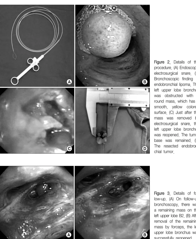
Figure 2. Datails of the procedure. (A) Endoscopic electrosurgical snare. (B) Bronchoscopic finding of endobronchial lipoma. The left upper lobe bronchus was obstructed with a round mass, which has a smooth, yellow colored surface. (C) Just after the mass was removed by electrosurgical snare, the left upper lobe bronchus was reopened. The tumor base was remained. (D) The resected endobron- chial tumor.
Figure 3. Details of fol- low-up. (A) On follow-up bronchoscopy, there was a remaining mass on the left upper lobe B2. (B) After removal of the remaining mass by forceps, the left upper lobe bronchus was successfully reopened.
alization, and, as seen our case report, electrosurgical snaring
9. Removal of endobronchial lipoma using flexi- ble bronchoscopic electrosurgical snaring has some ad- vantages. It does not require general anesthesia, can di- agnose pathology and remove the mass at the same
time, and is less invasive than surgical resection. But if there is massive bleeding during procedure, it is diffi- cult to control bleeding and protect the airway than rig- id bronchoscopy
10.
Muraoka et al.
1reviewed the cases of endobronchial
Tuberculosis and Respiratory Diseases Vol. 74. No. 2, Feb. 2013