• 검색 결과가 없습니다.

Successful Removal of Endobronchial Blood Clots Using Bronchoscopic Cryotherapy at Bedside in the Intensive Care Unit

N/A
N/A
Protected

Academic year: 2021

Share "Successful Removal of Endobronchial Blood Clots Using Bronchoscopic Cryotherapy at Bedside in the Intensive Care Unit"

Copied!
4
0
0

로드 중.... (전체 텍스트 보기)

전체 글

(1)

Successful Removal of Endobronchial Blood Clots Using Bronchoscopic Cryotherapy at Bedside in the Intensive Care Unit

Hongyeul Lee, M.D., Cho Sun Leem, R.N., Jae Ho Lee, M.D., Choon-Taek Lee, M.D. and Young-Jae Cho, M.D., M.P.H.

Division of Pulmonary and Critical Care Medicine, Department of Internal Medicine, Seoul National University College of Medicine, Seoul National University Bundang Hospital, Seongnam, Korea

Acute airway obstruction after hemoptysis occurs due to the presence of blood clots. These conditions may result in life- threatening ventilation impairment. We report a case of obstruction of the large airway by endobronchial blood clots which were removed using bronchoscopic cryotherapy at the bedside of intensive care unit. A 66-year-old female with endometrial cancer who had undergone chemotherapy, was admitted to the intensive care unit due to neutropenic fever. During mechanical ventilation, the minute ventilation dropped to inadequately low levels and chest radiography showed complete opacification of the left hemithorax. Flexible bronchoscopy revealed large blood clots obstructing the proximal left main bronchus. After unsuccessful attempts to remove the clots with bronchial lavage and forceps extraction, blood clots were removed using bronchoscopic cryotherapy. This report shows that cryotherapy via flexible bronchoscopy at the bedside in the intensive of intensive care unit is a simple and effective alternative for the removal of endobronchial blood clots.

Keywords: Bronchi; Bronchoscopy; Cryotherapy; Hemorrhage

resolve rapidly when blood clots are coughed up. Endobron- chial blood clot formation is not always preceded by hemop- tysis prior to the development of sudden airway obstruction particularly in patients on mechanical ventilation

4

. We report a case of airway obstruction due to endobronchial blood clots during mechanical ventilation. The clots were removed using bronchoscopic cryotherapy at bedside of intensive care unit (ICU).

Case Report

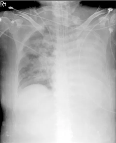
A 66-year-old female with endometrial cancer who had undergone chemotherapy with doxorubicin and cisplatin 8 days previously, presented to the hospital with neutropenic fever and pancytopenia. She was admitted to the hospital and treated with broad-spectrum antibiotics. Three days after admission, she developed hypoxic respiratory failure requiring intubation (endotracheal tube diameter, 8.0 mm) for acute pulmonary edema and septic shock. During mechani- cal ventilation, the minute ventilation suddenly dropped to Copyright © 2014

The Korean Academy of Tuberculosis and Respiratory Diseases.

All rights reserved.

Introduction

An endobronchial blood clot is an unsusal, albeit not rare cause of airway obstruction

1-3

. In spontaneously breathing pa- tients, the clinical features of endobronchial blood clots may

CASE REPORT

http://dx.doi.org/10.4046/trd.2014.77.4.193

ISSN: 1738-3536(Print)/2005-6184(Online) • Tuberc Respir Dis 2014;77:193-196

193

Address for correspondence: Young-Jae Cho, M.D., M.P.H.

Division of Pulmonary and Critical Care Medicine, Department of Internal Medicine, Seoul National University College of Medicine, Seoul National University Bundang Hospital, 82 Gumi-ro 173beon-gil, Bundang-gu, Seongnam 463-707, Korea

Phone: 82-31-787-7058, Fax: 82-31-787-4070 E-mail: [email protected]

Received: May 27, 2014 Revised: Jun. 10, 2014 Accepted: Jun. 25, 2014

cc

It is identical to the Creative Commons Attribution Non-Commercial

License (http://creativecommons.org/licenses/by-nc/3.0/).

(2)

H Lee et al.

194 Tuberc Respir Dis 2014;77:193-196 www.e-trd.org

significantly low levels and mildly blood-tinged material was obtained when suctioning the endotracheal tube. Chest radi- ography revealed no change from previous radiography. Next morning; 12 hours later, chest radiography showed a com- pletely opacified left hemithorax (Figure 1). On auscultation, there were no breath sounds over the left chest. The patient’s platelets were 38,000/μL. Flexible bronchoscopy (5.9 mm di- ameter, 2.8 mm channel diameter; BF-1T260; Olympus, Tokyo, Japan) at bedside of ICU revealed a large blood clot obstruct- ing the proximal left main bronchus (Figure 2). Because of the friable structure of the clot, bronchoscopic lavage and forceps extraction were unsuccessful. After repeated attempts to re- move the blood clot over one hour, a flexible cryoprobe (2.4 mm diameter; ERBE, Tubingen, Germany) was applied via flexible bronchoscopy to the blood clot and frozen for 10 sec- onds. The blood clot firmly attached to the probe and was suc-

cessfully removed in four large pieces (Figures 3, 4). A follow- up chest X-ray revealed significant improvement in aeration of the left lung (Figure 5). Fifteen minutes were required to remove the blood clots using bronchoscopic cryotherapy. No further obstructive events occurred. A repeat bronchoscopic evaluation 2 weeks later showed no evidence of the clot and all of the airways were patent. She recovered from septic shock and discharged from hospital on the 24th day.

Discussion

Acute airway obstruction due to the presence of blood clots occurs in a variety of clinical conditions, including complica- tions of respiratory disease, such as tuberculosis, bronchiecta- sis, bronchial carcinoma, and pulmonary arteriovenous mal- Figure 1. Chest radiography showed a completely opacified left

hemithorax.

Figure 2. Left main bronchus obstructed by blood clots.

Figure 3. (A, B) Bronchoscopic cryothera-

py.

(3)

Bronchoscopic cryotherapy of endobronchial blood clots at bedside of ICU

http://dx.doi.org/10.4046/trd.2014.77.4.193 195

www.e-trd.org

formations, as well as other causes, such as anticoagulation or pancytopenia

1-6

. These conditions may result in lifethreaten- ing impairment of ventilation. The initial efforts to remove an endobronchial clot should involve flexible bronchoscopic evaluation with saline irrigation and suction

7

. Forceps extrac- tion through the working channel or removal of the clot with the forceps and bronchoscope together may be necessary. If all of the aforementioned methods fail, the next step is rigid bronchoscopy. Rigid bronchoscopy provides greater advan- tages in terms of direct suctioning and forceps extraction with large rigid bore suction catheters and forceps. However, patients undergoing rigid bronchoscopy need to be extubated before the procedure. Although mechanical ventilation can be performed through a side-port channel, the previously im- paired ventilator status may be worse. Furthermore this tech- nique requires the expertise of specialists. Recently, alternative methods for removing endobronchial blood clots have been introduced, such as a Fogarty arterial embolectomy catheter or application of thrombolytics via a flexible bronchoscopy

8,9

. These methods are only recommended when severe respira- tory compromise is present and other alternatives have failed.

Bronchoscopic cryotherapy has been used for the biopsy of airway pathology and the ablation of endobronchial lesions, such as a malignant masses or benign tracheal stenosis

10-12

. Owing to its freezing characteristics, bronchoscopic cryother- apy can also be used to remove foreign bodies that contain sufficient water, a procedure referred to as cryoextraction

13

. During cryoextraction, tissue or foreign bodies are frozen to the tip of the probe and removed by pulling on the probe to- gether with the bronchoscopy. The development of a thin and flexible cryoprobe has allowed bronchoscopic cryotherapy without the risks associated with general anesthesia and rigid bronchoscopy

14

.

In this case of our patient, we removed blood clots that in-

duced ventilation dysfunction without extubation and general anesthesia, which are both required for rigid bronchoscopy.

Furthermore, we conducted bronchoscopic cryotherapy at bedside in ICU. It is foreseeable that removal of blood clots using rigid bronchoscopy may lead to a prolonged procedure time due to the removal of only small pieces of the clot with each forceps bite. In contrast, the bronchoscopic cryotherapy technique allows en-bloc removal of the blood clot. An ad- ditional advantage of this technique is that the learning curve to utilize the cryotherapy probe is quite short, and does not require the more extended training needed to master rigid bronchoscopy; something which is not available in many in- stitutions.

Bronchoscopic cryotherapy usually applied after blood clots formation. Previous study described the endoscopic use of cryotherapy to treat actively bleeding diffuse mucosal vas- cular lesions in gastrointestinal tract

15

. Argon plasma coagula- tion, laser, and other thermal modalities have the potential for deep tissue injury with ulceration and bleeding. Cryotherapy resulted in only superficial tissue damage that usually was completely healed by 3 months in the study. Bronchoscopic cryotherapy could be alternative modality to treat not only blood clots but also active bleeding mucosal lesions in bron- chus. One concern about removal endobronchial blood clots using bronchoscopic cryotherapy is that a large blood clot might be difficult to extract through the small sized endotra- cheal tube.

To our knowledge, this is the first case to report the utiliza- tion of bronchoscopic cryotherapy to remove endobronchial blood clots at bed side of ICU. It highlights the utility of cryo- therapy probes through flexible bronchoscopy to remove any object, with sufficient water content at bed side of ICU. We Figure 4. Four large blood clots removed by bronchoscopic cryo-

therapy.

Figure 5. Follow-up chest X-ray revealed significant improvement

in the aeration of the left lung.

(4)

H Lee et al.

196 Tuberc Respir Dis 2014;77:193-196 www.e-trd.org

conclude that these properties of the procedure assisted us in the safe and easy removal of endobronchial blood clots at the ICU bedside, and thus, avoided the use of more invasive ap- proaches such as rigid bronchoscopy.

Conflicts of Interest

No potential conflict of interest relevant to this article was reported.

References

1. Mindline J. Massive collapse of the lung complicating hae- moptysis. Br Med J 1935;2:1201-40.

2. Allen RP, Siefkin AD. Emergency airway clot removal in acute hemorrhagic respiratory failure. Crit Care Med 1987;15:985- 6.

3. Botnick W, Brown H. Endobronchial urokinase for dissolu- tion of massive clot following transbronchial biopsy. Chest 1994;105:953-4.

4. Foucher P, Merati M, Baudouin N, Reybet-Degat O, Camus P, Jeannin L. Fatal ball-valve airway obstruction by an extensive blood clot during mechanical ventilation. Eur Respir J 1996;9:

2181-2.

5. Sakr L, Dutau H. Massive hemoptysis: an update on the role of bronchoscopy in diagnosis and management. Respiration 2010;80:38-58.

6. Manzano-Fernandez S, Pastor FJ, Marin F, Cambronero F, Caro C, Pascual-Figal DA, et al. Increased major bleeding complications related to triple antithrombotic therapy usage

in patients with atrial fibrillation undergoing percutaneous coronary artery stenting. Chest 2008;134:559-67.

7. Arney KL, Judson MA, Sahn SA. Airway obstruction arising from blood clot: three reports and a review of the literature.

Chest 1999;115:293-300.

8. Bodenham AR. Removal of obstructing blood clot from the lower airway: an alternative suction technique. Anaesthesia 2002;57:40-3.

9. Vajo Z, Parish JM. Endobronchial thrombolysis with strepto- kinase for airway obstruction due to blood clots. Mayo Clin Proc 1996;71:595-6.

10. Roh JH, Park TS, Kim SY, Yoo JW, Park GM, Park SY, et al. En- dobronchial hamartoma treated bronchoscopically: report of two cases. Korean J Med 2009;77:625-9.

11. Lee SH, Choi WJ, Sung SW, Kim YK, Kim CH, Zo JI, et al.

Endoscopic cryotherapy of lung and bronchial tumors: a sys- tematic review. Korean J Intern Med 2011;26:137-44.

12. Jung JY, Lee SY, Kim DH, Lee KJ, Lee EJ, Kang EH, et al. Clini- cal benefits and complications of cryotherapy in advanced lung cancer with central airway obstruction. Tuberc Respir Dis 2008;64:272-7.

13. Rubio E, Gupta P, Ie S, Boyd M. Cryoextraction: a novel ap- proach to remove aspirated chewing gum. Ann Thorac Med 2013;8:58-9.

14. De Weerdt S, Noppen M, Remels L, Vanherreweghe R, Meysman M, Vincken W. Successful removal of a massive en- dobronchial blood clot by means of cryotherapy. J Bronchol 2005;12:23-4.

15. Kantsevoy SV, Cruz-Correa MR, Vaughn CA, Jagannath SB,

Pasricha PJ, Kalloo AN. Endoscopic cryotherapy for the treat-

ment of bleeding mucosal vascular lesions of the GI tract: a

pilot study. Gastrointest Endosc 2003;57:403-6.

수치

Figure 2. Left main bronchus obstructed by blood clots.
Figure 5. Follow-up chest X-ray revealed significant improvement  in the aeration of the left lung.

참조

관련 문서