서 론
해면(sponge)은 다양한 공생 미생물 군집을 함유 하는 다세포 무척추동물로써 고착성의 저서생물이 다. 또한 filter feeding을 하는 생물로 미생물을 먹이
로 하거나 미생물과 공생을 하며 살아간다. 이 과정 에서 풍부하고도 다양한 생물학적 활성을 갖는 천연 물을 생산하는 것으로 알려져 있다[8].
Petrosia corticata 해면은 항암, 항균, 세포독성 등 의 생리활성을 갖는 대사산물을 생산하며 이에 관한
* Corresponding author
Phone: 82-10-8799-7498 Fax: 82-42-629-8751 E-mail: [email protected]
This is an open-access journal distributed under the terms of the Creative Commons Attribution Non-Commercial License
(http://creativecommons.org/licenses/bync/3.0)
http://dx.doi.org/10.15433/ksmb.2015.7.2.042 ISSN 2383-5400 (Online)
제주도에 서식하는 Petrosia corticata 해면의 배양가능한 공생세균 군집구조의 계절적 차이
Seasonal Differences of Cultivable Bacterial Communities Associated with the Marine Sponge, Petrosia corticata, Collected from Jeju Island
정종빈, 박진숙*
Jong-Bin Jeong, Jin-Sook Park*
한남대학교 생명시스템과학과, 대전시 유성구 유성대로 1646, 34054, 대한민국
Department of Biological Science and Biotechnology, Hannam University, Daejeon 34054, Republic of Korea (Received 28 December 2015, Revised 1 January 2016, Accepted 4 January 2016)
Abstract The community structure of cultivable bacteria associated with the marine sponge, Petrosia corticata, collected from Jeju Island in summer (September) of 2012 and winter (January) of 2013, were compared by the PCR-ARDRA method. Bacterial strains were cultured for 4 days at 26℃ on Zobell medium and marine agar medium. After PCR amplification of 16S rRNA gene of individual strains, the restriction enzymes MspI and HaeIII were used to make restriction patterns. As a result, 24 ARDRA patterns from the summer sponge and 20 ARDRA patterns from the winter sponge were obtained. The sequencing result of 1-3 selected strains from each pattern showed over 98% similarities with the known sequences from the public database. At the phylum level, the bacterial community structures of both sponges (summer and winter) were identical qualitatively and composed of 4 phyla : Proteobacteria, Actinobacteria, Bacteroidetes, and Firmicutes. Alphaproteobacteria accounted for 42.5% of total in summer sponge and 25.2%
in winter, decreasing in the winter sample. Gammaproteobacteria accounted for 27.5% of total in summer sponge and 35.2% in winter, increasing in the winter sample. At the genus and species level, summer sponge had more diverse bacterial communities than winter sponge. Actinobacteria, Bacteroidetes, and Firmicutes increased in the winter sample.
Keywords : 16S rRNA gene, ARDRA, Bacteria, Petrosia corticata, Sponge
많은 연구가 진행되고 있다. 특히 이들로부터 분리 된 corticatic acids, petrocortynes, petrotetraydiols, pet- rosiacetyenes 등의 구조 및 약리 작용에 대한 많은 연구 보고가 이루어지고 있다[9, 13, 14, 17, 19]. 우리 나라 거문도에서 채집한 Petrosia 해면 종으로부터 세포독성을 갖는 polyacetylene이 보고되기도 하였다 [7, 11]. 이들 해면 유래 생리 활성물질이 해면 공생 세균에 의해 생산되는 것으로 알려지면서[4, 8], 해 면 공생미생물의 다양성과 천연물 profiles에 관한 많 은 연구가 이루어지고 있다[16, 18, 22]. 해면공생세 균의 군집구조 및 다양성에 관한 연구는 배양되지 않는 공생세균까지 검출 가능한 pyrosequencing, de- naturing gradient gel electrophoresis (DGGE) 등 분자 적 방법이 주로 이용되고 있으나 천연물 profiles에 관련되는 세균다양성에 관한 연구는 천연물의 산업 적 생산 등을 고려하여 배양 가능한 세균을 포함하 여 이루어지고 있다[12, 16].
본 연구에서는 제주도에서 P. corticata 해면을 각 각 여름과 겨울에 채집하여 이들의 배양 가능한 공 생세균 다양성을 조사하고 계절에 따른 차이를 파악 하고자 하였다.
재료 및 방법
해면 시료의 채집 및 환경조건
해양 해면 P. corticata에 공생하는 세균 군집의 계절 적 변화를 조사하기 위하여 2012년 9월(여름)과, 2013 년 1월(겨울)에 각각 제주도 서귀포시 대정읍 무릉아 치에서 스쿠버 다이빙을 이용하여 해면을 채집하였 다. 채집한 해면은 멸균된 인공해수(ASW)로 3회 세척 후 4℃에서 운반하여 12시간 이내에 실험에 사용하였 다. 여름철과 겨울철의 채집 장소는 동일지역으로 각각의 GPS는 33° 15′08″N, 126° 11′36″E와 33° 15′03″N, 126° 11′44″E이었으며, 여름철 해 면 채집 일의 수온은 22.7℃, 염도 32.1 PSU이었으 며, 겨울철 채집 일의 수온은 14.4℃, 염도 33.3 PSU 이었다.
해면 공생세균의 분리 및 배양
해면 조각을 멸균된 인공해수로 3회 세척 후, 해면 의 안쪽을 1 cm3 크기로 잘라 인공해수 3 ml 넣어
균질화시킨 다음 10분간 초음파 처리하였다. 10-1부 터 10-4까지 순차 희석한 후 변형된 ZoBell 배지 (peptone 5 g, yeast extract 1 g, FePO4·4H2O 0.01 g, NaCl 23.6 g, MgSO4·7H2O 5.94 g, MgCl2·6H2O 4.53 g, CaCl2 1.3 g, KCl 0.64 g, agar 15 g, DW 1 L, pH 7.2)와 MA 배지 (marine agar 2216, Difco, USA)에 100 μl씩 도말하여 26℃에서 4일간 배양하였다. 여름 과 겨울에 각각 채집된 해면으로부터 여름 해면에서 129개, 겨울 해면에서 130개의 세균 균주를 각각 분 리하고 분리된 균주들의 순수 분리를 위하여 동일한 배지에서 계대 배양하여 단일 콜로니를 얻었다.
DNA 추출 및 16S rRNA gene의 PCR 증폭 염색체 DNA는 분리된 세균 균주의 각 콜로니로 부터 gDNA Isolation kit (Qiagen, Germany)를 사용하 여 분리하였으며 분리된 DNA는 PCR 반응의 주형으 로 사용하였다. 16S rRNA gene의 증폭에는 27f (AGA GTT TGA TCC TGG CTC AG)와 1492r (TAC GGY TAC CTT GTT ACG AC)의 universal primer 쌍을 사용하였다. PCR 반응은 100 ng의 주형 DNA와 5U e-Taq polymerase (Solgent, Korea), 10 mM dNTP, 10× e-Taq buffer, 10 pmol primer를 최종 반응량이 50 μl가 되도록 혼합하여 수행하였다. GeneAmp PCR system 2700 thermal cycler (Applied Biosystems, Version 2.0, USA)를 이용하여 94℃에서 5분간 초기 변성시킨 후, 94℃에서 40초간 변성, 55℃에서 40초 간 냉각, 72℃에서 1분간 신장, 이 과정을 30 cycle 반복 수행한 후 최종적으로 72℃에서 10분간 신장시 켰다. 증폭된 DNA의 확인을 위해서 PCR 반응액 3 μl를 취하여 1% agarose gel (Bio-Rad, USA)을 이용하 여 Mupid-ex (ADVANCE, Japan)로 100 V, 25분간 1×TAE buffer (40 mM Tris-acetate, 1 mM EDTA, pH 8.0)에서 전기영동 하였다. 전기영동 후, EtBr (ethidium bromide, 50 ng/ml)에 10분간 염색하여 Gel Logic 200 (Kodak, USA)을 이용하여 UV하에서 약 1.5 kb 단편을 확인하였다.
ARDRA 분석
Amplified ribosomal DNA restriction analysis (ARDRA) 분석을 위해 2종의 제한효소 Fast Digest HaeIII (Thermo Scientific, Lithuania)와 Fast Digest
MspI (Thermo Scientific, Lithuania)을 사용하였다.
PCR 산물에 각각의 제한 효소를 첨가하여 37℃에서 5분간 반응시켰다. 반응물은 3% agarose gel을 사용 하여 1×TAE buffer로 100 V, 30분간 전기 영동한 후 EtBr로 염색하여 Gel Logic 200을 이용하여 UV하에 서 관찰하여 각 균주의 밴드유형을 확인하였다.
ARDRA type의 패턴 분석을 위해 FPQuestTM (Bio-Rad, Belgium) software를 이용하였다.
염기서열 결정 및 계통수 작성
16S rRNA gene-ARDRA fingerprinting type에 따라 각각의 type별로 1-3개의 분리 균주를 선택하여 여름 해면에서 33균주, 겨울해면에서 28균주, 총 61균주 의 부분 염기 서열(500 bp 이상)을 분석하였다. 증폭 된 PCR 산물은 Wizard SV Gel and PCR Clean-Up System (Promega, USA)을 이용하여 정제한 후 염기 서열 분석(Macrogen, Korea)을 의뢰하였다. 결정된 16S rRNA 유전자의 염기서열은 기 등록된 염기서열 을 대상으로 Blast search를 수행하였다. 각 염기서열 의 alignment는 CLUSTAL W [23]를 이용하여 정렬 하였고 Molecular Evolutionary Genetics Analysis
(MEGA) software version 6.0 [20]로 neighbor-joining 방법을 이용하여 각각의 계통수를 작성하였다.
1,000회 반복 bootstrap 분석에 의해 계통수의 견고성 을 확인하였다.
결과 및 고찰
해면공생세균의 분리 및 ARDRA 분석
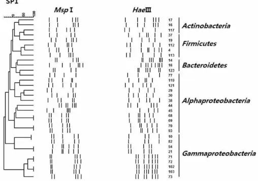
해면 P. corticata의 계절에 따른 배양 가능한 공생 세균 군집 구조의 변화를 관찰하기 위하여 각각 여 름과 겨울에 채집된 P. corticata로부터 배양 가능한 균주를 총 259균주를 분리하여 16S rRNA gene의 ARDRA 분석을 수행하였다. 두 종의 제한 효소를 사용하여 16S rRNA gene-ARDRA 분석을 수행한 결 과(Fig. 1 and 2), 여름 해면의 경우 MspI에 의해 17개 의 type , HaeIII에 의해 24 type을 얻었으며, 이를 근거로 분석한 결과, 총 24개의 서로 다른 ARDRA type으로 구분되었다(Table 1). 겨울 해면의 경우 MspI에 의해 15개의 type, HaeIII에 의해 20개의 type 을 얻었으며, 이를 근거로 분석한 결과, 총 20개의 서로 다른 ARDRA type을 얻을 수 있었다(Table 2).
Figure 1. Dendrogram showing the relationship among bacterial isolates based on the 16S rRNA gene-ARDRA profiles from the marine sponge P. corticata SP1 collected in summer.
Figure 2. Dendrogram showing the relationship among bacterial isolates based on the 16S rRNA gene-ARDRA profiles from the marine sponge P. corticata WP7 collected in winter.
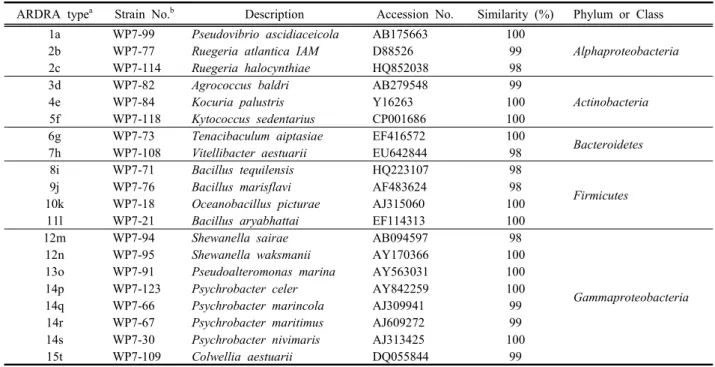
Table 1. ARDRA fingerprinting types and sequence similarities of 16S rRNA gene from bacterial strains isolated from the marine sponge P. corticata SP1 collected in summer.
ARDRA typea Strain No.b Description Accession No. Similarity (%) Phylum or Class 1a SP1-16 Microbacterium esteraromaticum Y17231 99
Actinobacteria
2b SP1-117 Janibacterhoylei FR749912 99
3c SP1-7 Erythrobacter vulgaris AY706935 99
Alphaproteobacteri a
4d SP1-29 Pseudovibrio ascidiaceicola AB175663 100
5e SP1-63 Roseovarius crassostreae AF114484 98
6f SP1-68 Ruegeria atlantica D88526 99
6g SP1-70 Ruegeria atlantica D88526 99
7h SP1-77 Phaeobacter inhibens AY177712 98
8i SP1-119 Labrenzia alba AJ878875 100
9j SP1-14 Spongiibacterium flavum FJ348473 98 Bacteroidetes
10k SP1-4 Bacillus nanhaiensis GU477780 100
Firmicutes
11l SP1-19 Bacillus aryabhattai EF114313 100
12m SP1-37 Planococcus donghaensis EF079063 99
13n SP1-112 Oceanobacillus profundus DQ386635 100
14o SP1-113 Bacillus oceanisediminis GQ292772 99
15p SP1-10 Pseudoalteromonas spongiae AY769918 100
Gammaproteobacte ria
16r SP1-21 Vibrio harveyi X74706 99
17s SP1-54 Microbulbifer epialgicus AB266054 99
16t SP1-71 Vibrio fortis AJ514916 99
16u SP1-72 Vibrio pelagius AJ293802 99
16v SP1-73 Vibrio harveyi X74706 99
15q SP1-82 Pseudoalteromonas phenolica AF332880 99
16w SP1-102 Vibrio chagasii AJ316199 100
16x SP1-103 Marinobacter vinifirmus DQ235263 98
a, Arabian number and alphabetic abbreviation present ARDRA types with the restriction enzymes MspI and HaeIII respectively.
b, Strains used in phylogenetic tree constructed.
Table 2. ARDRA fingerprinting types and sequence similarities of 16S rRNA gene from bacterial strains isolated from the marine sponge P. corticata WP7 collected in winter.
ARDRA typea Strain No.b Description Accession No. Similarity (%) Phylum or Class 1a WP7-99 Pseudovibrio ascidiaceicola AB175663 100
Alphaproteobacteria
2b WP7-77 Ruegeria atlantica IAM D88526 99
2c WP7-114 Ruegeria halocynthiae HQ852038 98
3d WP7-82 Agrococcus baldri AB279548 99
Actinobacteria
4e WP7-84 Kocuria palustris Y16263 100
5f WP7-118 Kytococcus sedentarius CP001686 100
6g WP7-73 Tenacibaculum aiptasiae EF416572 100
Bacteroidetes
7h WP7-108 Vitellibacter aestuarii EU642844 98
8i WP7-71 Bacillus tequilensis HQ223107 98
Firmicutes
9j WP7-76 Bacillus marisflavi AF483624 98
10k WP7-18 Oceanobacillus picturae AJ315060 100
11l WP7-21 Bacillus aryabhattai EF114313 100
12m WP7-94 Shewanella sairae AB094597 98
Gammaproteobacteria
12n WP7-95 Shewanella waksmanii AY170366 100
13o WP7-91 Pseudoalteromonas marina AY563031 100
14p WP7-123 Psychrobacter celer AY842259 100
14q WP7-66 Psychrobacter marincola AJ309941 99
14r WP7-67 Psychrobacter maritimus AJ609272 99
14s WP7-30 Psychrobacter nivimaris AJ313425 100
15t WP7-109 Colwellia aestuarii DQ055844 99
a, Arabian number and alphabetic abbreviation present ARDRA types with the restriction enzymes MspI and HaeIII respectively.
b, Strains used in phylogenetic tree constructed.
염기서열 분석 및 계통학적 분석
ARDRA 분석에 의해 여름 해면의 24개의 type, 겨 울 해면의 20개의 type으로 나뉜, P. corticata 해면으 로부터 분리된 세균들에 대하여 각 ARDRA type 별 로 1-3개의 분리 균주를 선별하여 16S rRNA gene 부분 염기서열을 분석하였다. 여름 해면의 경우 33 균주, 겨울 해면의 경우 28개의 균주에 대하여 염기 서열을 분석하였으며 염기서열이 분석된 총 61개의 분리 균주는 모두 기존에 보고된 세균 종과 98% 이 상의 유사도를 나타내었다(Tables 1 and 2). 염기서열 이 분석된 균주들의 동정 결과와 이들의 ARDRA type을 근거로 하여 처음 ARDRA 분석에 쓰여 졌던 259개의 균주들을 분석하고 이들의 분석 결과에 근 거하여 군집의 차이를 분석하고 계통수를 작성하였 다(Fig. 3).
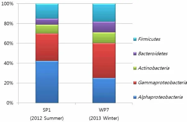
두 계절의 해면으로부터 분리된, 배양 가능한 공 생세균은 모두 Alphaproteobacteria, Gammaproteobacteria, Actinobacteria, Bacteroidetes, Firmicutes, 4문(phylum) 5강(class)으로 동일하게 나타났다(Fig. 4).
Alphaproteobacteria는 여름에 42.5%가 관찰되어 여 름해면에 가장 우점 하는 세균 군집으로 나타났으
며, Gammaproteobacteria는 겨울에 35.2%가 관찰되 어 겨울 해면에 가장 우점 하는 세균군집이었다 (Table 3). 여름에 비해 겨울에 Alphaproteobacteria는 감소하고(25.2%) Gammaproteobacteria는 증가(35.2%) 하는 차이를 나타내었으나 전체적으로 Proteobacteria 는 이 해면에서 우점종이었다. 이는 Li 등[10]에 의해 남중국해에 서식하는 4종 해면의 혼합배양에 의한 공생세균 군집구조 분석에서 Proteobacteria가 우점 한다는 결과와 일치하였으며, 일반적으로 해면공생 세균 군집구조에서 가장 높은 비율을 차지하는 것으 로 알려져 있다[6, 26]. Alphaproteobacteria의 종 구성 에서는 두 계절 모두 Pseudovibrio와 Rugeria 속의 종 이 우점 하였다. 반면 Gammaproteobacteria의 종 구 성에서 겨울철에 Psychrobacter 속의 세균이 우점하 며 여름철에는 Vibrio 속의 세균이 우점 하는 경향을 나타내었다(Table 3). 일반적으로 Psychrobacter 속의 균주는 극지방의 영구동토층과 얼음에서 자주 발견 되는 속으로 추운 환경에서 자주 발견되는 것을 볼 수 있다[15]. Vibrio 속의 균주들은 주로 여름철에 증 가하는 경향을 나타내며[2] 이 실험에서도 동일한 결과를 볼 수 있었다. Bacteroidetes 문에 속하는
Figure 3. Phylogenetic relationships were determined by the 16S rRNA gene sequences of the bacterial isolates from the marine sponge P. corticata. Numbers above branches indicate bootstrap values of neighbor-joining analysis (> 50%) from 1,000 replicates. The scale bar represents 0.05 substitutions per nucleotide position. Aquifex pyrophilus was used as an out group. Filled circle denotes summer sponges; opened diamond denotes winter sponges.
Table 3. Relative abundance of bacterial species in marine sponges of P. corticata.
Phylum or Class Bacterial species SP1 WP7
(Summer) (Winter)
Actinobacteria 9.0a 10.8
Agrococcus baldri 3.6b
Kocuria palustris 3.6
Kytococcus sedentarius 3.6
Microbacterium esteraromaticum 6.0
Janibacter hoylei 3.0
Bacteroidetes 6.0 10.8
Spongiibacterium flavum 6.0
Vitellibacter aestuarii 7.2
Tenacibaculum aiptasiae 3.6
Firmicutes 15.0 18.0
Bacillus aryabhattai 3.0 7.2
Bacillus marisflavi 3.6
Bacillus nanhaiensis 3.0 Bacillus oceanisediminis 3.0
Bacillus tequilensis 3.6
Oceanobacillus profundus 3.0
Oceanobacillus picturae 3.6
Planococcus donghaensis 3.0
Alphaproteobacteria 42.5 25.2
Erythrobacter vulgaris 3.0
Labrenzia alba 7.1
Phaeobacter inhibens 3.0
Pseudovibrio ascidiaceicola 16.2 14.4
Roseovarius crassostreae 3.0
Ruegeria atlantica 10.1 7.2
Ruegeria halocynthiae 3.6
Gammaproteobacteria 27.5 35.2
Marinobacter vinifirmus 3.0 Microbulbifer epialgicus 3.0
Pseudoalteromonas marina 3.6
Pseudoalteromonas phenolica 3.0 Pseudoalteromonas spongiae 3.0
Psychrobacter celer 10.0
Psychrobacter marincola 3.6
Psychrobacter maritimus 3.6
Psychrobacter nivimaris 3.6
Psychrobacter piscatorii 6.5
Vibrio chagasii 3.0
Vibrio fortis 3.0
Vibrio harveyi 3.0
Shewanella waksmanii 3.6
Shewanella sairae 3.6
Colwellia aestuarii 3.6
a, The number represents percentage (%) of each phylum in total bacterial community.
b, The number represents percentage (%) of each species in total bacterial community.
Figure 4. Seasonal comparison among the major bacterial groups of the cultivable bacterial communities from the marine sponge, P. corticata.
Spongiibacterium flabum 종은 제주도에서 채집한 Halichondria oshor 해면으로부터 분리 되었으며, 국 내 연구진이 처음 발견한 신속, 신종 균주이다[28].
이번 실험을 통해 이 균주가 P. corticata에서도 분리 되어 해면 특이적으로 존재하는 균주임을 알 수 있 었다. Actinobacteria와 Firmicutes 문의 경우 여름에 채집한 해면에 비해 겨울에 채집한 해면에서 증가되 는 것으로 나타났는데 두 세균 그룹은 세포벽이 두 껍거나 환경이 변화함에 따라 포자를 생성하여 내성 을 가지는 그람 양성 세균 그룹으로 세균이 잘 자라 는 여름철에 비해 온도가 낮아지고 염도가 높아지는 겨울철에 높은 비율을 나타낸 것으로 추정된다.
전체적으로 Alphaproteobacteria를 제외한 나머지 세균 그룹은 모두 겨울에 비율이 증가하고 여름에는 감소하는 패턴을 나타내었으나(Fig. 4), 종 수준과 속 수준에서 분석한 결과 여름 해면이 겨울 해면에 비 해 좀 더 다양한 종으로 구성된 것을 알 수 있었다 (Table 3). 따라서 계절에 따른 P. corticata 두 개체를 PCR-ARDRA 방법을 이용하여 배양 가능한 공생세 균 군집구조를 비교한 결과 문 수준에서는 동일하였 으나 종 수준에 있어서는 다양하였으며 각 세균 그
룹별로 차지하는 비율이 계절적으로 차이가 있음을 알 수 있었다.
이는 DGGE 방법을 이용하여 온도 변화에 따른 Rhopaloeides odorabile 해면의 공생세균 군집구조를 비교하였을 때 공생세균의 군집구조에서 변화가 관 찰되었으며[25], pyrosequencing 방법을 이용하여 2009년부터 2010년까지 미국 플로리다에서 채집한 Axinella corrugata 해면의 계절에 따른 공생세균 군 집구조 변화를 연구한 결과에서 계절에 따른 변화가 관찰된다는 보고[27]와도 일치하는 결과이다. 또한 한국의 조간대에 서식하는 주황해변해면 (Hymeniacidon sinapium)의 경우에도 계절에 따른 공 생세균 군집구조의 변화가 보고된 바 있다[6].
그러나 북서지중해에서 채집한 Ircinia spp. 해면으 로부터 1년 6개월간 T-RFLP방법을 이용하여 계절에 따른 공생세균 군집구조를 분석한 결과 주기적으로 매우 안정적인 군집구조를 나타낸다는 결과가 보고되 었으며, 남극해면과 열대해면을 이용한 해면공생세균 군집구조의 분석 결과에 의하면 동종 해면의 공생세 균 군집구조는 숙주 특이적이며 일정한 환경변화에도 안정성을 유지하는 것으로 나타나 본 실험결과와는
차이를 나타내었다[3, 26]. 또한 pyrosequencing 방법 을 이용하여 뉴질랜드에서 채집한 해면 Ecionemia alata와 Tethya bergquistae해면의 온도에 따른 공생 세균 군집구조 변화를 관찰한 결과 숙주 특이적인 균주가 있어 환경변화에도 안정적인 결과를 보이는 것으로 나타났다[1].
해면과 공생세균 사이의 상호작용에 관한 연구는 충분히 이루어지지 않고 있다[1, 12]. 더욱이 계절변 화에 따른 해면공생세균 군집구조 변화에 대해서는 오랜 시간 추적 조사가 필요하다. 또한 공생세균의 군집구조는 같은 해면종이라도 지리적 분포에 따라 달라지거나[21], 혹은 실험방법에 따라 달라지는 경 향이 있다[5]. 또한 배양법에 근거한 ARDRA 방법을 통해 해면 공생세균의 전체 군집구조를 파악하는 경 우 자연계에서 배양 가능한 세균은 0.1%-1%정도로 국한되므로 전체 세균 군집구조의 파악에는 한계가 있으며[6], 이론적으로 배양이 어려운 세균 종까지 파악이 가능한 것으로 알려진 DGGE 방법 역시 샘플 에서 세포가 차지하는 비율이 전체의 1% 이상을 구 성하는 세균종인 경우에만 검출 가능한 것으로 알려 져 있다[10]. 해면에 존재하는 전체 공생세균 군집구 조의 규명은 장기간에 걸쳐 다양한 방법을 조합하여 군집구조를 분석하고 파악할 필요가 있을 것으로 사 료된다.
결 론
여름과 겨울에 채집한 2 개체 해면 P. corticata의 배 양 가능한 공생세균 군집구조를 PCR-ARDRA 방법에 의해 분석한 결과 여름에 채집한 해면에서는 24개, 겨 울에 채집한 해면에서는 20개의 ARDRA type을 얻을 수 있었다. 각 type별로 1-3개의 분리균주를 선정하여 16S rRNA gene의 부분 염기서열을 분석한 결과, 공생 세균 군집구조는 문 수준에서 두 개체의 해면 모두 Alphaproteobacteria, Gammaproteobacteria, Actinobacteria, Bacteroidetes, Firmicutes, 4문(phylum) 5강(class)으로 동 일하게 나타났으나 여름에 채집한 해면에서는 Alphaproteobacteria가 우점 하는 반면 겨울에 채집한 해 면에서는 Gammaproteobacteria가 우점하는 패턴을 보 였다. 또한 속 및 종 수준에서의 공생세균 군집구조는 겨울 해면에 비해 여름 해면에서 다양하였으며 Actinobacteria, Bacteroidetes, Firmicutes문의 세균 그룹
은 겨울에 증가하였다.
감사의 글
2015년 한남대학교 학술연구조성비 지원에 의해 수행되었으며 이에 감사드립니다.
References
1. Cardenas, C. A., Bell, J. J., Davy, S. K., Hoggard, M., and Taylor, M. W. 2014. Influence of environmental var- iation on symbiotic bacterial communities of two tem- perate sponges. FEMS Microbiol. Ecol. 88, 516-527.
2. Davis, J. W. and Sizemore, R. K. 1982. Incidence of Vibrio species associated with blue crabs (Callinectes sapidus) collected from Galveston Bay, Texas. Appl.
Environ. Microbiol. 43, 1092-1097.
3. Erwin, P. M., Pita, L., Lopez-Legentil, S., and Turon, X. 2012. Stability of sponge-associated bacteria over large seasonal shifts in temperature and irradiance. Appl.
Environ. Microbiol. 78, 7358-7368.
4. Imhoff, J. F. and Stöhr, R. 2003. Sponge-associated bac- teria: general overview and special aspects of bacteria associated with Halichondria panicea. In Sponges (Porifera). Springer Berlin Heidelberg. 37, 35-57.
5. Jeong, I. H. and Park, J. S. 2012. Phylogenetic analysis of bacterial diversity in the marine sponge, Asteropus simplex, collected from Jeju island. Korean J. Microbiol.
48, 275-283.
6. Jeong, J. B. and Park, J. S. 2012. Seasonal differences of bacterial communities associated with the marine sponge, Hymeniacidon sinapium. Korean J. Microbiol.
48, 262-269.
7. Kim, J. S., Lim, Y. J., Im, K. S., Jung, J. H., Shim, C.
J., Lee, C. O., Hong, J., and Lee, H. 1999. Cytotoxic polyacetylenes from the marine sponge Petrosia sp. J.
Nat. Prod. 62, 554-559.
8. Lee, Y. K., Lee, J. H., and Lee, H. K. 2001. Microbial symbiosis in marine sponges. J. Microbiol. 39, 254-264.
9. Li, H. Y., Matsunaga, S., and Fusetani, N. 1994. Corticatic acids A-C, antifungal acetylenic acids from the marine sponge, Petrosia corticata. J. Nat. Prod. 57, 1464-1467.
10. Li, Z., Hu, Y., Liu, Y., Huang, Y., He, L., and Miao, X. 2007. 16S rDNA clone library-based bacterial phylo- genetic diversity associated with three South China Sea
sponges. World J. Microbiol. Biotechnol. 23, 1265-1272.
11. Lim, Y. J., Park, H. S., Im, K. S., Lee, C. O., Hong, J., Lee, M. Y., Kim, D. K., and Jung, J. H. 2001.
Additional cytotoxic polyacetylenes from the marine sponge Petrosia species. J. Nat. Prod. 64, 46-53.
12. Montalvo, N. F., Davis, J., Vicente, J., Pittiglio, R., Ravel, J., and Hill, R. T. 2014. Integration of cul- ture-based and molecular analysis of a complex sponge-associated bacterial community. PloS one. 9, e90517.
13. Nishimura, S., Matsunaga, S., Shibazaki, M., Suzuki, K., Harada, N., Naoki, H., and Fusetani, N. 2002. Corticatic acids D and E, polyacetylenic geranylgeranyl transferase type I inhibitors, from the marine sponge Petrosia corticata. J. Nat. Prod. 65, 1353-1356.
14. Noda, A., Sakai, E., Kato, H., Losung, F., Mangindaan, R. E., de Voogd, N. J., Yokosawa, H., and Tsukamoto, S. 2015. Strongylophorines, meroditerpenoids from the marine sponge Petrosia corticata, function as protea- some inhibitors. Bioorg. Med. Chem. Lett. 25, 2650-2653.
15. Rodrigues, D. F., da C Jesus, E., Ayala-del-Rio, H. L., Pellizari, V. H., Gilichinsky, D., Sepulveda-Torres, L., and Tiedje, J. M. 2009. Biogeography of two cold-adapt- ed genera: Psychrobacter and Exiguobacterium. ISME.
J. 3, 658-665.
16. Santos, O. C. S., Soares, A. R., Machado, F. L. S., Romanos, M. T. V., Muricy, G., Giambiagi‐deMarval, M., and Laport, M. S. 2015. Investigation of bio- technological potential of sponge‐associated bacteria collected in Brazilian coast. Lett. Appl. Microbiol. 60, 140-147.
17. Sasaki, S., Tozawa, T., Van Wagoner, R. M., Ireland, C. M., Harper, M. K., and Satoh, T. 2011.
Strongylophorine-8, a pro-electrophilic compound from the marine sponge Petrosia (Strongylophora) corticata, provides neuroprotection through Nrf2/ARE pathway.
Biochem. Biophys. Res. Commun. 415, 6-10.
18. Sun, W., Zhang, F., He, L., Karthik, L., and Li, Z. 2015.
Actinomycetes from the South China Sea sponges: iso- lation, diversity, and potential for aromatic polyketides discovery. Front. Microbiol. 6, doi: 10.3389/fmicb.
2015.01048
19. Takada, K., Okada, S., and Matsunaga, S. 2014.
Structural reappraisal of corticatic acids, biologically ac- tive linear polyacetylenes, from a marine sponge of the genus Petrosia. Fish. Sci. 80, 1057-1064.
20. Tamura, K., Stecher, G., Peterson, D., Filipski, A., and Kumar, S. 2013. MEGA6: molecular evolutionary genet- ics analysis version 6.0. Mol. Biol. Evol. 30, 2725-2729.
21. Taylor, M. W., Schupp, P. J., De Nys, R., Kjelleberg, S., and Steinberg, P. D. 2005. Biogeography of bacteria associated with the marine sponge Cymbastela concentrica. Environ. Microbiol. 7, 419-433.
22. Thoms, C., Horn, M., Wagner, M., Hentschel, U., and Proksch, P. 2003. Monitoring microbial diversity and natural product profiles of the sponge Aplysina cav- ernicola following transplantation. Mar. Biol. 142, 685-692.
23. Thompson, J. D., Higgins, D. G., and Gibson, T. J. 1994.
CLUSTAL W: improving the sensitivity of progressive multiple sequence alignment through sequence weight- ing, position-specific gap penalties and weight matrix choice. Nucleic Acids Res. 22, 4673-4680.
24. Wang, G. 2006. Diversity and biotechnological potential of the sponge-associated microbial consortia. J. Ind.
Microbiol. Biotechnol. 33, 545-551.
25. Webster, N. S., Cobb, R. E., and Negri, A. P. 2008.
Temperature thresholds for bacterial symbiosis with a sponge. ISME J. 2, 830-842.
26. Webster, N. S., Negri, A. P., Munro, M. M., and Battershill, C. N. 2004. Diverse microbial communities inhabit Antarctic sponges. Environ. Microbiol. 6, 288-300.
27. White, J. R., Patel, J., Ottesen, A., Arce, G., Blackwelder, P., and Lopez, J. V. 2012. Pyrosequencing of bacterial symbionts within Axinella corrugata sponges: diversity and seasonal variability. PLoS One. 7, e38204.
28. Yoon, B. J. and Oh, D. C. 2012. Spongiibacterium fla- vum gen. nov., sp. nov., a member of the family Flavobacteriaceae isolated from the marine sponge Halichondria oshoro, and emended descriptions of the genera Croceitalea and Flagellimonas. Int. J. Syst. Evol.
Microbiol. 62, 1158-1164.