다양한 배양 조건에서 제대혈 유래 CD34+ 조혈 세포의 체외 증식
최용운·오덕재† 세종대학교공과대학생명공학과
143-747 서울시광진구군자동 98 (2005년 12월 8일접수, 2006년 1월 6일채택)
Ex vivo Expansion of CD34+ Hematopoietic Cells from Cord Blood in Various Culture Environments
Yong Woon Choi and Duk Jae Oh†
Department of Bioscience and Biotechnology, Sejong University, 98, Gunja-dong, Gwangjin-gu, Seoul 143-747, Korea (Received 8 December 2005; accepted 6 January 2006)
요 약
본연구에서는제대혈유래의조혈줄기세포를효과적으로배양하기위한선행연구로서세포배양환경에따른조 혈줄기세포증식능의변화를관찰하였다. 제대혈의단핵구세포에서분리한 CD34+세포를성장인자조성-I(이하, coc-I) (EPO, GM-CSF, SCF, IL-3) 및성장인자조성-II(이하, coc-II) (TPO, G-CSF, SCF, IL-6, Flt3/Flk-2 ligand)가포함되어 있는 IMDM(Iscove’s modified Dulbecco’s medium) 및무혈청배지(serum free media, SFM)에서배양하였으며, 우태 아혈청(FBS)의첨가영향, 2차원및 3차원배양후각조건에서의세포증식및콜로니형성능을비교하였다. 일반적 으로 coc-I에서의세포증식및콜로니증식이 coc-II에서보다높았다. 3차원배양(methocult)에서는가장높은세포증 식(2,258±456배)을나타냈으며, 같은조성의 2차원배양(IMDM + coc-I + FBS)에서는가장높은콜로니증식(BFU-E:
652±19, CFU-GM: 520±58, CFU-GEMM: 339±100배)이나타났다. 배지를기준으로보면, coc-II 조성에우태아혈청 이포함되지않은경우를제외한모든경우에서세포증식및콜로니증식이무혈청배지에서보다 IMDM에서높았다. 결 론적으로, 모든배양조건중에서 ‘IMDM + coc-I + FBS’ 및 ‘IMDM + coc-I’에서가장좋은콜로니증식을보였으 며, 우태아혈청의첨가및 2차원배양조건이콜로니증식에더효과적인것으로확인되었다. 본연구결과는앞으로 조혈줄기세포의체외증식에필요한공정개발이나생물반응기설계에유용한정보를제공할수있을것으로사료된다.
Abstract- This study compared cell expansion and colony forming ability in human cord blood stem cells cultured ex vivo with two kinds of cytokine combinations, two kinds of media, presence or absence of fetal bovine serum (FBS) and two or three dimensional (2D or 3D) culture environments. Purified CD34+ cells were cultured in the IMDM (Iscove’s Modified Dulbecco’s Medium) and SFM (Serum Free Medium) containing a cytokine cocktail–I (coc-I) (EPO, GM- CSF, SCF, and IL-3) or a cytokine cocktail–II (coc-II) (TPO, G-CSF, SCF, IL-6, and Flt3/Flk-2 ligand) with or without FBS. Generally, higher cellular and clonogenic expansion were observed in the coc-I cytokine condition, compared to coc-II cytokine condition. 3D (Methocult) and 2D (IMDM + coc-I + FBS) conditions gave the greatest cell (2,258±456 fold) and CFU (BFU-E: 652±19, CFU-GM: 520±58, CFU-GEMM: 339±100 fold) expansions, respectively. In aspect of medium, IMDM was better than SFM, except for coc-II condition without FBS. In conclusion, ‘IMDM + coc-I + FBS’
and ‘IMDM + coc-I’ were the best CFU expansions on the occasion of all culture conditions. FBS and 2D conditions had affirmative effect on CFU expansion, generally. These data might provide a variety of notions about ex vivo expan- sion of hematopoietic stem cells.
Key words: Ex vivo Expansion, Cord Blood, CD34+ Cell
1. 서 론
조혈줄기세포(hematopoietic stem cells, HSC)는모든종류의혈 구세포로분화할수있음과동시에자가증식능력을가지고있는
세포이기때문에여러종류의혈액및면역질환에서이식치료제 로써사용될수있다[1].
제대혈(cord blood(CB))은골수나말초혈액을대체할수있는이 식용조직으로서, 그중에포함되어있는조혈줄기세포의광범위한 적용성에기초하여세포치료제로서의중요성이강조되고있으며, 1900년대후반부터조혈줄기및조혈전구세포의특별한공급원으로
†To whom correspondence should be addressed.
E-mail: [email protected]
써알려져왔다. 1980년대초반에는인간의태아혈액에서조혈전구 세포의정량화가이루어졌으며[2, 3], 1980년대후반에는 fanconi anemia 환자에게동종이형(allogenic)의제대혈이식이최초로성공
하였다[4]. 현재는혈액세포중에서줄기세포후보군의분리가가능
하며, 여러종류의생물반응기의개발및상업화되어있는여러종 류의성장인자를사용해서기질세포가없는환경에서도배양할수 있게되었다. 또한, 제대혈은이식편대숙주반응(graft versus host
disease, GVHD)의위험이낮기때문에조직적합성이완전히일치하
지않더라도이식이가능하다. 그러나한번채취할수있는제대혈 에포함되어있는조혈세포의양으로는소아나청소년기의사람에 게만부분적으로이식할수있는정도이기때문에성인에게적용하 기위해서는제대혈에있는조혈줄기세포뿐만아니라조혈전구및
분화된세포를동시에증식해야할필요가있다[5]. 분화된세포들은
이식후부작용으로발생할수있는호중구감소및혈소판감소를 막을수있으며, 조혈줄기및조혈전구세포는이식후장기적인생 존율을높이는역할을한다. 조혈세포의체외증식은재조합유전자 에의한인공적인성장인자의생산과더불어발전할수있었다. 1960
년대에이미여러종류의성장인자가제대혈에포함되어있음이알
려졌고[6], 이후계속적인연구과정을통하여여러가지성장인자
의기능이규명되었는데, 그 중에서대표적인성장인자들로서
EPO(erythropoietin)는적혈구전구세포의성장및분화를촉진하며 더욱분화된적혈구전구세포로의증식및분화를촉진하는데관여 함이알려졌고[7, 8], IL-3(interleukin-3)는백혈구전구세포의증식 및세포자살을막는역할을하는것으로보고되었다[9-11]. 또한, GM-CSF(granulocyte, macrophage colony stimulating factor)는과립 백혈구, 대식세포, 호산구전구세포의증식을자극하며[12-14], G- CSF(granulocyte colony stimulating factor)는과립백혈구전구세포 의증식을자극하고[15, 16], TPO(thrombopoietin)는거핵구전구세 포의증식을자극하는것으로보고되었다[17, 18]. Flt3/Flk2 ligand[19, 20] 및 IL-6(interleukin-6)[21-23]는조혈세포의증식및분화에는관 여하는정도가낮지만, 다른성장인자와함께조혈전구세포의증 식에영향을미치는것으로알려졌으며, SCF(stem cell factor)는조 혈전구세포의생존에직접적으로영향을미치는것으로알려졌다[24,
25]. 한편, 혈청은일반적으로동물세포배양에서성장을지원하는
역할로서많이사용되는데, 배양조건에따라다른결과가나타나는 경우가있으며, 표준화된연구결과를위해서는광범위한검증이요 구된다. 또한, 동물에서유래한혈청의사용으로인하여동물유래 감염원에의한감염의위험이존재하기때문에체외배양에서혈청 을제외한배지의사용은임상적용을위해서배양조건을표준화하 는것뿐만아니라이종유래의감염원의위험을제거할수있다는
측면에서매우유리한면이있으며[26], 현재이러한목적을위해서
특수한조성의무혈청배지가개발되고있다. 본연구에서는상기에 서제시한다양한배양조건에서조혈줄기세포증식능의변화를관 찰하기위해 8가지성장인자(EPO, TPO, G-CSF, GM-CSF, IL-3, IL-6, Flt3/Flk2 ligand, SCF)를두종류의조성군(coc-I, coc-II)으로나 누었는데, 성장인자조성-I(coc-I)은콜로니분석에일반적으로사용 되고있는 MethocultTM의조성을기본으로하였고, 성장인자조성- II(coc-II)는성장인자조성-I에포함되지않으면서조혈세포의체외 증식에많이사용되는성장인자들을사용하였다. 또한, 두종류의배 지, 우태아혈청의첨가여부, 2차원및 3차원의체외배양조건에서 포도당소모, 젖산생성, 세포의증식및콜로니형성능을비교하였다.
2. 실험 재료 및 방법 2-1. 세포분리
기증동의를받아확보한제대혈은 C.P.D.A1 항응고액이포함되
어있는혈액 bag(Green Cross, KOREA)에서 4oC의조건에서보 관후 24시간이내에분리하였다. 세포분리는제대혈을 IMDM (Iscove’s modified Dulbecco’s medium) (Gibco, USA)과 1:1 (v/v)
로혼합후, Ficoll-paque (밀도: 1.077 g/mL) (Pharmacia, Sweden)
을사용한밀도구배법으로 400-g의조건에서 30분동안원심분리 를하여단핵구세포(mononuclear cells)를분리하였고, 이를다시
PBE(PBS/0.5% bovine serum albumin/2 mM EDTA) 300µl와혼 합 후 FcR blocking reagent 100µl및 CD34 microbead(Miltenyi Biotec, Germany) 100µl를혼합하여 30분동안냉장보관하였다.
상기의세포분산액을 PBE로 1회세척하고, MidiMACS(Miltenyi Biotec, Germany)를사용하여 CD34+세포를분리하였다. 생존율은
트리판블루용액으로염색후 hemocytometer를이용하여측정하
였으며, 생존율은 95%이상이었다.
2-2. 배지및 첨가물
배지는 IMDM(Gibco, USA), 무혈청배지(Ultraculture)(Cambrex,
USA) 및 3차원배양을위한반고형배지로콜로니증식분석에사
용하는 MethocultTM(Stemcell Technologies, Canada)를사용하였다.
성장인자의조성은두종류를사용하였다. 성장인자조성-I(coc-I)
은 3 U/mL rh (recombinant human) EPO (erythropoietin) (LG PhD, Korea), 10 ng/mL rh GM-CSF (granulocyte, macrophage colony stimulating factor) (LG PhD, Korea), 50 ng/mL rh SCF (stem cell factor) (Biosource, USA), 10 ng/mL rh IL-3 (interleukin-3) (Biosource, USA)을포함하고있고, 성장인자조성-II (coc-II)는 50 ng/mL rh TPO (thrombopoietin) (R&D system, USA), 40 ng/mL rh G-CSF (granulocyte colony stimulating factor) (Choongwae, Korea), 50 ng/mL rh SCF (Biosource, USA), 100 ng/mL rh IL-6 (interleukin-6) (Biosource, USA), 80 ng/mL rh Flt3/Flk-2 ligand (R&D system, USA)를 포함하고있는데, 모든배지에 0.1 mM 2-mercaptoethanol (Sigma, USA), 1% (v/v) BSA (bovine serum albumin) (Sigma, USA)의 최종농도로첨가하였다. 우태아혈청
(fetal bovine serum, FBS) (JBI, Korea)이첨가된경우에는 30%
(v/v) FBS의농도로하였다.
2-3. 세포농도 측정
크리스털바이올렛(crystal violet) 용액으로염색후 hemocytometer를 사용하여유핵세포의수를측정하였다. 12 well culture plate(SPL, Korea)의 well에 3.1×10³개의 CD34+세포를 2배수 (n=2)로접종하였 으며, 1 ml의 working volume으로 37oC의 CO2인큐베이터에서배
양하였다. 배지중의포도당 및젖산농도는 YSI-2700 SELECT
Biochemistry Analyzer (YSI, USA)를사용하여측정하였으며, 배 양 14일째에배지의절반을새배지로교환하였다. 2차원및 3차원 배양환경에서의차이를비교하기위하여같은성장인자조성에서 액체배지(IMDM + coc-I + FBS) 및반고형배지(MethocultTM)에 세포를배양하였다. 증식된세포는트리판블루및크리스털바이 올렛용액으로분석하여생존율및유핵세포의수를확인하였고,
콜로니분석에도사용하였다.
2-4. 콜로니 분석
증식능력이있는세포를정량적으로측정할수있는콜로니의 수는반고형배지인 MethocultTM를 사용하여 CFU-GM(colony- forming units granulocyte/macrophage), BFU-E(burst-forming units erythroid), CFU-GEMM(colony-forming units granulocyte/erythrocyte, macrophage, and megakaryocyte)을측정하였다. 단핵구세포에서 분리된 CD34+세포및증식된세포를 MethocultTM 배지 1 ml당
2,000개의비율로 35 mm plastic dish(SPL, Korea)에 4배수(n=4)로 접종한후습한조건에서 37oC의 CO2인큐베이터에서배양하였으 며, 14일후에위상차현미경을사용하여형성된콜로니의수를측 정하였다. 콜로니를형성하는세포의수가 50개이상일때 1개의 콜로니로개수하였다.
3. 결과 및 고찰
배양기간동안생존세포(viable cell) 및유핵세포(nucleus cell)
수의변화를 Fig. 1에나타내었다. 일반적으로 coc-I 및 coc-II 조건 에서모두배양 6일째이후에세포가급격하게증가하는것을확 인할수있었다. 유핵세포수를기준으로했을때, coc-I 조건에서
는배양 12~14일경에최대증식을보인후감소하다가배지를교
환한후 3일째부터다시증가하는경향을나타내었고, coc-II 조건 에서는배양 17일경에최대로증식한후배지교환에관계없이감 소하는경향을나타내었다. 세포의증식은 coc-II 조건에서보다 coc-I
조건에서더빠르게일어났으며, 이는특히 IMDM 배지조성에서 두드러지게나타나는것을확인할수있었다. Daley 등[27]은 EPO
가세포증식에효과적인영향을주는것으로보고하였고, Piacibello
등[28]은 Flt3 + TPO의성장인자조성이 LTC-IC(long term culture- initiating cell) 증가에큰영향을준다고보고하였는데, 이는 EPO가
포함되어있는 coc-I 조건에서세포의증식이더많이발생하는본
실험의결과와부합하는것이었다. coc-I 조건에서우태아혈청은세
포의증식및콜로니형성에긍정적인영향을주었으며, coc-II 조
건에서우태아혈청은세포의증식에는긍정적인영향을주었으나 콜로니형성에는큰영향을주지않았다. Balducci 등[29]은혈청이 포함되어있는배지에서의유핵세포증식및 CFU-GM의증식이 혈청이포함되어있지않은배지에서보다더좋은경향을나타냈 다고보고하였는데, 이결과는혈청이포함되어있지않은배지에 서더나은경향을보였다는이전의보고들과는상반되는결과였 다[30-32]. Shadduck등[33]은조혈줄기세포의배양에서배양 1~2
Fig. 1. Time courses of viable cell and nucleus cell expansions in different culture conditions. For viable cell count, trypan blue dye exclusion method was used. For determining nucleus cell expansion, nucleus staining using crystal violet was applied. One milliliter of 3.1×10³ CD34+ nucleated cell suspensions was seeded in a well (12 well plate; n=2). Arrow represents time point of media exchange. At day-14, half of the culture medium was replaced with fresh medium. When FBS was supplemented, its concentration was 30% (v/v). (a), (b): Cytokine cock- tail-I (coc-I) was used.(c), (d): Cytokine cocktail-II (coc-II) was used.
주까지혈청이포함된배지에서배양을한후배지가포함되지않 은배지로세포를옮겨서배양하는것이더욱긍정적인결과를나 타낸다고보고하였는데, 본실험의결과와종합해볼때성장인자의
조성및배양배지의조건에따라혈청이세포및콜로니증식에 서로다른영향을줄수있음을확인할수있었다. 우태아혈청만이 포함된배지에서는세포가증식되지않았다.
Fig. 2. The expansion of total colony forming units in different culture conditions. Total CFU expansion fold was calculated at colony counts by nucleus cell expansion fold. When FBS was supplemented, its concentration was 30% (v/v).(a), (b), (c): Cytokine cocktail-I (coc-I) was used.(d), (e), (f): Cytokine cocktail-II (coc-II) was used.
Fig. 2는배양 9일, 14일, 27일째에콜로니분석을한결과를보
여주고있다. 2,000개의단위세포당콜로니형성및전체세포당
콜로니형성은우태아혈청의첨가여부에관계없이배양시간이지 남에따라감소하였는데, 이러한현상은조혈줄기세포및조혈전구 세포가배양기간동안지속적으로감소함을의미한다. 3차원배양 에서는콜로니증식이 2차원배양에서보다매우적었는데, 이는본 실험의 3차원조건은조혈줄기혹은전구세포의증식보다는말초 혈액으로의분화에영향을주는것으로사료된다. 우태아혈청만이 포함된배지에서와배양 27일째의모든배지조건에서는콜로니형 성이관찰되지않았다.
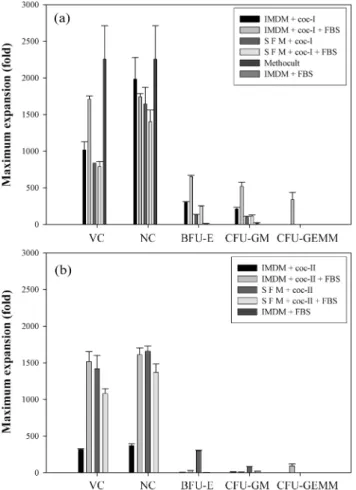
Fig. 3은배양기간동안각각의배지조성에서의최대생세포수,
유핵세포수, BFU-E, CFU-GM 및 CFU-GEMM의수를나타낸 그래프이다. 일반적으로 coc-II 조건보다 coc-I 조건에서세포증식 및콜로니증식이더많았다. 3차원배양(Methocult)에서는가장높 은세포증식(2,258±456배)을나타냈으며, 같은조성의 2차원배양
(IMDM + coc-I + FBS)에서는가장높은콜로니증식(BFU-E: 652±19, CFU-GM: 520±58, CFU-GEMM: 339±100배)이나타났다.
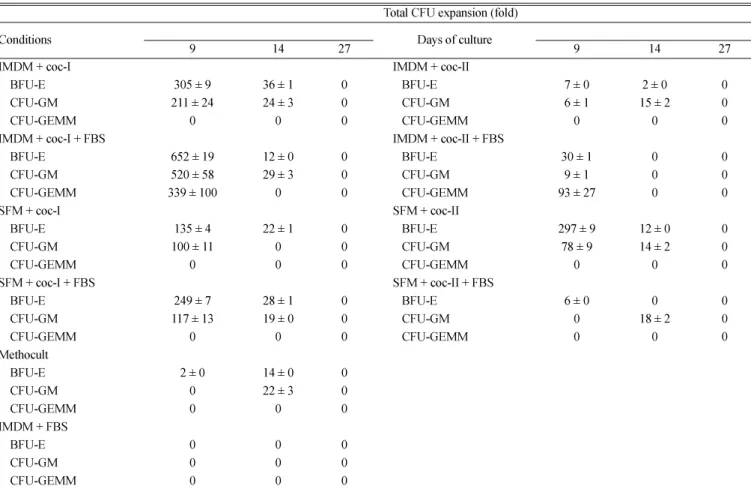
Table 1과 2는배양기간동안각각의배지조성에서의유핵세
포수및콜로니수를나타낸표이다. 배지를기준으로보면, 우태
아혈청이포함되지않은 coc-II 조건을제외하고는세포증식및
콜로니증식이무혈청배지에서보다 IMDM에서높았다. 혈청이포 함되어있는조건인 ‘IMDM + coc-I + FBS’ 와 ‘SFM + coc-I + FBS’에서세포의증식은각각 1,741±46, 1,403±160배였으며콜로 니증식은각각 BFU-E: 652±19, 249±7; CFU-GM: 520±58, 117±13;
CFU-GEMM: 339±100, 0배였고, ‘IMDM + coc-II + FBS’와 ‘SFM + coc-II + FBS’에서세포증식은각각 1,613±91, 1,371±114배였 으며콜로니증식은각각 BFU-E: 30±1, 6±0; CFU-GM: 9±1, 18±2;
CFU-GEMM: 93±27, 0배였다. 혈청이 포함되지 않은 조건인
‘IMDM + coc-I’ 및 ‘SFM + coc-I’에서세포증식은각각 1,983±297, 1,645±228배였으며콜로니 증식은각각 BFU-E: 305±9, 135±4;
CFU-GM: 211±24, 100±11배였고, ‘IMDM+coc-II’ 및 ‘SFM+coc-II’에
서 세포증식은각각 371±23, 1,661±68배였으며콜로니증식은
BFU-E: 7±0, 297±9; CFU-GM: 15±2, 78±9배였다. 2차원및 3차 원배양에서, 세포의증식은 3차원배양에서가더높았지만콜로니
Fig. 3. Maximum expansion fold of viable cells (VC), nucleus cells (NC), and CFU during culture time. When FBS was supple- mented, its concentration was 30% (v/v).(a): Cytokine cocktail-I (coc-I) was used.(b): Cytokine cocktail-II (coc-II) was used.
Table 1. Time courses of nucleus cell expansion
Nucleus cell expansion (fold)
Conditions Days of culture
3 6 9 11 14 17 20 27
IMDM + coc-I 1 ± 0 11 ± 0 1,693 ± 23 1,209 ± 68 1,241 ± 68 1,645 ± 46 1,354 ± 274 1,983 ± 297 IMDM + coc-I + FBS 1 ± 0 28 ± 0 1,129 ± 461 1,741 ± 46 1,483 ± 182 1,048 ± 342 1,193 ± 137 1,677 ± 0 SFM + coc-I 1 ± 0 12 ± 0 1,564 ± 68 1,806 ± 91 1,064 ± 228 1,204 ± 349 1,112 ± 160 1,645 ± 228 SFM + coc-I + FBS 1 ± 0 23 ± 0 1,661 ± 23 1,774 ± 46 1,951 ± 160 1,806 ± 228 1,758 ± 205 1,403 ± 160 Methocult 6 ± 0 12 ± 0 1,241 ± 23 2,258 ± 456 1,129 ± 228 1,225 ± 91 1,774 ± 456 1,290 ± 456
IMDM + FBS 0 11 ± 0 1,1,1,0 1,1,1, 0 1,1,1, 0 1,1,1, 0 1,1,1, 0 1,1,1, 0
IMDM + coc-II 1 ± 0 17 ± 0 1,084 ± 5 1,123 ± 9 1,255 ± 5 1,371 ± 23 1,235 ± 55 1,166 ± 16 IMDM + coc-II + FBS 1 ± 0 25 ± 0 1,310 ± 9 1,839 ± 91 1,242 ± 68 1,613 ± 91 1,226 ± 46 1,339 ± 68 SFM + coc-II 2 ± 0 55 ± 0 1,887 ± 114 1,048 ± 114 1,419 ± 182 1,661 ± 68 1,239 ± 27 1,323 ± 91 SFM + coc-II + FBS 1 ± 0 29 ± 5 1,355 ± 46 1,839 ± 46 1,903 ± 91 1,226 ± 91 1,371 ± 114 1,935 ± 319 Purified CD34+ cord blood cells were cultured in 12 well plates at 37oC, 5% CO2 in air.
Results represent mean ± SD from two separate experiments. When FBS was supplemented, its concentration was 30% (v/v).
coc-I: EPO(3 U/mL)+GM-CSF(10 ng/mL)+SCF(50 ng/mL)+IL-3(10ng/mL).
coc-II: TPO(50ng/mL)+G-CSF(40ng/mL)+SCF(50ng/mL)+IL-6(100ng/mL)+Flt3/Flk-2 ligand(80ng/mL).
증식은 2차원배양에서월등히좋은것으로확인되었다. 2차원및
3차원배양에서세포증식은각각 1,741±46, 2,258±456배였으며콜 로니증식은각각 BFU-E: 652±19, 14±0; CFU-GM: 520±58, 22±3;
CFU-GEMM: 339±100, 0배였다.
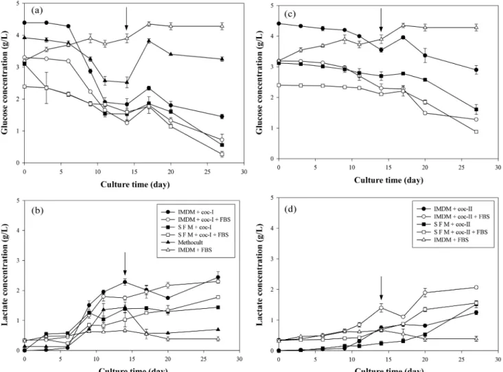
배양기간동안포도당소모및젖산생성정도를 Fig. 4에나타
내었다. 포도당소모및젖산생성은세포의급격한증가와더불어 증가하였으나, 증식한세포의수에비해서포도당소모및젖산생 성정도는크지않았으며, 혈청의첨가여부는포도당소모및젖
산생성에큰영향을미치지않는것으로나타났다. coc-I 조건의
IMDM 배지에서는배양 6일이후에포도당소모량이급격히증가
하는반면 SFM 배지에서는완만한소모경향이나타났으며, coc-II
조건에서는 IMDM 및 SFM 배지모두에서포도당소모량이완만
한경향이나타났다. 포도당소모및젖산생성은 coc-II 조건보다
coc-I 조건에서더많았는데, 이는 coc-I 조건에서의세포증식이
coc-II 조건에서보다더빠르게일어났기때문이며, 특히 IMDM 배 지조성에서뚜렷하게나타나는것을확인할수있었다. 2차원및
3차원배양비교에서는 2차원배양에서의포도당소모및젖산생 산량이높은것으로나타났다.
생존율은배양 6일째이후부터지속적으로감소하였으며, coc-II
조건에서보다 coc-I 조건에서감소정도가더급격하게일어나는
것을확인할수있었다. coc-I 조건에서생존율은배양 6일째이후
부터감소하기시작하여배양 11~14일에급격히감소하였으며, 배
양 27일때에는 40%정도가되었고, 우태아혈청의첨가여부는생 존율변화경향에영향을주지않는것으로확인되었다. coc-II 조
건에서생존율은배양 6~14일째이후부터감소하기시작하여배양
27일때에는 30~60%정도였으며, 우태아혈청이포함된경우생존
율의감소가우태아혈청이포함되지않은경우보다완만하게진행 되는것을확인할수있었다. 배양 14일째에배지교환후 coc-I 조 건에서는생존율이증가한후감소하였으나 coc-II 조건에서는생 존율의증가가나타나지않았다. 2차원및 3차원배양에서의생존 율감소경향은유사하게나타났다(data not shown).
4. 결 론
본연구에서는다양한배양조건에서의체외배양을통해서제 대혈유래의조혈세포의증식및배양환경의변화를살펴보았다. 세 포증식및콜로니증식은 coc-I 조건이 coc-II 조건보다더긍정적 으로나타났으며, 우태아혈청의첨가여부는포도당소모및젖산 생성정도에영향을주지않았다. 배지를기준으로보면우태아혈
청이포함된경우 IMDM이무혈청배지에서보다긍정적인결과를
Table 2. Time courses of progenitor cell expansion
Total CFU expansion (fold)
Conditions 9 14 27 9Days of culture 14 27
IMDM + coc-I IMDM + coc-II
BFU-E 305 ± 9 36 ± 1 0 BFU-E 7 ± 0 2 ± 0 0
CFU-GM 211 ± 24 24 ± 3 0 CFU-GM 6 ± 1 15 ± 2 0
CFU-GEMM 0 0 0 CFU-GEMM 0 0 0
IMDM + coc-I + FBS IMDM + coc-II + FBS
BFU-E 652 ± 19 12 ± 0 0 BFU-E 30 ± 1 0 0
CFU-GM 520 ± 58 29 ± 3 0 CFU-GM 9 ± 1 0 0
CFU-GEMM 339 ± 100 0 0 CFU-GEMM 93 ± 27 0 0
SFM + coc-I SFM + coc-II
BFU-E 135 ± 4 22 ± 1 0 BFU-E 297 ± 9 12 ± 0 0
CFU-GM 100 ± 11 0 0 CFU-GM 78 ± 9 14 ± 2 0
CFU-GEMM 0 0 0 CFU-GEMM 0 0 0
SFM + coc-I + FBS SFM + coc-II + FBS
BFU-E 249 ± 7 28 ± 1 0 BFU-E 6 ± 0 0 0
CFU-GM 117 ± 13 19 ± 0 0 CFU-GM 0 18 ± 2 0
CFU-GEMM 0 0 0 CFU-GEMM 0 0 0
Methocult
BFU-E 2 ± 0 14 ± 0 0
CFU-GM 0 22 ± 3 0
CFU-GEMM 0 0 0
IMDM + FBS
BFU-E 0 0 0
CFU-GM 0 0 0
CFU-GEMM 0 0 0
Cultures were initiated with 2,000 nucleus cells in 35mm plastic dishes at 37oC, humidified atmosphere of 5% CO2 in air. Results represent mean ± SD from four separate experiments.
coc-I: EPO(3U/mL)+GM-CSF(10 ng/mL)+SCF(50 ng/mL)+IL-3(10 ng/mL).
coc-II: TPO(50 ng/mL)+G-CSF(40 ng/mL)+ SCF(50 ng/mL)+IL-6(100 ng/mL)+Flt3/Flk-2 ligand(80 ng/mL).
나타냈으며, 우태아혈청이포함되지않은경우에는무혈청배지가
IMDM과같거나좀더좋은경향을나타내었다. 또한, 분화된세 포로의증식면에서는 3차원배양이유리하였으나, 조혈전구세포와 관련된콜로니증식면에서는 2차원배양이유리하였다. 본연구 결과는앞으로제대혈뿐만아니라골수및말초혈조혈줄기세포의 체외증식에필요한공정개발이나생물반응기설계에유용한정보 를제공할수있을것으로사료된다.
참고문헌
1. Zon, L. I., “Developmental Biology of Hematopoiesis,”Blood,
86(8), 2876-2891(1995).
2. Hann, I. M., Bodger, M. P. and Hoffbrand, A. V., “Development of Pluripotent Hematopoietic Progenitor Cells in the Human Fetus,”Blood, 62(1), 118-123(1983).
3. Linch, D. C., Knott, L. J., Rodeck, C. H. and Huehns, E. R.,
“Studies of Circulating Hemopoietic Progenitor Cells in Human Fetal Blood,”Blood, 59(5), 976-979(1982).
4. Gluckman, E., Broxmeyer, H. A., Auerbach, A. D., Friedman, H.
S., Douglas, G. W., Devergie, A., Esperou, H., Thierry, D.,
Socie, G. and Lehn, P., “Hematopoietic Reconstitution in a Patient with Fanconi’s Anemia by Means of Umbilical-cord Blood from an HLA-identical Sibling,”N Engl J Med, 321(17), 1174- 1178(1989).
5. Kogler, G., Radke, T. F., Lefort, A., Sensken, S., Fischer, J., Sorg, R.V. and Wernet, P., “Cytokine Production and Hematopoiesis Supporting Activity of Cord Blood-derived Unrestricted Somatic Stem Cells,”Exp Hematol, 33(5), 573-583(2005).
6. Ende, M., “Lymphangiosarcoma. Report of a Case,”Pac Med Surg, 74(2), 80-82(1966).
7. Kubanek, B., Ferrari, L., Tyler, W. S., Howard, D., Jay, S. and Jr.
Stohlman, F., “Regulation of Erythropoiesis. 23. Dissociation Between Stem Cell and Erythroid Response to Hypoxia,”Blood,
32(4), 586-596(1968).
8. Jr.Stohlman, F., “Some Aspects of Erythrokinetics,”Semin Hematol,
4(4), 304-314(1967).
9. Metcalf, D., “Control of Granulocytes and Macrophages: Molec- ular, Cellular, and Clinical Aspects,”Science, 254(5031), 529-533 (1991).
10. Ihle, J. N., “Interleukin-3 and Hematopoiesis,”Chem Immunol,
51, 65-106(1992).
Fig. 4. Time courses of glucose and lactate concentrations in different culture conditions. Arrow represents time point of media exchange. At day- 14, half of the culture medium was replaced with fresh medium. When FBS was supplemented, its concentration was 30% (v/v). (a), (b):
Cytokine cocktail-I (coc-I) was used.(c), (d): Cytokine cocktail-II (coc-II) was used.
11. Leary, A. G., Wong, G. G., Clark, S. C., Smith, A. G. and Ogawa, M., “Leukemia Inhibitory Factor Differentiation-inhibiting Activity/
human Interleukin for DA Cells Augments Proliferation of Human Hematopoietic Stem Cells,”Blood, 75(10), 1960-1964(1990).
12. Socinski, M. A., Cannistra, S. A., Elias, A., Antman, K. H., Schnipper, L. and Griffin, J. D., “Granulocyte-Macrophage Col- ony Stimulating Factor Expands the Circulating Haemopoietic Progenitor cell Compartment in Man,”Lancet, 1(8596), 1194- 1198(1988).
13. Migliaccio, G., Migliaccio, A. R. and Adamson, J. W., “In Vitro Differentiation of Human Granulocyte/macrophage and eRyth- roid Progenitors: Comparative Analysis of the Influence of Recombinant Human Erythropoietin, G-CSF, GM-CSF, and IL-3 in Serum-supplemented and Serum-deprived Cultures,”Blood,
72(1), 248-256(1988).
14. Tomonaga, M., Golde, D. W. and Gasson, J. C., “Biosynthetic (recombinant) Human Granulocyte-macrophage Colony-stimu- lating Factor: Effect on Normal Bone Marrow and Leukemia Cell Lines,”Blood, 67(1), 31-36(1986).
15. Clark, S. C. and Kamen, R., “The Human Hematopoietic Col- ony-stimulating Factors,”Science, 236(4806), 1229-1237(1987).
16. Ohara, A., Suda, T., Saito, M., Miura, Y., Okabe, T. and Takaku, F., “Effect of Recombinant Human Granulocyte Colony-stimu- lating Factor on Hemopoietic Cells in Serum-free Culture,”Exp Hematol, 15(6), 695-699(1987).
17. de Sauvage, F. J., Hass, P. E., Spencer, S. D., Malloy, B. E., Gurney, A. L., Spencer, S. A., Darbonne, W. C., Henzel, W. J., Wong, S. C., Kuang, W. J., Karl, J. O., Bruce, H., Lawrence, A. S., Jr., David, V. G. and Dan, L. E., “Stimulation of Megakaryocytopoiesis and Thrombopoiesis by the c-Mpl Ligand,”Nature, 369(6481), 533- 538(1994).
18. Bartley, T. D., Bogenberger, J., Hunt, P., Li, Y. S., Lu, H. S., Mar- tin, F., Chang, M. S., Samal, B., Nichol, J. L., Swift, S., Johnson, M.
J., Hsu, R.-Y., Parker, V. P., Suggs, S., Skrine, J. D., Merewether, L.
A., Clogston, C., Hsu, E., Hokom, M. M., Hornkohl, A., Choi, E., Pangelinan, M., Sun, Y., Mar, V., McNinch, J., Simonet, L., Jacobsen, F., Xie, C., Shutter, J., Chute, H., Basu, R., Selander, L., Trollinger, D., Sieu, L., Padilla, D., Trail, G., Elliott, G., Izumi, R., Covey, T., Crouse, J., Garcia, A., Xu, W., Castillo, J. D., Biron, J., Cole, S., Hu, M. C.-T., Pacifici, R., Ponting, I., Saris, C., Wen, D., Yung, Y. P., Lin, H. and Rosselman R. A., “Identification and Cloning of a Megakaryocyte Growth and Development Factor that is a Ligand for the Cytokine Receptor Mpl,”Cell, 77(7), 1117-1124 (1994).
19. Hannum, C., Culpepper, J., Campbell, D., McClanahan, T., Zurawski, S., Bazan, J. F., Kastelein, R., Hudak, S., Wagner, J., Mattson, J., Luh, J., Duda, G., Martina, N., Peterson, D., Menon, S., Shanafelt, A., Muench, M., Kelner, G., Namikawa, R., Ren- nick, D., Roncarolo, M.-G., Zlotnik, A., Rosnet, O., Dubreuil, P., Birnbaum, D. and Lee, F., “Ligand for FLT3/FLK2 Receptor Tyrosine Kinase Regulates Growth of Haematopoietic Stem Cells and is Encoded by Variant RNAs,”Nature, 368(6472), 643-648 (1994).
20. Lyman, S. D., James, L., Vanden Bos, T., de Vries, P., Brasel, K., Gliniak, B., Hollingsworth, L. T., Picha, K. S., McKenna, H. J., Splett, R. R., Fletcher, F. A., Maraskovsky, E., Farrah, T., Foxworthe, D., Williams, D. E. and Beckmann, M. P., “Molecular Cloning of
a Ligand for the flt3/flk-2 Tyrosine Kinase Receptor: a Prolifer- ative Factor for Primitive Hematopoietic Cells,”Cell, 75(6), 1157-1167(1993).
21. Rennick, D., Jackson, J., Yang, G., Wideman, J., Lee, F. and Hudak, S., “Interleukin-6 Interacts with Interleukin-4 and Other Hematopoietic Growth Factors to Selectively Enhance the Growth of Megakaryocytic, Erythroid, Myeloid, and Multipotential Pro- genitor Cells,”Blood, 73(7), 1828-1835(1989).
22. Hodgkin, P. D., Bond, M. W., O’Garra, A., Frank, G., Lee, F., Coffman, R. L., Zlotnik, A. and Howard, M., “Identification of IL-6 as a T Cell-derived Factor that Enhances the Proliferative Response of Thymocytes to IL-4 and Phorbol Myristate Ace- tate,”J. Immunol, 141(1), 151-157(1988).
23. Caracciolo, D., Clark, S. C. and Rovera, G., “Human Interleukin- 6 Supports Granulocytic Differentiation of Hematopoietic Pro- genitor Cells and Acts Synergistically with GM-CSF,”Blood,
73(3), 666-670(1989).
24. Brandt, J., Briddell, R. A., Srour, E. F., Leemhuis, T. B. and Hoffman, R., “Role of c-kit Ligand in the Expansion of Human Hematopoietic Progenitor Cells,”Blood, 79(3), 634-641(1992).
25. Brandt, J. E., Bhalla, K. and Hoffman, R., “Effects of Interleu- kin-3 and c-kit Ligand on the Survival of Various Classes of Human Hematopoietic Progenitor Cells,”Blood, 83(6), 1507-1514(1994).
26. Lebkowski, J. S., Schain, L. R. and Okarma, T. B., “Serum-free Culture of Hematopoietic Stem Cells: a Review,”Stem Cells,
13(6), 607-612(1995).
27. Daley, J. P., Daley, B. M., Wysocki, M. G., Caligiuri, M. A. and Biddle, W. C., 1996. Ex Vivo Expansion of Human Hematopoietic Progenitor Cells in Serum-free STEMPROTM-34 Medium, pp.
62-67. vol. 18. Life Technologies, Gaithersburg, U. S. A.
28. Piacibello, W., Sanavio, F., Garetto, L., Severino, A., Dane, A., Gammaitoni, L. and Aglietta, M., “Differential Growth Factor Requirement of Primitive Cord Blood Hematopoietic Stem Cell for Self-renewal and Amplification vs Proliferation and Differ- entiation,”Leukemia, 12(5), 718-727(1998).
29. Balducci, E., Azzarello, G., Valenti, M. T., Capuzzo, G. M., Pap- pagallo, G. L., Pilotti, I., Ausoni, S., Bari, M., Rosetti, F., Sartori, D., Ciappa, A., Porcellini, A. and Vinante, O., “The Impact of Progenitor Enrichment, Serum, and Cytokines on the Ex Vivo Expansion of Mobilized Peripheral Blood Stem Cells: a Con- trolled Trial,”Stem Cells, 21(1), 33-40(2003).
30. Mobest, D., Mertelsmann, R. and Henschler, R., “Serum-free Ex Vivo Expansion of CD34(+) Hematopoietic Progenitor Cells,”Bio- technol Bioeng, 60(3), 341-347(1998).
31. Poloni, A., Giarratana, M. C., Firat, H., Kobari, L., Gorin, N. C.
and Douay, L., “The Ex Vivo Expansion Capacity of Normal Human Bone Marrow Cells is Dependent on Experimental Con- ditions: Role of the Cell Concentration, Serum and CD34+ Cell Selection in Stroma-free Cultures,”Hematol Cell Ther, 39(2), 49- 58(1997).
32. Gilmore, G. L., DePasquale, D. K., Lister, J. and Shadduck, R.
K., “Ex vivo Expansion of Human Umbilical Cord Blood and Peripheral Blood CD34(+) Hematopoietic Stem Cells,”Exp Hema- tol, 28(11), 1297-1305(2000).
33. Shadduck, R. K., Gilmore, G. L. and Lister, J., “Role of Serum- free Medium in the Ex Vivo Expansion of Human Cord Blood Hematopoietic Stem Cells,”Stem Cells, 18(2), 154-155(2000).