Portulaca oleracea L. Extract Lowers Postprandial Hyperglycemia by Inhibiting Carbohydrate-digesting Enzymes
Jae-Eun Park and Ji-Sook Han*
Department of Food Science and Nutrition, Pusan National University, Busan 46241, Korea Received October 27, 2017 /Revised December 4, 2017 /Accepted December 5, 2017
Postprandial hyperglycemia plays an important role in the development of Type 2 Diabetes and dia- betic complications. Controlling postprandial hyperglycemia is the most important factor for reducing the risks of diabetic complications in Type 2 diabetic patients. This study was designed to determine whether Portulaca oleracea L. extract suppresses the activation of carbohydrate-digesting enzymes, and lowers postprandial hyperglycemia in diabetic mice through streptozotocin. P. oleracea was extracted with either 80% ethanol (PEE) or water (PWE), and the extract solutions were concentrated. The α -glucosidase and α-amylase inhibition assays were performed using the chromogenic method. Normal mice and STZ-induced diabetic mice were orally treated with PEE, PWE (300 mg/kg of body weight) or acarbose (100 mg/kg of body weight), with soluble starch (2 g/kg of body weight). The α -glucosidase and α-amylase inhibitory effectiveness by PEE were markedly more effective than PWE, and both extracts indicated a higher effectiveness than the acarbose (positive control). The rise in post- prandial blood glucose due to starch loading was markedly inhibited in the PEE group when com- pared to the control group in diabetic and normal mice. Furthermore, the area under the concentration –time curve values were markedly declined by the PEE injection in the diabetic group when com- pared to that exerted for the control group. These results demonstrate that P. oleracea extracts lower postprandial hyperglycemia by inhibiting carbohydrate-digesting enzymes, and that the ethanol ex- tract is more efficacious than the water extract.
Key words : α-amylase, α-glucosidase, diabetes, Portulaca oleracea L., postprandial hyperglycemia
*Corresponding author
*Tel : +82-51-510-2836, Fax : +82-51-583-3648
*E-mail : [email protected]
This is an Open-Access article distributed under the terms of the Creative Commons Attribution Non-Commercial License (http://creativecommons.org/licenses/by-nc/3.0) which permits unrestricted non-commercial use, distribution, and reproduction in any medium, provided the original work is properly cited.
Journal of Life Science 2018 Vol. 28. No. 4. 421~428 DOI : https://doi.org/10.5352/JLS.2018.28.4.421
Introduction
Approximately 4.4% of people were diagnosed as having diabetes in 2,000 and this is estimated to increase to 9.7%
by 2,050[11]. The worldwide increase in the prevalence of diabetes in adults will considerably affect not only public health systems, but also patient health, quality of life, and longevity [24]. Diabetic mellitus is a metabolic disease with chronic hyperglycemia. Particularly, postprandial symptoms include rapidly increasing blood glucose levels, and post- prandial blood glucose has recently been implicated in the pathophysiology of diabetic complications [4]. Acute af- ter-meal hyperglycemia causes endothelial functional dis- turbance and oxidative stress [15]. A study reported a corre- lation of the incidence of atherosclerotic lesions with post-
prandial blood glucose levels [32]. Long-duration post- prandial hyperglycemia can lead to the advance of mac- ro-vessel and microvascular diseases such as angina, neuro- retinopathy, diabetic nephropathies and peripheral neuro- pathies [24]. Therefore, managing postprandial hyper- glycemia is one effective way for diabetic patients.
The inhibitory actions of carbohydrate hydrolases, α-glu- cosidase and α-amylase, postpone carbohydrate digestion and absorption, reducing glucose uptake and slowing post- prandial plasma glucose intake [3]. To manage hyper- glycemia makes frequent use of oral pharmacotherapeutic α-glucosidase inhibitors, acarbose and voglibose [27, 29].
However, many oral antidiabetic medications have several harmful aftereffects, including body weight gain, abdominal pain, and hypoglycemia [10, 28]. Thus, studies are needed to develop more effective and safer hypoglycemic agents from natural sources.
Portulaca oleracea L. is a perennial plant belonging to Portu- lacaceae and is widely distributed from temperate to tropical regions worldwide and consumed as a nutritious vegetable abundant in α-linolenic acid and β-carotene [36]. The plant has been traditionally used to treat inflammation, eczema,
and diarrhea, as well as for detoxification, and has been used for a long time as a folk remedy [6, 23, 35, 37]. Recent studies reported that P. oleracea extract has various bioactivities, in- cluding antimicrobial, antioxidant, anti-atherogenic, renopro- tective, and immunomodulatory activities [13, 20].
However, no studies have determined whether P. oleracea alleviates postprandial blood glucose levels by suppressing carbohydrate digestive enzymes. Accordingly, the potent of ethanol extracts and water extracts of P. oleracea on inhibition of carbohydrate digestive enzymes were investigated for this study. Furthermore, alleviation of postprandial hyperglyce- mia through administration of P. oleracea extracts was inves- tigated in diabetic animal models.
Materials and Methods
Materials and preparation of P. oleracea extract P. oleracea were obtained from Hongcheon Hyosung Food (Hongcheon Hyosung Food Inc., Gangwon, Hongcheon, Korea) in March 2017. They were lyophilized using a vac- uum freeze dryer (Samwon Freezing Engineering Co., Busan, Korea) and pulverized a powder form with a grinder (Shinhan Science & Technology Co., Kyunggi, Korea). The powdered samples (3 kg) were extracted with decuple of 80% ethanol or water for 3 days at ordinary temperature.
The filtered sample was vaporized by vacuum (BUCHI Co., Flawil, Switzerland) to give ethanol (PEE) and water (PWE) extracts. The dried PEE and PWE were stored at -80°C.
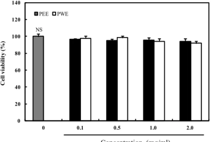
Measurement of cytotoxicity
Cell viability was analyzed using 3T3-L1 cells (KCLB, Seoul, Korea) and 3-(4,5-dimethylthiazol-2-yl)-2,5-diphenylte- trazolium bromide (MTT, Sigma, St. Louis, MO, USA). 3T3- L1 cells were seeded into 96-well plates at 1×10⁴ cells/well and preincubated at 37°C under with 5% CO2 for 24 hr. After 24 hr, the medium was made a replacement with medium containing diverse consistencies of PEE and PWE: absence of PEE or PWE, 0.1, 0.2, 0.5, 1.0 and 2.0 mg/ml, respectively, for 24 hr. Following treatment, a filtered MTT solution (100 μL) was superinduced to each well and incubated for an additional 4 hr at 37°C. Formazan was carefully aspirated and DMSO (100 μl) was added to each well. The absorbance of the DMSO solution was assayed at 540 nm with a micro- plate reader (Model 680, Bio-Rad Laboratories Inc., Hercules, CA, USA).
Inhibitory activities of P. oleracea extract on α-glu- cosidase in vitro
The inhibition assay for α-glucosidase activity was ana- lyzed by a chromogenic process using a yeast enzyme [33].
Simply, yeast α-glucosidase (100 U; Sigma, St. Louis, MO, USA) was lysed in 100 mM phosphate buffer (pH 7.0) in- cluded 0.2 g/l sodium azide. A 5 mM solution of p-nitro- phenyl-α-D-glucopyranoside (≥99%; Sigma, St. Louis, MO, USA) was dissolved in the same PBS (pH 7.0) to obtain the substrate solution. Then, the α-glucosidase solution (50 μl) and 10 μl of PEE or PWE, lysed in dimethyl sulfoxide (DMOS, Bio Basic Inc., KCMO, Canada), were blended in a microtiter plate, and the absorbance was analyzed at 405 nm using a microplate reader at 0 min. The mixture was incubated for 5 min and the substrate solution (50 μl) was supplemented and incubated at room temperature for 5 min.
Then, an increase was measured in the absorbance from the zero-time point. The inhibitory activities of PEE and PWE at various concentrations were indicated as absorbance changes caused by the test extracts relative to those in the vehicle control (%). The IC50 values (i.e., the concentrations of PEE and PWE resulting 50% inhibition of the maximum activity) were measured.
Inhibitory activities of P. oleracea extract on α- amylase in vitro
The α-amylase inhibitory activity were assayed equiv- alently using the method presented in α-glucosidase in- hibitory activity assay excluding α-amylase from porcine pancreas (100 U; Sigma, St. Louis, MO, USA) and p-nitro- phenyl-α-D-maltopentoglycoside (≥99%; Sigma, St. Louis, MO, USA).
In vivo experiments
ICR male mice (4-week-old; purchased from Orient, Inc., Seoul, Korea) used for the study were kept under a 12 hr on/12 hr off cycle and controlled room temperature. The mice were fed a pelleted feed (5L79, Orient, Inc., Seoul, Korea), and water was freely provided. After an adaptation period of 2 weeks, diabetes was induced as described below.
All procedures involving the handling and care of mice com- plied with the current international laws and policies (National Institutes of Health Guide for the Care and Use of Laboratory Animals) and were permitted by the uni- versity's Animal Ethics Committee (PNU-2017-1496).
NS
0 20 40 60 80 100 120 140
0 0.1 0.5 1.0 2.0
Cell viability (%)
PEE PWE
Concentration (mg/ml)
Fig. 1. Cytotoxic effects of Portulaca oleracea L. extracts. 3T3-L1 cells were treated with various concentrations (0.1, 0.5, 1.0, and 2.0 mg/ml) of PEE and PWE for 24 hr, and cyto- toxic effects was measured by the MTT assay. Each value is expressed as the mean ± SD of triplicate experiments.
NS: not significant. PEE: P. oleracea 80% ethanol extract;
PWE: P. oleracea water extract; Acarbose: positive control.
Concentration (mg/ml)
Fig. 2. Inhibitory activities of Portulaca oleracea L. extracts on α- glucosidase. Each value is expressed as the mean±SD of triplicate experiments. Values with different superscript letters are significantly different (p<0.05) based on Dun- can’s multiple range tests. The concentration of acarbose, used as a positive control, was 0.50 mg/ml. PEE: P. oler- acea 80% ethanol extract; PWE: P. oleracea water extract;
Acarbose: positive control.
Induction of experimental diabetes model
Diabetes was induced by an intraperitoneal injection of streptozotocin (STZ; 60 mg/kg, Sigma, St. Louis, MO, USA) dissolved in citrate buffer (0.1 M, pH 4.5). Fasting blood glu- cose level was checked after 7th day of injection to confirm diabetes induction from tail vein using a glucose meter and glucose strips. Mice with fasting blood glucose level above 250 mg/dL were regarded to be diabetes.
Measurement of blood glucose level
The mean blood glucose level in each group (Normal mice and diabetic mice) was similar, and each group was divided into four groups of seven mice. A total of 8 groups were used and orally administered the following after overnight fasting: 1) control: soluble starch (2 g/kg of body weight [BW]), 2) PEE: soluble starch with PEE (300 mg/kg of BW), 3) PWE: soluble starch with PWE (300 mg/kg of BW), 4) acarbose: soluble starch with acarbose (100 mg/kg of BW).
Blood samples were collected from the tail vein at 0, 30, 60, and 120 min, and blood glucose was checked using a blood glucose meter (Roche Diagnistics GmbH, Germany). Areas under the concentration-time curves (AUCs) were identified using the trapezoidal rule [16].
Statistical analysis
Data are represented as the mean ± standard deviation (SD). Statistical analysis was conducted using SAS software ver. 9.1 (SAS Institute, Inc., Cary, NC, USA). Dissimilarity between groups were assessed by one-way analysis of var- iance, followed by Duncan’s post-hoc multiple range tests.
The p-value<0.05 was regarded statistically significant.
Results and Discussion
Cytotoxic effect of P. oleracea
To evaluate whether PEE and PWE have toxic effect to the cells, PEE and PWE were treated in 3T3-L1 cells for 24 hr, and cytotoxic effect was measured using MTT assay. PEE and PWE were not exerting any cytotoxic effect at the vari- ous concentrations (0.1, 0.5, 1.0 and 2.0 mg/ml) in 3T3-L1 cells (Fig. 1).
Inhibitory activities of P. oleracea extracts against α-glucosidase and α-amylase in vitro
The inhibitory activities of PEE and PWE against α-gluco- sidase are shown in Fig. 2. PEE inhibited the α-glucosidase
activity concentration-dependently by 39.08, 44.64, 56.34, and 75.42% at concentrations of 0.05, 0.10, 0.25, and 0.50 mg/ml, respectively. The inhibitory effect of PWE against α-glucosi- dase was also dose-dependent, and the levels of inhibition were 34.20, 39.56, 48.45, and 64.55% at concentrations of 0.05, 0.10, 0.25, and 0.50 mg/ml, respectively. However, the in- hibitory activity of PEE against α-glucosidase was markedly
Table 1. IC50 values of Portulaca oleracea L. extracts for α-glucosi- dase and α-amylase
Sample IC50 (mg/ml)1)
α-glucosidase α-amylase
PEE PWE Acarbose
0.168±0.023c 0.274±0.011b 0.295±0.017a
0.212±0.044c 0.241±0.033b 0.334±0.021a
1)IC50 value is the concentration of a sample required for 50%
inhibition. Each value is expressed as the mean ± SD of tripli- cate experiments. Values with different superscript letters with- in a column are significantly different (p<0.05) based on Duncan’s multiple range tests. PEE: P. oleracea 80% ethanol ex- tract; PWE: P. oleracea water extract; Acarbose: positive control.
Concentration (mg/ml)
Fig. 3. Inhibitory activities of Portulaca oleracea L. extracts on α- amylase. Each value is expressed as the mean±SD of trip- licate experiments. Values with different superscript let- ters are significantly different (p<0.05) based on Dun- can’s multiple range tests. The concentration of acarbose, used as a positive control, was 0.50 mg/ml. PEE: P. oler- acea 80% ethanol extract; PWE: P. oleracea water extract;
Acarbose: positive control.
higher than PWE. Acarbose, a commercial hypoglycemic pharmaceutical product, inhibited enzyme activity by 60.03
% at a concentration of 0.50 mg/ml. Thus, at the same con- centration (0.50 mg/ml), PEE showed markedly higher in- hibitory activities than acarbose.
The inhibitory activities of PEE and PWE against α-amy- lase are shown in Fig. 3. PEE inhibited α-amylase activity concentration-dependently by 18.25, 38.52, 55.24, and 62.22%
at concentrations of 0.05, 0.10, 0.25, and 0.50 mg/ml, respectively. The inhibition effect of PWE against α-amylase was also concentration-dependent, and the levels of in- hibition were 15.15, 30.01, 51.23, and 58.22% at concen- trations of 0.05, 0.10, 0.25, and 0.50 mg/ml, respectively. The
inhibition effect of PEE against α-amylase was markedly higher than acarbose at the same concentration (0.50 mg/
ml). The IC50 values of PEE against α-glucosidase and α- amylase were 0.16±0.02 and 0.21±0.04 mg/ml, and those of PWE were 0.27±0.01 and 0.24±0.03 mg/ml, respectively, in- dicating that the extracts had significantly higher inhibitory activities than acarbose (0.29±0.01 and 0.33±0.02 mg/ml, re- spectively) (Table 1).
Postprandial hyperglycemia is a major initial symptom of diabetes and is involved in the occurrence of diabetes-related complications [2]. Postprandial hyperglycemia was reported to have a stronger correlation than fasting hyperglycemia with the mortality and morbidity rate of cardiovascular dis- ease [9]. Therefore, alleviation of postprandial hyperglycemia is an important goal when treating diabetic patients. Starch is mainly digested in the small intestine by pancreatic α- amylase action, which produces maltose and isomaltose oli- gosaccharides; these are hydrolyzed by intestinal α-glucosi- dase to produce absorbable monosaccharides. The enzyme α-glucosidase subserves the last step of carbohydrate diges- tion, and therefore α-glucosidase inhibitors can block carbo- hydrate absorption and lower postprandial blood glucose level [33]. Thus, by delaying the absorption of glucose via inhibition of carbohydrate hydrolases reduction of post- prandial hyperglycemia may be achieved, greatly facilitating diabetes control [3, 25].
In present study, the inhibition activities of PEE and PWE against α-glucosidase and α-amylase were examined to de- termine the ability of the extracts as a functional food to reduce postprandial hyperglycemia. The results indicated that both PEE and PWE had inhibition effects on α-glucosi- dase and α-amylase. The strong inhibitory effects of PEE and PWE may be related to the presence of active compounds such as polyphenols in the P. oleracea extracts. Polyphenol compounds can bind to proteins and other macromolecules via their phenolic hydroxyl groups [7]. Binding is induced through a hydrogen bond between the hydroxyl group in polyphenol and the carboxylate group of Asp197 and Glu233 at the active sites of α-glucosidase and α-amylase, on the basis of the relationship between inhibition activity and mo- lecular interactions [14]. A previous study showed that the hydroxyl groups of polyphenols may be important in en- zyme inhibition [30].
It has also been reported that active polyphenol com- pounds such as proanthocyanidin, rutin, and quercetin, which are present in P. oleracea, are better eluted with etha-
a
a
a
c
b
c b
b
c b
c
c 250
300 350 400 450 500
0 30 60 120
Blood glucose (mg/dL)
Time (min) Control
PEE PWE Acarbose
Fig. 4. Blood glucose levels after administration of Portulaca oler- acea L. extracts to streptozotocin-induced diabetic mice.
Control (distilled water), PEE (300 mg/kg), PWE (300 mg/kg), and acarbose (100 mg/kg) were orally co-ad- ministered with starch (2 g/kg). Each value is expressed as the mean±SD of seven mice per each group. Values with different superscript letters are significantly differ- ent (p<0.05) based on Duncan’s multiple range tests. PEE:
P. oleracea 80% ethanol extract; PWE: P. oleracea water extract; Acarbose: positive control.
a
a
a b
b
c b
a
b
c
c
c
50 100 150 200
0 30 60 120
Blood glucose (mg/dL)
Time (min) Control
PEE PWE Acarbose
Fig. 5. Blood glucose levels after administration of Portulaca oler- acea L. extracts to normal mice. Control (distilled water), PEE (300 mg/kg), PWE (300 mg/kg), and acarbose (100 mg/kg) were orally co-administered with starch (2 g/
kg). Each value is expressed as the mean±SD of seven mice per each group. Values with different superscript letters are significantly different (p<0.05) based on Dun- can’s multiple range tests. PEE: P. oleracea 80% ethanol extract; PWE: P. oleracea water extract; Acarbose: positive control.
nol than with water [19, 18]. Proanthocyanidin reportedly inhibits the activity of carbohydrate digestive enzymes in carbohydrate metabolism [22]. Quercetin reversibly and slowly binds to glucosidase and shows non-competitive in- hibition [5]. The total phenol contents of PEE and PWE were 58.16 and 49.09 mg/g, respectively, and the flavonoid con- tents were 20.08 and 14.98 mg/g, respectively [17]. Both PEE and PWE efficiently inhibited α-glucosidase and α-amylase because both contained polyphenol compounds. However, because the content of polyphenol components in an extract is correlated with its inhibitory activity against α-glucosi- dase, PEE, which contains more polyphenol compounds than PWE, can retard the digestive absorption of carbohy- drates by hindering α-glucosidase and α-amylase in the small intestine more effectively than PWE.
Effects of P. oleracea extracts on blood glucose levels in vivo
The effects of P. oleracea on postprandial blood glucose levels were assayed in diabetic and normal mice. In diabetic mice, postprandial blood glucose levels were significantly lower the PEE-treatment group than the control group (Fig.
4). The blood glucose levels in diabetic control mice in- creased to 383.25±14.8 mg/dL at 30 min and 429.2±21.9 mg/
dL at 60 min after a meal and then declined to 360.2±24.5 mg/dL at 120 min. However, when PEE was administered with starch, the increase in postprandial blood glucose levels was significantly suppressed (337.3±20.1, 368.5±22.8, and 290.1±21.6 mg/dL at 30, 60, and 120 min, respectively;
p<0.05). When PWE was administered with starch, post- prandial hyperglycemia also decreased (365.6±20.2, 379.2±
18.15, and 324.2±17.3 mg/dL at 30, 60, and 120 min, re- spectively; p<0.05), but the decrease was not as pronounced as that observed with PEE. Peak postprandial blood glucose was also markedly lower in normal group following starch and PEE consumption than in those administered starch alone (Fig. 5). This confirms that PEE suppresses post- prandial hyperglycemia due to starch in normal group. The AUC in diabetic group treated PEE (656.3±42.6 mg·h/dL) was significantly lower (p<0.05) than that in diabetic control group (761.8±59.4 mg·h/dL) (Table 2).
Hyperglycemia is a serious problem in diabetes, with rap- id postprandial hyperglycemia and prolonged hyper- glycemia leading to chronic complications such as visual im- pairment, renal dysfunction, neurological disorders, and car- diac dysfunction [31, 34]. Thus, maintaining nearly normal- ity level of fasting and postprandial glucose is important for
Table 2. Areas under the concentration–time curves (AUCs) of postprandial glucose responses in normal and strepto- zotocin-induced diabetic mice
Group1) AUC (mg・h/dL)
Normal mice Diabetic mice Control
PEE PWE Acarbose
268.1±14.7a 233.8±29.1c 245.4±18.2b 195.6±18.5d
761.8±59.4a 656.3±42.6c 697.0±38.2b 608.2±47.8d
1)PEE (300 mg/kg), PWE (300 mg/kg), acarbose (100 mg/kg), and distilled water (control) were orally co-administered with starch (2 g/kg). Each value is expressed as the mean ± SD of seven mice. Values with different superscript letters within a column are significantly different (p<0.05) based on Duncan’s multiple range tests. PEE: Portulaca oleracea L. 80% ethanol ex- tract; PWE: P. oleracea water extract; Acarbose: positive control.
managing diabetes [26]. This study confirmed the anti- hyperglycemic effect of PEE and PWE in diabetic and nor- mal group after starch ingestion by administering PEE or PWE. In diabetic mice administered PEE, postprandial hy- perglycemia significantly decreased. This result suggests that PEE delays the absorption of dietary starch, alleviating in post-prandial hyperglycemia. These mitigation effects can be inferred from the inhibition of carbohydrate digestion en- zymes by PEE in diabetic mice. Acarbose, which flattened the maximum of postprandial blood glucose, decreased the AUC value [12]. PEE also significantly decreased not merely the maximum blood glucose level but also the AUC value.
Patients of diabetes experience postprandial hyperglyce- mia, leading to a variety of disturbances of metabolism, in- cluding cardiac dysfunction [8]. Thus, controlling post- prandial hyperglycemia is prominent in the management of diabetic mellitus and prevention of diabetic complications.
Plenty of pharmaceutical products are used as postprandial hypoglycemic agents. However, they are generally asso- ciated with significant toxicity or potential detrimental effect [10, 21]. Therefore, studies of natural compounds that can reduce postprandial hyperglycemia are currently ongoing.
Our results indicate that P. oleracea improves postcibal hy- perglycemia and staves off complications of diabetes melli- tus.
In conclusion, P. oleracea extracts strongly suppressed α- glucosidase and α-amylase activities. Comparison of two ex- tracts showed that PEE had stronger inhibiting effects than PWE. Administration of PEE with starch to diabetic mice reduced postprandial hyperglycemia by more than admin- istration of PWE with starch. These results suggest that PEE
is a better candidate than PWE as a functional food for con- trolling postprandial hyperglycemia.
Acknowledgments
This research was supported by the Basic Science Research Program through the National Research Foundation of Korea (NRF) funded by the Ministry of Science, ICT &
Future Planning (2017R1A2B4005323).
References
1. Barakat, L. A. and Mahmoud, R. H. 2011. The anti- atherogenic, renal protective and immunomodulatory ef- fects of purslane, pumpkin and flax seeds on hyperchole- sterolemic rats. N. Am. J. Med. Sci. 3, 351-357.
2. Baron, A. D. 1998. Postprandial hyperglycemia and α-gluco- sidase inhibitors. Diabetes Res. Clin. Pract. 40, 51-55.
3. Bhandari, M. R., Anurakkun, N. J., Hong, G. and Kawabata, J. 2008. α-Glucosidase and α-amylase inhibitory activities of Nepalese medicinal herb Pakhanbhed (Bergenia ciliata, Haw.). Food Chem. 106, 247-252.
4. Ceriello, A. 2005. Postprandial hyperglycemia and diabetes complications: is it time to treat? Diabetes 54, 1-7.
5. Chai, T. T., Kwek, M. T., Ong, H. C. and Wong, F. C. 2015.
Water fraction of edible medicinal fern Stenochlaena palustris is a potent α-glucosidase inhibitor with concurrent anti- oxidant activity. Food Chem. 186, 26-31.
6. Chen, J., Shi, Y. P. and Liu, J. Y. 2003. Determination of noradrenaline and dopamine in Chinese herbal extracts from Portulaca oleracea L. by high-performance liquid chro- matography. J. Chromatogr. A. 1003, 127-132.
7. Cuvelier, M. E., Richard, H. and Berset, C. 1996. Antioxida- tive activity and phenolic composition of pilot-plant and commercial extracts of sage and rosemary. J. Am. Oil. Chem.
Soc. 73, 645-652.
8. Dennis, J. W., Laferte, S., Waghorne, C., Breitman, M. L.
and Kergel, R. S. 1987. Beta 1-6 Branching of asn-linked oli- gosaccharides is directly associated with metastasis. Science 236, 582-585.
9. Haller, H. 1998. The clinical importance of postprandial glucose. Diabetes Res. Clin. Pract. 40, 43-49
10. Hanefeld, M. 1998. The role of acarbose in the treatment of non-insulin-dependent diabetes mellitus. J. Diabetes Com- plicat. 12, 228-237.
11. Honeycutt, A. A., Boyle, J. P., Broglio, K. R., Thompson, T.
J., Hoerger, T. J., Geiss, L. S. and Narayan, K. M. 2003. A dynamic Markov model for forecasting diabetes prevalence in the United States through 2050. Health Care Manag. Sci.
6, 155-164.
12. Inoue, I., Takahashi, K., Noji, S., Awata, T., Negishi, K. and Kataya-ma, S. 1997. Acarbose controls postprandial hyper- proinsulinemia in non-insulin-dependent diabetes mellitus.
Diabetes Res. Clin. Pract. 36, 143-151.
13. Kamal, U., Abdul, S. J., Eaqub, A. and Mohd, R. I. 2012.
Evaluation of Antioxidant properties and mineral composi- tion of purslane (portulaca oleracea) at different growth stages. Int. J. Mol. Sci. 13, 10257-10267.
14. Kawamura-Konishi, Y., Watanabe, N., Saito, M., Nakajima, N., Sakaki, T., Katayama, T. and Enomoto, T. 2012. Isolation of a new phlorotannin, a potent inhibitor of carbohydrate- hydrolyzing enzymes, from the brown alga Sargassum patens. J. Agric. Food Chem. 60, 5565-5570.
15. Kawano, H., Motoyama, T., Hirashima, O., Hirai, N., Miyao, Y., Sakamoto, T., Kugiyama, K., Ogawa, H. and Yasue, H.
1999. Hyperglycemia rapidly suppresses flow-mediated en- dothelium-dependent vasodilation of brachial artery. J. Am.
Coll. Cardiol. 34, 146-154.
16. Kim, J. S. 2004. Effect of Rhemanniae radix on the hyper- glycemic mice induced with streptozotocin. J. Kor. Soc. Food Sci. Nutr. 33, 1133-1138.
17. Kim, M. J. 2011. Studies on the Biological Activities of Purslane(Portulaca oleracea), MS thesis, University of Gyeongsang, Jinju, Gyeongnam, korea.
18. Kin, J. E., Joo, S. I., Seo, J. H. and Lee, S. P. 2009. Antioxidant and α-glucosidase inhibitory effect of Tartary Buckwheat ex- tract obtained by the treatment of different solvents and enzymes. J. Kor. Soc. Food Sci. Nutr. 38, 989-995.
19. Kwon, Y. R., Cho, S. M., Hwang, S. P., Kwon, G. M., Kim, J. W. and Youn, K. S. 2014. Antioxidant, physiological activ- ities, and acetylcholinesterase inhibitory activity of Portulaca oleracea extracts with different extraction methods. J. Kor. Soc.
Food Sci. Nutr. 43, 389-396.
20. Lei, X., Li, J., Liu, B., Zhang, N. and Liu, H. 2015. Separation and identification of four new compounds with antibacte- rial activity from Portulaca oleracea L. Molecules 20, 16375- 16387.
21. Li, Y., Wen, S., Kota, B. P., Peng, G., Li, G. Q., Yamahara, J. and Roufogalis, B. D. 2005. Punica granatum flower extract, a potent α-glucosidase inhibitor, improves postprandial hy- perglycemia in Zucker diabetic fatty rats. J. Ethnopharmacol.
99, 239-244.
22. Lu, Y., Demleitner, M. F., Song, L., Rychlik, M. and Huang, D. 2016. Oligomeric proanthocyanidins are the active com- pounds in Abelmoschus esculentus Moench for its α-amylase and α-glucosidase inhibition activity. J. Funct. Foods 20, 463- 471.
23. Palaniswamya, U. R., Bible, B. B. and McAvoy, R. J. 2004.
Oxalic acid concentrations in Purslane (Portulaca oleraceae L.) is altered by the stage of harvest and the nitrate to ammo- nium ratios in hydroponics. Sci. Hortic. 102, 267-275.
24. Pirart, L. 1978. Diabetes mellitus and its degenerative com- plieations: a prospective study of 4400 patients observed be-
tween 1947 and 1973. Diabetes Care 1, 168-172.
25. Puls, W., Keup, U., Krause, H. P., Thomas, G. and Hoffmeis- ter, F. 1997. Glucosidase inhibition. A new approach to the treatment of diabetes, obesity, and hyperlipoproteinaemia.
Naturwissenschaften 64, 536-537.
26. Ratner, R. E. 2001. Controlling postprandial hyperglycemia.
Am. J. Cardiol. 88, 26-31.
27. Saito, N., Sakai, H., Suzuki, S., Sekihara, H. and Yajima, Y.
1998. Effect of an α-glucosidase inhibitor (voglibose), in combination with sulphonilureas, on glycaemic control in type 2 diabetes patients. J. Int. Med. Res. 26, 219-232.
28. Scott, L. J. and Spencer, C. M. 2000. Miglitol: a review of its therapeutic potential in type 2 diabetes mellitus. Drugs 59, 521-549.
29. Standl, E., Baumgartl, H. J., Fuchtenbusch, M. and Stemplin- ger, J. 1999. Effect of acarbose on additional insulin therapy in type 2 diabetic patients with late failure of sulphonylurea therapy. Diabetes Obes. Metab. 1, 215-220.
30. Stern, J. L., Hagerman, A. E., Steinberg, P. D. and Mason, P. K. 1996. Phlorotannins–protein interactions. J. Chem.
Ecol. 22, 1877-1899.
31. Tai, E. S., Lim, S. C., Tan, B. Y., Chew, S. K., Heng, D. and Tan, C. E. 2000. Screening for diabetes mellitus-a two-step approach in individuals with impaired fasting glucose im- proves detection of those at risk of complications. Diabet.
Med. 17, 771-775.
32. Temelkova-Kurktschiev, T. S., Koehler, C., Henkel, E., Leon- hardt, W., Fuecker, K. and Hanefeld, M. 2004. Postchallenge plasma glucose and glycemic spikes are more strongly asso- ciated with atherosclerosis than fasting glucose or HbA1c level. Diabetes Care 23, 1830-1834.
33. Watanabe, J., Kawabata, J., Kurihara, H. and Niki, R. 1997.
Isolation and identification of alpha-glucosidase inhibitors from tochucha (Eucommia ulmoides). Biosci. Biotechnol. Bio- chem. 61, 177-178.
34. West, I. C. 2000. Radicals and oxidative stress in diabetes.
Diabet. Med. 17, 171-180.
35. Yazici, I., Turkan, I., Sekmen, A. H. and Demiral, T. 2007.
Salinity tolerance of purslane (Portulaca oleracea L.) is ach- ieved by enhanced antioxidative system, lower level of lipid peroxidation and proline accumulation. Environ. Exp. Bot.
61, 49-57.
36. Yen, G. C., Chen, H. Y. and Peng, H. H. 2001. Evaluation of the cytotoxicity, mutagenicity and antimutagenicity of emerging edible plants. Food Chem. Toxicol. 39, 1045-1053.
37. Zhang, J. Y., Chen, X. G. and Hu, Z. D. 2002. Quantification of noradrenaline and dopamine in Portulaca oleracea L. by capillary electrophoresis with laser-induced fluorescence detection. Anal. Chim. Acta. 471, 203-209.
초록:쇠비름(Portulaca oleracea L.) 추출물의 탄수화물 소화 효소 저해와 식후 고혈당 완화 효과
박재은․한지숙*
(부산대학교 식품영양학과)
식후고혈당은 제 2형 당뇨병의 발병에 부정적인 영향을 미치고 미세혈관 및 대혈관 질환 등의 당뇨병 합병증 유발과 밀접한 관계가 있다. 따라서 식후고혈당을 조절하는 것이 당뇨병 합병증의 위험을 줄이는 가장 중요한 요소이다. 식후고혈당은 소장에서 α-글루코시다아제와 같은 탄수화물 소화 효소를 저해함으로써 조절될 수 있다.
이에 본 연구는 쇠비름 에탄올 추출물(PEE)과 물 추출물(PWE)이 탄수화물 소화 효소를 저해하고, 당뇨병 마우스 에서 식후 고혈당을 강하시키는 효과에 대해 조사하였다. α-글루코시다아제와 α-아밀라아제에 대한 저해효과는 두 추출물 모두 양성대조군인 acarbose보다 더 효과적이었으며, PEE에 의한 α-글루코시다아제 저해 효과가 PWE 보다 더 효과적이었다. Diabetic mice에 전분(2 g/kg)을 투여한 후의 혈당 증가는 30, 60, 120분에 각각 383.7, 429.3, 360.2 mg/dL로 나타났고, 전분(2 g/kg)과 PEE 또는 PWE 추출물(300 mg/Kg)을 투여한 후의 혈당 증가는 30, 60, 120분에 각각 337.0, 368.5, 290.1 mg/dL과 365.8, 379.2, 324.3 mg/dL로 나타나, PEE 추출물 투여군이 대조 군에 비해 식후 혈당 강하가 효과적으로 나타남을 알 수 있었다. 이러한 결과는 쇠비름 추출물이 탄수화물 소화 효소를 저해함으로써 식후 고혈당을 완화시키고, 특히 쇠비름 에탄올 추출물(PEE)이 쇠비름 물 추출물(PWE) 보 다 식후 고혈당을 완화시키는데 더욱 효과가 있는 것으로 나타났다.