Cucurbitacin-I, a Naturally Occurring Triterpenoid, Inhibits the CD44 Expression in Human Ovarian Cancer Cells
Hee Won Seo and Jin-Kyung Kim*
Department of Biomedical Science, Catholic University of Daegu, Gyeongsan-Si 38430, Korea Received January 12, 2018 /Revised March 7, 2018 /Accepted June 4, 2018
Cucurbitacin-I, a natural triterpenoid derived from Cucurbitaceae family plants, exhibits a number of potentially useful pharmacological and biological activities. Indeed, the previous study demonstrated that cucurbitacin-I reduced the proliferation of colon cancer cells by enhancing apoptosis and causing cell cycle arrest at the G2/M phase. CD44, a type I transmembrane protein with the function of adher- ing to cells, mediates between the extracellular matrix and other cells through hyaluronic acid. Recent studies have demonstrated that an overexpression of the CD44 membrane receptor results in tumor initiation and growth, specific behaviors of cancer stem cells, the development of drug resistance, and metastasis. The aim was to examine the effect of cucurbitacin-I on CD44 expression human ovarian cancer cells because the effect of cucurbitacin-I on CD44 expression has not been reported. The ex- pressions of CD44 mRNA and protein were detected using a quantitative real-time reverse-tran- scription polymerase chain reaction and a Western blot analysis, respectively. Treatment with cucurbi- tacin-I inhibited the expression of CD44 mRNA and protein. A subsequent analysis revealed that cu- curbitacin-I blocked the phosphorylation of activator protein-1 (AP-1) and nuclear factor kappa-B (NF- κB), which are key regulators of CD44 expression. Taken together, the data demonstrate that cucurbi- tacin-I regulates the AP-1 and NF-κB signaling pathways, leading to decreased CD44 expression.
Key words : CD44, Cucurbitacin-I, ovarian cancer, SKOV-3 cell
*Corresponding author
*Tel : +82-53-850-3774, Fax : +82-53-850-3774
*E-mail : [email protected]
This is an Open-Access article distributed under the terms of the Creative Commons Attribution Non-Commercial License (http://creativecommons.org/licenses/by-nc/3.0) which permits unrestricted non-commercial use, distribution, and reproduction in any medium, provided the original work is properly cited.
Journal of Life Science 2018 Vol. 28. No. 6. 733~737 DOI : https://doi.org/10.5352/JLS.2018.28.6.733
Introduction
Ovarian cancer is an abnormal cell growth arising in the ovary. It is one of the three major cancers contracted by women along with breast and cervical cancer. The majority of ovarian cancers are epithelial and develop in women over 50. Although the progress has been made in the treatment of ovarian cancer, the mortality rate remains high. The ma- jority of ovarian cancer patients have tumor metastasis at the time-point of diagnosis, and tumor invasion and meta- stasis are important causes of treatment failure [6]. There- fore, investigation of the molecular mechanisms involved in tumor proliferation, adhesion and invasion is of great im- portance for improving the therapeutic effectiveness of ovar- ian cancer treatments.
CD44 is a transmembrane and cell surface glycoprotein involved in cell-cell interactions, cell adhesion, and migra-
tion [8]. It is considered a major cell surface marker for meta- stasis and progression in certain types of cancers, including ovarian carcinoma. CD44 is a receptor for hyaluronic acid [1, 2]. It interacts with hyaluronic acid to activate Nanog- Stat-3 and ankyrin based signaling pathways that is consid- ered to be responsible for conferring tumor cell growth in ovarian and breast cancers [4]. In addition, CD44 expression is a prognostic indicator of shorter survival time in patients with ovarian cancer [13]. Based on the aforementioned find- ings, we hypothesized that suppression of CD44 expression could enhance the efficiency of chemotherapy and prevent the development of metastases.
Cucurbitacin-I is a naturally occurring triterpenoid de- rived from Cucurbitaceae family plants that exhibits a num- ber of potentially useful pharmacological and biological ac- tivities [3, 5, 9, 10, 12, 16, 17]. It was originally identified to be a potent selective inhibitor of the Janus kinase 2/signal transducer and activator of transcription 3 (JAK2/STAT3) signaling pathway with antiproliferative and antitumor properties [3]. Upon inhibition of STAT3-dependent gene transcription, cucurbitacin-I elicits antiproliferative effects various cancer cells with activated STAT3 signaling [5, 12, 16]. However, the effect of cucurbitacin-I on CD44 expre- ssion in ovarian cancer was not investigated. Therefore, the
- Note -
present study aimed to investigate the effects of cucurbita- cin-I on the expression of CD44 of SKOV-3 ovarian cancer cells.
Materials and Methods
Chemicals and reagents
Cucurbitacin-I was purchased from Santa Cruz (Santa Cruz, CA, USA). RPMI-1640 medium, fetal bovine serum (FBS), penicillin, streptomycin, and trypsin/EDTA were pur- chased from Hyclone (Logan, UT, USA). Protein detection BCA kit was obtained from Thermo Scientific Pierce (Rock- ford, IL, USA). Antibody against CD44 standard form were purchased from Abcam (Cambridge, MA, USA). Anti-phos- pho-p65, anti-phospho-cJun, and anti-phospho-cFos were obtained from Cell Signaling Technology (Danvers, USA).
Anti-β-actin antibody and other materials were purchased from Sigma Aldrich (St. Louis, MO, USA).
Cell and cell culture
The ovarian cancer cell line SKOV-3 was obtained from the Korean Cell Bank (Seoul, Korea). Cells were cultured in RPMI-1640 medium supplemented with 10% FBS, 100 U/ml penicillin and 100 μg/ml streptomycin at 37℃ in a humidi- fied atmosphere of 5% CO2 and 95% air.
Cell proliferation assay
SKOV-3 cells were seeded into wells of 96-well plates at a density of 1×10⁴ cells/well. The rate of cell proliferation was determined at 24 hr with CellTiter 96® AQueous One Solution (Promega, Madison, WI, USA). Cell Proliferation as- say according to the manufacturer’s instruction.
Flow Cytometry
The expression levels of surface CD44 protein were de- termined by immunostaining with anti-human CD44-FITC antibody (BD Biosciences, San Diego, CA, USA) for 30 min on ice. Cells stained with the appropriate isotype-matched control antibody were used as negative controls. After stain- ing, cells were analyzed with a FACSCalibur instrument equipped with CellQuest software (BD Biosciences). The mean fluorescence intensity (MFI) of samples was analyzed by FlowJo software (TreeStar Inc., Ashland, OR, USA).
Quantitative real-time reverse-transcription poly- merase chain reaction (qRT-PCR)
Total RNA was isolated from SKOV-3 cells using Trizol Reagent (Invitrogen, Carlsbad, CA, USA). DNA was elimi- nated from total RNA using RNA Qualified RNase-Free DNase (Promega) and cDNA was synthesized by GoScript™
Reverse Transcription System (Promega). qRT-PCR assay was carried out with LightCycler (Roche Diagnostics, Basel, Switzerland) using LightCycler FastStart DNA Master SYBR Green I (Roche Diagnostics). All the experiments were re- peated twice in triplicate each time. Transcripts of glycer- aldehyde-3-phosphate dehydrogenase (GAPDH) as a house- keeping gene were quantified as endogenous RNA of refer- ence to normalize each sample. Relative quantities were esti- mated by the -ΔΔCt method. CD44 and GAPDH primer pairs are as follows: CD44, forward 5’-CCTTTGATGGACCA ATTACCATAAC-3’, reverse 5’-TCAGGATTCGTTCTGTAT CCT-3’; GAPDH, forward 5’-TGAACGGGAAGCTCACTGG- 3’, reverse 5’-TCCACCACCCTGTTGCTGTA-3’.
Western blot analysis
Cells were lysed in Pro-prep protein extraction solution (iNtRon, Sungnam, Korea). The protein concentrations were determined using a BCA protein assay kit. Whole cell ex- tracts were separated by 10% sodium dodecyl sulfate-poly- acrylamide gel electrophoresis and transferred to a PVDF membrane. After blocking with 3% skim milk for 1 hr, the membranes were incubated with primary antibody at 4℃
overnight. The membranes were then washed with PBST (1×
PBS, 0.1% Tween 20) and incubated for 1 hr with anti-rabbit IgG or anti-mouse IgG conjugated with HRP. After washing, the protein bands were then visualized using a WEST- One
™ Western Blot Detection Spray (iNtRon) and DAVINCH- Chemi CAS-400SM (Davinch-k, Seoul, Korea). Dates were assessed using Total Lab software (Davinch-k).
Statistical analysis
Values are expressed as the mean ± SEM of the results of at least three experiments. The values were then evaluated by one-way analysis of variance (ANOVA) with Bonferroni multiple comparison post tests using the GraphPad Prism 4.0 software (GraphPad Software, San Diego, CA, USA).
Differences with p values <0.05 were considered statistically significant.
Results and Discussion
Cucurbitacin-I inhibits ovarian cancer cell growth in a dose-dependent manner
A B
Fig. 1. Effects of Cucurbitacin-I on cell viability in SK-OV-3 cells.
(A) Chemical structure of cucurbitacin-I. (B) SKOV-3 cells were treated with indicated concentrations of cu- curbitacin-I for 24 hr and determined the proliferation by MTS assay. The results are means ± SEM of three independent experiments in triplicate. **p<0.01, ***p<0.001 versus 0 nM cucurbitacin-I-treated cells.
A B
C D
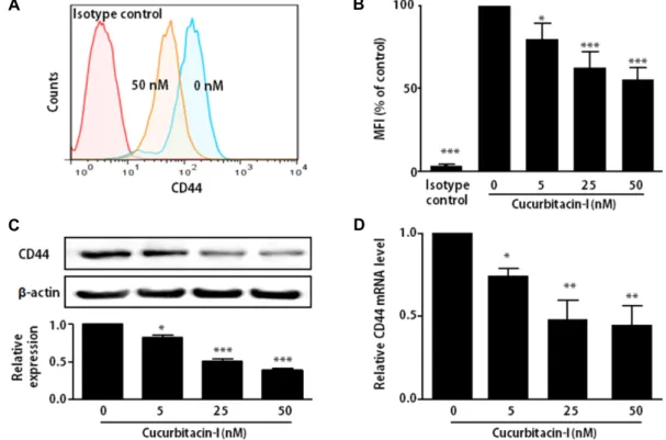
Fig. 2. Effect of cucurbitacin-I on the expression of CD44. SKOV-3 cells were treated with indicated concentration of cucurbitacin-I for 24 hr. (A) Flow cytometry analysis of cell surface CD44 expression by CD44 and isotype control antibodies. (B) Representative mean fluorescence intensity (MFI) for cell surface CD44 expression in SKOV-3 cells. (C) Western blot analysis of CD44 (upper) and the band intensities were quantified by densitometry (lower). The relative expression normalized with β-actin in whole cell lysates of SKOV-3 cells. β-actin was used as the loading control. (D) CD44 mRNA levels were quantified by qRT-PCR and normalized with the housekeeping gene, GAPDH. The results are means ± SEM of four independent experiments. *p<0.05, **p<0.01, ***p<0.001 versus 0 nM cucurbitacin-I-treated cells.
To determine the effect of cucurbitacin-I on the pro- liferation of ovarian cancer cells, the SKOV-3 cells were treat- ed with various concentrations of cucurbitacin-I (0, 25, 50, 100, and 250 nM) for 24 hr prior to an MTS cell viability assay. Treatment with 100 and 200 nM cucurbitacin-I led to
significant decreases in cell viability (Fig. 1B). Although, sev- eral previous studies reported the antiproliferative effect of cucurbitacin-I on human cancer cells including glioblastoma [17], colon cancer cells [9, 16], and nasopharyngeal carcino- ma cells [10], ovarian cancer cells has not been investigated.
Our data demonstrate for the first time that cucurbitacin-I inhibits the proliferation of ovarian cancer cells.
Cucurbitacin-I inhibits the expression of CD44 in ovarian cancer cell
To examine the effect of cucurbitacin-I on CD44 ex- pression, SKOV-3 cells were cultured with various concen- tration of cucurbitacin-I for 24 hr. We used up to 50 nM of cucurbitacin-I in the rest of the experiments, because cel- lular toxicity of cucurbitacin-I can affect in protein and mRNA expression.
Surface CD44 levels in SKOV-3 cells were evaluated using flow cytometry. Cucurbitacin-I dramatically reduced the ex- pression of surface CD44 molecule as shown in Fig. 2A and 2B. Consistent with surface CD44 levels, cellular protein lev-
A B
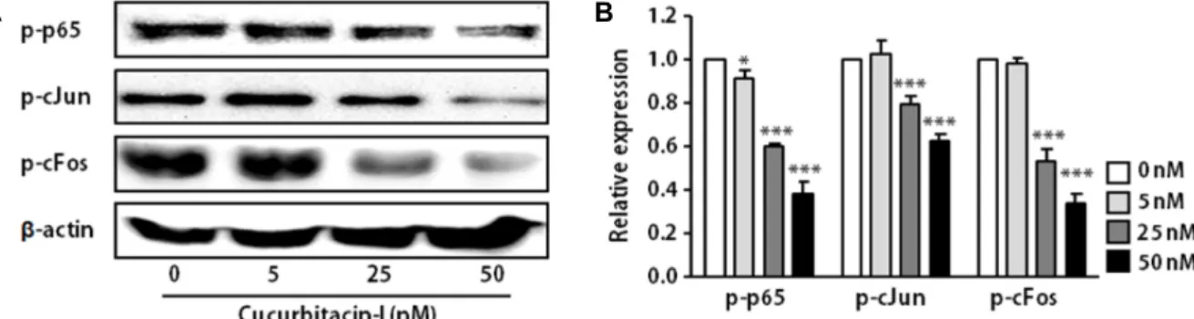
Fig. 3. Cucurbitacin-I inhibited the phosphorylation of AP-1 and NF-kB in SK-OV-3 cells. SKOV-3 cells were treated with indicated concentration of cucurbitacin-I for 24 hr. Western blot analysis of phospho-p65, -cFos and –c-Jun (A) and quantified results using densitometric analysis (B) in SKOV-3 cells. β-actin was used as the loading control. The results are means ± SEM of four independent experiments. *p<0.05, ***p<0.001 versus 0 nM cucurbitacin-I-treated cells.
els of CD44 by measured Western blot analysis also de- creased in SKOV-3 cells treated with cucurbitacin-I (Fig. 2C).
In addition, CD44 mRNA expression was also significantly lower in cucurbitacin-I-treated SKOV-3 cells compared to non-treated cells (Fig. 2D). These results suggest that cu- curbitacin-I can suppress CD44 expression on mRNA and protein levels.
Cucurbitacin-I inhibits the activation of NF-κB and AP-1 in ovarian cancer cell
Previous study demonstrated that activator protein-1 (AP-1) and nuclear factor kappa-B (NF-κB) are key trans-act- ing factors that interact with cis-element, CR1, located up- stream of the CD44 promoter [15]. In addition, inhibition of NF-κB resulted in a reduction in CD44 expression. CD44 repression via NF-κB inhibition consequently decreased pro- liferation and invasiveness of breast cancer cells [15].
The activation of NF-κB and AP-1 occurs via phosphor- ylation, resulting in the translocation of these molecules into the nucleus. Nuclear translocated NF-κB and AP-1 can in- duce the expression of their target genes, such as CD44.
Therefore, we determined the effect of cucurbitacin-I on NF- κB and AP-1 activation by measuring phosphorylated NF-κB (p65) and AP-1 (cFos and cJun). As shown in Fig. 3, cucurbi- tacin-I significantly reduced the levels of phodphory- lated-p65, -cFos and –cJun. These findings provide new in- sight into the molecular mechanism of cucurbitacin-I under- lying the regulation of CD44 expression.
CD44 was reported to be involved in proliferation, adhe- sion and invasion of ovarian cancer cells [11]. In addition, an ovarian cancer tissue microarray analysis of paired pri- mary, metastatic and recurrent tumor tissues from 26 pa- tients, demonstrated that the metastatic as well as recurrent
ovarian cancer tissues expressed higher levels of CD44 than the primary tumor [7]. A meta-analysis unveiled that pa- tients with CD44 was associated with poor prognosis in ovarian cancer patients, and that a CD44 status was asso- ciated with common clinicopathological features and poor prognostic factors [14]. These findings suggest that the regu- lation of CD44 expression may a possible therapy of ovarian cancer. The present study highlighted the crucial role of cu- curbitacin-I on reducing the expression of CD44 in ovarian cancer cells. Although further studies on the underlying de- tailed molecular mechanisms are required, the present re- sults may provide a possibility of cucurbitacin-I as a phar- maceutical agent for ovarian cancer treatment.
References
1. Aruffo, A., Stamenkovic, I., Melnick, M., Underhill, C. B.
and Seed, B. 1990. CD44 is the principal cell surface receptor for hyaluronate. Cell 61, 1303-1313.
2. Bajorath, J., Greenfield, B., Munro, S. B., Day, A. J. and Aruffo, A. 1998. Identification of CD44 residues important for hyaluronan binding and delineation of the binding site.
J. Biol. Chem. 273, 338-343.
3. Blaskovich, M. A., Sun, J., Cantor, A., Turkson, J., Jove, R.
and Sebti, S. M. 2003. Discovery of JSI-124 (cucurbitacin I), a selective Janus kinase/signal transducer and activator of transcription 3 signaling pathway inhibitor with potent anti- tumor activity against human and murine cancer cells in mice. Cancer Res. 63, 1270-1279.
4. Bourguignon, L. Y., Peyrollier, K., Xia, W. and Gilad, E.
2008. Hyaluronan-CD44 interaction activates stem cell marker Nanog, Stat-3-mediated MDR1 gene expression, and ankyrin-regulated multidrug efflux in breast and ovarian tu- mor cells. J. Biol. Chem. 283, 17635-17651.
5. Chen, Y. W., Chen. K. H., Huang, P. I., Chen, Y. C., Chiou, G. Y., Lo, W. L., Tseng, L. M., Hsu, H. S., Chang, K. W.
and Chiou, S. H. 2010. Cucurbitacin I suppressed stem-like
초록:난소암 세포주의 CD44 발현에 미치는 Cucurbitacin-I의 효과
서희원․김진경*
(대구가톨릭대학교 생명화학부 의생명과학전공)
박과 작물에 함유되어 있는 tetracyclic triterpene 성분 중 하나인 쿠쿠르비타신(cucurbitacin)-I는 대장암, 유방 암, 간암세포에서의 항종양 활성이 밝혀져 있으나 난소암에서의 쿠쿠르비타신-I의 역할은 보고된 바 없다. CD44 는 세포막에 존재하는 당단백질로 생체 내 리간드인 glycosaminoglycan hyaluronic acid를 통해 세포 외부 매트릭 스와 다른 세포와의 접촉을 매개한다. 최근 연구에 의해 CD44의 발현이 난소암세포의 증식 및 세포 부착과 침윤 을 증가시키는 주요 원인이라는 것이 보고되었다. 이러한 결과는 CD44의 발현을 억제함으로써 난소암의 진행을 조절할 수 있음을 시사하고 있다. 본 연구에서는 쿠쿠르비타신-I가 난소암세포의 CD44의 발현을 억제할 수 있는 지의 여부를 조사하였다. 인간의 난소암 세포인 SKOV-3를 이용한 MTS assay를 수행한 결과, 쿠쿠르비타신-I는 100 nM이상의 농도에서 세포독성을 나타내었다. 세포독성을 나타내지 않는 농도의 쿠쿠르비타신-I를 SKOV-3 세 포에 처리하여 Western blot 분석과 qRT-PCR을 수행한 결과, 쿠쿠르비타신-I에 의해 CD44의 단백질과 mRNA의 발현이 유의적으로 감소되는 것을 확인하였다. 또한 쿠쿠르비타신-I에 의한 CD44의 발현 억제가 NF-κB와 AP-1의 인산화 감소에 기인하고 있음을 밝혔다. 이러한 결과는 쿠쿠르비타신-I가 CD44 발현을 억제하는 기능을 가지며, 이는 난소암 치료에 도움을 줄 수 있는 제재로서 쿠쿠르비타신-I의 가능성을 제시하는 것이다.
property and enhanced radiation-induced apoptosis in head and neck squamous carcinoma-derived CD44+ALDH1+cells.
Mol. Cancer Ther. 9, 2879-2892.
6. Cortez, A. J., Tudrej, P., Kujawa, K. A. and Lisowska, K. M.
2018. Advances in ovarian cancer therapy. Cancer Chemother.
Pharmacol. 81, 17-38.
7. Gao, Y., Foster, R., Yang, X., Feng, Y., Shen, J. K., Mankin, H. J., Hornicek, F. J., Amiji, M. M. and Duan, Z. 2015.
Up-regulation of CD44 in the development of metastasis, recurrence and drug resistance of ovarian cancer. Oncotarget 6, 9313-9326.
8. Jia, Q., Feng, M., Wang, Y. and Xue, S. 2008. Gastric cancer cells in collagen gel matrix: Three-dimensional growth and differential expression of adhesion molecules (CD44s, CD54, E-cadherin). J. Biomed. Mater. Res. A. 84, 917-925.
9. Kim, H. J., Park, J. H. and Kim, J. K. 2014. Cucurbitacin-I, a natural cell-permeable triterpenoid isolated from Cucurbi- taceae, exerts potent anticancer effect in colon cancer. Chem.
Biol. Interact. 219, 1-8.
10. Lui, V. W., Yau, D. M., Wong, E. Y., Ng ,Y. K., Lau, C.
P., Ho, Y., Chan, J. P., Hong, B., Ho, K., Cheung, C. S., Tsang, C. M., Tsao, S. W. and Chan, A. T. 2009. Cucurbitacin I elicits anoikis sensitization, inhibits cellular invasion and in vivo tumor formation ability of nasopharyngeal carcinoma cells. Carcinogenesis 30, 2085-2094.
11. Mao, M., Zheng, X., Jin, B., Zhang, F., Zhu, L. and Cui, L.
2017. Effects of CD44 and E-cadherin overexpression on the proliferation, adhesion and invasion of ovarian cancer cells.
Exp. Ther. Med. 14, 5557-5563.
12. Ren, Y., Yu, K., Sun, S., Li, Z., Yuan, J., Han, X. D., Shi, J. and Zhen, L. 2014. JSI124 inhibits breast cancer cell growth by suppressing the function of B cells via the down- regulation of signal transducer and activator of transcription 3. Oncol. Lett. 8, 928-932.
13. Sacks, J. D. and Barbolina, M. V. 2015. Expression and func- tion of CD44 in epithelial ovarian carcinoma. Biomolecules 5, 3051-3066.
14. Shi, Y. Y. and Jiang, H. 2016. Prognostic role of the cancer stem cell marker CD44 in ovarian cancer: A meta-analysis.
Genet. Mol. Res. 15, doi: 10.4238/gmr.15038325.
15. Smith, S. M., Lyu, Y. L. and Cai, L. 2014. NF-κB affects pro- liferation and invasiveness of breast cancer cells by regulat- ing CD44 expression. PLoS One 9, e106966.
16. Song, J., Liu, H., Li, Z., Yang, C. and Wang, C. 2015. Cucur- bitacin I inhibits cell migration and invasion and enhances chemosensitivity in colon cancer. Oncol. Rep. 33, 1867-1871.
17. Su, Y., Li, G., Zhang, X., Gu, J., Zhang, C., Tian, Z. and Zhang, J. 2008. JSI-124 inhibits glioblastoma multiforme cell proliferation through G(2)/M cell cycle arrest and apoptosis augment. Cancer Biol. Ther. 7, 1243-1249.