Journal of Korean Arthroscopy Soc.
V
이
ume 13, Number 2, June, 2009전방십자인대 재건술 - 잔류조직 보존술식 -
순천향대학교 의과대학 정형외과학교 실
이병일 •천동일
ACL Reconstruction
-Remnant Preserving Technique -
Byung-Ul Lee, M.D., Dong-11 Chun, M.D.
Department of Orthopaedic Surgery ,Soonch니nhyang University Hospital, Seoul, Korea
Optimal treatment of the tom anterior cruciate ligament (ACL) remains controversial. The complexity of surgically reproducing the natural biomechanical and anatomical function of the ACL has led to a diversity of reconstructive procedures. Controversy continues to exist regarding the best reconstructive procedure for the ACL deficient knee, but currently, there is no ideal method. Because of the increased frequency of ACL injury and the functional impairment resulting from that, the role of mechanoreceptors in the ACL recent
ly has attracted considerable attention. Proper reconstruction of the ruptured ACL does not always have good results. Success after operation may depend not only on the mechanical stability but also on the quality of recovery of proprioception. It is well known that most ACL are ruptured in proximal half and most mechanoreceptors have been reported to be located in the subsynovial layer and near the tibial insertion of the ACL. Expected roles of tibial remnant is to enhance the revascularization and cellular proliferation of the graft, to preserve proprioceptive function, and to be able to acquire anatomical placement of the graft without roof impingement. The remnant of the ruptured ACL has been removed to clearly visualize the ACL footprint or decrease the risk of impingement and Cyclops lesion in most current techniques for ACL reconstruction. Therefore it seems reasonable to assume that preserving the tibial remnant as much as possible as a source of reinnervation, if technically possible without causing impingement, would be of potential benefit to the patient. In addition, it will facilitate the vascular ingrowth and ligamentization of the grafted ACL.
서 론
슬관절의 전방 십자인대 파열시 관절의 안정성을 확보하여 기능을회복시키고, 퇴행 변화로의 예방을 위하여 수술적 치 료로 재건술을 시행하며 대부분만족할 만한 결과를보고하 고있다. 그러나 지금까지 전방 십자인대 재건술은기계적 안 정성에 주로그초점이 맞추어져 발전해 왔으나, 그 결과 성공 적인 재건술로 기계적으로 안정화되 었음에도 불구하고기능 적 향상 정도가 기계적 안정성 회복 정도에 미치지 못하게 되
* Address reprint request to Dong II Chun, M.D.
Department of Orthopaedic Suigery, College ofMedicine, Soonchunhyang University, 22 Daesagwan-gil, Yongsan-gu, Seoul, Korea
Tel: 82-2-709-9250, F宓:82-2-794-9414 E-mail: [email protected]
는 경우들을경험하게 된다.따라서 근래 기계적 안정성 뿐 아 니라 생물학적 측면을 고려한 재건술식에 대한 다양한 연구 가시도되고 있다. 그 중 관절내 고유수용감각(propriocep
tion) 에 대한
관심
및 이에 대한 연구가 활발히 이루어지고 있고 이러한 연구에서 기계적 안정성과 함께 고유수용감각의 회복정도가 재건술후 기능적 결과에 영향을 미칠 것으로추 측하고 있다Sherman 등"은대부분의 전방십자인대 파열은 근위부에서 발생된다고보고하였고, Schutte 등些은 전 방십자인대의 기계적 수용체(mechanoreceptor)가 경골부 착부 인대주위의 활액막하층에서 발견된다고 하였다.
사람의 파열된 전방십자인대를 대상으로 한 기계적 수용체 에 대흐卜여는 Denti 등이 gold chloride 염색을이용하여 최 초로 보고하였고, Georgoulis 등58은 파열된 전방십자인대가 후방십자인대조직에 유착되어 있는 경우 약 3년이 지난 후에 도기계적 수용체가 존재한다고 하였다. Adachi 등a은전방
—97—
대한관절경학회지 제
13권 제
2호
2 009년
십자인대의 부분손상시 손상받지 않은 다발 내에 기계적 수 용체의 존재와 이러한 기계적 수용체의 숫자와 고유수용감각 의 기능과의 연관성을 보고하였다. 이 등Mg은고유수용체를 보존하기 위하여 전방십자인대 재건술 시 파열된 인대의 활 액막을 포함한잔류조직을가능한 많이 보존하는 술 식을 고 안하고경골 부착부 잔류조직의 보존 여부에 따른임상 결과 를 분석하여 그 유용성에 대한보고를 한 바 있다.
이에 저자들은이 술 식에 대한 수술 기법, 임상결과, 면역 조직화학염색법을 이용한 잔류조직내 기계적 수용체의 존재 여부,추시 관절경 소견, 잔류조직의경골터널 확장에 대한 영 향 등에 대하여문헌 고찰과 함께 기술하고자한다.
1.수술방법
기존수술방법은 술 후 Cyclops 병변의 발생을 방지하기 위해 파열된 인대의 잔류조직을 완전히 제거한후 이식건을 고정하는 것이 일반적으로 되어 있으며 그로 인해 잔류조직 내의 기계적 수용체는 보존되지 못하였다. Ochi 등®은전방
십자인대 부분파열시 손상받지 않은다발내의 기계적 수용체
가
남아있다는 Adachi 등沏
의 보고를근거로부분파열인 환 자에서 전내측다발 손상시는 전내측다발 보강술을,후외측 다발손상시는후외측다발보강술을시행하였다.그러나 적 응증이 부분파열인 경우에 한하며 전체 재건술의 약 10%에 서 해당이 된다 하였다.하지만 과간 절흔에 감입되지 않도록 잔류조직을 보존하며 시행되는 완전 파열된 전방십자인대 재Fig. 1. Anterior circles are the point of tib- Fig. 2. Note the probe tip through the center Fig. 3. The sutures are tied and staple fixa- ial tunnel of remnant preserving of the tibial remnant. tion is performed under proper ten- technique. And posterior circle is sion in a belt-buckle fashion. The point of the tibial tunnel of conven- remnant is retracted medially with a
tional technique. probe.
Fig. 4. If the tibial remnant is torn, an arthroscopic suture repair can be added.
Fig. 5. The schema and arthroscopic finding of ACL recon
struction using absorbable cross pin system (RIGIDfix) and quadruple hamstring autograft with tibial remnant preserving technique. The probe demonstrates the bor
der of graft & remnant.
건술에 대한보고는 아직 없었으나 이 등
项
은 슬괵건을 사용하면서 완전파열시 활액막을포함한 경골부착부잔류조직의
중심으로 이식건을 통과시켜 잔류조직을효과적으로보존함
으로써 고유수 용체의 기능을 유지시키며 활액막으로부터의 혈관재형성을 촉진시켜 이식건의 인대화에 도움을 주는 술기 를보고하였다.
A
Tension
Fig. 6. Histological appearance of mechanoreceptors in the remnants of the ruptured ACL specimens; (A) round Ruffini corpuscle, (B) fusiform Golgi corpuscle, (C) elongated Golgi corpuscle with degeneration (immunohistochemistry for neurofilament, x200).
Synovial coverage
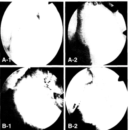
Fig. 7. (A) Arthroscopic classification of transplanted grafts based on graft tension. (A-l) A taut graft is evaluated as good, (A-2) a mild lax graft is fair, and (A-3) a lax graft is poor. (B) Arthroscopic 이assification of transplanted grafts based on synovial coverage. (B-l) A graft of complete covered synovium is evaluated as good, (B-2) a graft of incomplete covered synovium is fair, and (B-3) a graft of no covered synovium is poor. (C) Arthroscopic classification of transplanted grafts based on status of fiber bundle. (C-l) A graft with invisible graft bundle is evaluated as good, (C-2) a graft with visible graft bundle without rup
ture is fair, and (C-3) a graft with visible bundle with mpture is poor.
—99—
대한관절경학회지 제
13권 제
2호
2009년
Fig. 8. 2nd look arthroscopic findings of the grafts with (A) conventional technique, it shows taut graft with visible fiber bundle돊 in thin synovium, and (B) remnant preserving 也:hnique, it 마sws taut graft with invisible fiber bundle due to thick synovium and remnant.
Fig. 9. Arthroscopic findings of regeneration of something similar to PL bundles. (A-l, B-l) Just post. Op. arthroscopic findings show no PL bundle, (A-2, B-2) 2nd look arthroscopic findings show regeneration of something similar to PL bundle.
이 등"'의 방법은 관절경 검사로관절내 병변과전방 십자 인대의 파열 양상을 확인하고경골근위부전내측 피부를 절 개하여 자가 반건양건 또는 반건양건과 박건을 채취하여 두 가닥또는 네 가닥의 이식물로 준비하고 직경을측정하였다.
경골터널은 일반적으로 과간절흔의 충돌을피하기 위해 후방 에 위치시키나 본술식은 가능하면 foot print.의 중앙에 유도 강선을 위치시키며 터널이 foot print안에만 있으면 되므로 다소간의 오차는 허용된다(Fig. 1). 경골터널은 채취된 슬괵 건의 직경과 같은크기로 만들었으며, 확공기로원위 피질골 관통과 동시 에 확공기를 멈추어 가능한 한잔류조직을 손상시 키지 않도록하면서 잔류조직의 중심부를 탐침 등을 통해 통 과시켜 확인한다(Fig. 2). 대퇴부는 전내측 입구(anterome
dial portalX 통해 이식건의 직경과 동일한 curved curette을넣어 대퇴 부착부의 유도강선을 따라 약 2 cm 이 상 깊이의 소켓(socket)을 만든
후
봉합사를 통해 이식건을 잔류조직의 중심을 통과시켜 고정시킨다(Fig. 3). 그리고 경우에따라서 잔류조직이 파열되거나 불안정한경우봉합술을
동반할 수있다(Fig. 4).
본 술식의 기대되는장점으로는잔류조직의 중심부로 이식 건을 통과시켜 잔류조직의 활액막증식과 재혈관화를 촉진시 킬 수 있어 인대화 기간을 단축시키고, 잔류조직내의 기계적 수용체를보존하여술후환자의고유수용감각 및 만족도를 높 일 수 있을 것으로 사료된다.그러 나 본 술식의 제한점은 봉합 사의 이완에 의해 이식건과 골터널사이의 유합이 지연되거나 불안정할 수 있고% 인대-골 유합이 완전히 형성되기 전에 골 터널의 확장이 일어나 고정력이 약화될 가능성이 있으며
气
대 퇴소켓을만들 때 curette을 이용하기 때문에 일정한 직경 및 깊이의 소켓을 만들기 어렵다는 점이 있고, 이식건을 경골터 널에 고정할 때 일정한 긴장도를 유지하지 못하여, 증례에 따 라 다른 강도로 고정되므로 다소간 객관적이지 못한 문제가있
어근래에는 객관성을얻기 위해 대퇴부의고정은 흡수성 횡 고정핀 (RIGIDfix ACL cross pin system, Mitek8, USA) 을 사용하였고, 대퇴 소켓의 깊이는 대퇴골 터널 유도활을 이 용하여3 cm으로 일정하게 유지하였으며, 이식건의 경골 터널고정 시 긴장도를 객관화하기 위하여 긴장기를30 lbs로 일정 하게유지하면서 고정하는술식을 사용하고있다(Fig. 5).
2. 임상결과
현재 사용중인 이식건을 이용한 재건술은 기계적 안정성을
회복시켜줄 수는 있으나고유수용감각기능에 대해서는 이식
건의 재신경형성에 의한 자연치유에만 의존하는 상태이다.
재건술 시행 후 인대화과정중 기계적 수용체의 재생에 대한 보고는 있으나 대부분 동물실험 에 국한되어 있으며是"V Ochi 등何加은 인체에서somatosensory evoked poten
tials (SEPs)를 이용하여 그 존재를 확인하였으나, 기능적 유용성에 대해서는 아직 논란의 여지가많다.Kennedy 등",
은 전방십자인대 손상시 고유수용감각의 소실에 따른 관절불 안정성 및 이차적 손상에 대해 강조하였고 일부 저자들은 전 방십자인대 재건술후 관절의 이완도나 평가점수가 환자의 만족도나 슬관절의 기능 회복정도와 항상 일치하지는 않는다 고보고하였다心.
즉 재건술의
성공여부는 기계적 요소뿐
아니라 고유수용감각 기능의 회복 정도와도밀접한 관련이 있다*". 이 에 이 등은 전 방십자인대 재건술시 파열된 인대의 활액막을포함한 경골 부 착부 잔류조직을 보존하는 술기를시행하고수술 소견상 잔류 조직의 보존 정도에 따른임상결과를 분석하여,고유수용감각 검사상운동역치검사에서 30도시작시(P=0.031), 위치재생 검사에서는 15도(P=0.032) 및 30도 재생시 (P=0.024)에, 7 mm(전방십자인대 길이의 약 20%)이상의 잔류조직 보존군 에서 통계학적 유의성이 있는 양호한 결과를 얻었고, 기능적 검사인 single leg hop test에서도 잔류조직 보존군에서 좀 더 양호한 결과를 얻어(P=0.017), 전방십자인대 재건술시 가 능하면 잔류조직을 최대한 보존하여 상대적으로 고유수용감 각의 회복과만족스러운임상결과를 얻을 수 있다고 하였다.Fig. 10. Arthroscopic photographs of the grafts (A) without ACL remnant. The arrow shows the leakage of synovial fluid through the tunnel. Arthroscopic photograph of the graft (B) within ACL remnant. The arrow shows that the remnant prevents the leak
age of synovial fluid.
—101 —
대한관절경학회지 제
13권 제
2호
2009년
3. 잔류조직 내 기계적 수용체에대한 연구
사람의 전방십자인대 내 기계적 수용체의 존재는여러 연 구자들이 보고하였으며321例, 고유감각을 담당하는 기계적 수 용체의 위치는대퇴 및 경골 부착부의 활액막하층에 주로분 포하는 것으로 알려져 있다. 임상적으로전방 십자인대 파열
은
대퇴 부착부邸> 또는 인대 실질 내에서의 파열이 많으므로 전방십자인대의 경골 부착부의 잔류조직 내 에는 기계적 수 용체가 남아 있을 것으로 추정 된다.고유수용감각은 그 기능을 측정하는 임상적 검사들山,이 다 양하게 고안되어 있으나, 고유수용감각 기전 자체의 복잡성 때문에 측정시 변수가많고결과의 분석에도 문제가 있는 것 으로알려져있다&
硕
.따라서 보다 직접적인 방법으로 조직 내 에서고유수용감각 기능을담당하는 기계적 수용체의 존재를 규명하는것이 보다객관적인증거가 될 수 있을 것이다. 이 등",,은 사람의 전방십자인대 파열 후 남아있는 잔류조직을표 본으로 하여 기계적 수용체의 존재를 규명하고자 하였으며, 기존의 많은 연구자들이 이용하였던 도금 염색법物은 신경섬 유 이외의 섬유도비특이적으로 염색되어 신빙성이 높지 않 아* 신경섬유에 대한특이적 항체를 이용하는, 면역조직화학 염색법 등을 이용하여 분석하였다.그 결과 잔류조직 총 36예 중 12예(33.3%)에서
총 17개의
기계적 수용체를 관찰하였으며, 17개중 12개는 방추형의 Golgi소체 (fusiform Golgi Corpuscles) 였고, 5개는 난원 형의 Ruffini소체 였다(Fig. 6). 그러나12예 이외의 잔류조 직에는 기계적 수용체가 없다는 뜻이 아니며 파열된잔류조 직의 양이 적고 표본채취시 손상되 었거나또는수상후시간 이 경과함에 따라 소멸되기 때문에 기계적 수용체가 관찰되 지 않았을 가능성도 있다고 하였다. 결과적으로 외상에 의한 전방십자인대 손상 후경골 부착부잔류조직 내에 기계적 수 용체가 일부 소멸되지 않고 잔존함을 볼 때, 재건술 시 경골 부착부 잔류조직을 보존해 주는 것이 고유수용감각 기능의 회복에 도움이 될 것으로 생각된다.4.추시 관절경 소견
전방십자인대 재건술후2차 추시관절경 소견에 대한 연구 에서, Otsubo 등°破은 재건인대의 형태에 따라 tension, synovialization등으로 나누어 good, fair, poor로평가흐]"
였으며, 안 등»은 2차 추시 관절경 소견을 형태적으로 ten
sion^ 경우mildly lax, nearly normal로,synovializa- tion의
경우 fair, poor로
분류 평가하였다.저자들은 전방십자인대재건술시 잔류조직 보존유무에 따른 2차 추시 관절경소견을비교분석하였으며, 이식건의 형태를 tension, synovial coverage, status of fiber bundle의 3항 목으로 분석하였다. Tension은 관절경검사 상 경골을 전방전 위한상태에서 긴장된 이식건을probe로 긴장상태를 확인하
였으며, synovial coverage는 이식건에대한 활액막의 피복 정도로, status of fiber bundle의 경우는 이식건 bundle의 상태를 관절경검사로 평가하여 각각의 상태를 그양상에따라 good, fair, poor로나누었다(Fig. 7). 그리고 추시 기간과 이 식건의 형태와의 관계는 good을 3점,fairt- 2점, poor를1점 으로 하여 각 기간에 해당하는 점수의 합을 평균하여 3항목을 비교평가 하였으며 tension을 제외한 synovial coverage와 status of fiber bundle에서는 잔류조직 보존군에서 술 후 회 복이 빠른결과를 얻었다.
추시 관절경 소견상 일반적으로 잔류조직 보존군이 활액막 피복, 혈관 재형성 등인대화 과정이 잔류조직 제거군에 비하 여 빠른 소견이 있었으며,활액막 피복의 양상이 잔류조직 보 존군에서는 두꺼워 활액막을 통해 섬유다발의 모양을볼수
없으나, 잔류조직 제거군에서는 활액막 피복이 얇아 섬유다
발의 모양을 쉽게 볼 수 있는 경향이 있었다(Fig. 8). 특히 이 연구에서 아직까지는 명확하지는 않지만, 추시관절경 소견에 서술후에는 볼 수 없었던 후외측다발과 상당히유사한 소견 이 보이는경우가있어(Fig. 9) 인대화 과정에서 후외측다발 의 재생가능성이 있어 이에 대한 더 많은연구가필요할것으 로 생각된다
5. 경골터널 확장에 대한 잔류조직 보존의 효과
Hoher 등은골터널 확장의 원인을 생물학적 인자와 생역 학적 인자로 구분하였는데,생물학적 인자로는이식물에 대한 면역반응, cytokine등에 의한비특이적 염증반응, 터널 내독 성 잔류물질로 인한 세포괴사,확공시 발열 반응에 의한 세포 괴사, 이식건 재형성 과정에서의 세포괴사등을기술하였고, 생역학적 인자로는 터널 벽의 반복적 자극에의한박탈, 이식 건-터널 간 미세운동,과도한 초기의 적극적 재활치료, 부적 절한 이식건위치에 따른과도한 긴장부하 등을기술 하였다.
따라서 전방 십자인대 재건술후 활액이 골 터널내로 유입 되어 활액 내에 포함되어 있는 cytokine에 의해
골
흡수가일 어날 수 있는 이론적 배경의 근거가 될 수 있다(synovial bathing effect)7'11'17'. 생물학적 인자인 cytokine에 대하여 최근Cameron 등*은 전방십자인대 재건술 직 후 및 수주 동안 활액에서 TNF-a, IL-1A IL-6와 같은 cytokine의증
가를 보고하였고, Van den Berg洞는 IL-1 이 관절 염 에서 연 골파괴의 중요 매개체이며 이는 연골세포의 proteopglycan 합성을 억제하고destructive protease를 분비하여 연골세 포를 자극한다고 하였는데 이와 비슷한기능을하는 인자로 는TNF-a, LIF (leukemia inhibitory factor), ILT7이 포함된다고 하였다이에 저자들은 경골 부착부잔류조직 보존 술식을 사용시 이식건은 잔류조직 내경을 통과 함으로써 잔류조직과 이식건 간의 밀착 효과를 얻을 수 있어 잔류조직 자체가 일종의 check-valve 역할을 하여 경골 터널 내로의 활액 유입을 차
단함으로써 cytokine의 골 흡수 작용을감소시킬수 있다는 가정을 하게 되었고, 경골터널 확장 정도에 대한 비교 연구에 서 단순 방사선학적 계측결과를 분석하여 본바 잔류조직 보
존술식에서 경골터널의 확장이 감소되 었음을 확인하였으며
이는 전방십자인대 파열시 남아 있는 잔류조직은 생물학적 인자인 활액의 유입을차단하는 역할을 하여 골흡수를 억제 하고 따라서 이식건과 경골터널 간의 치유를 촉진하는 매개 역할을 하는것으로 보인다(Fig. 10). 따라서 잔류조직 보존 술식은 기계적 안정성 뿐 만아니라 생물학적 및 기능적 결과 를 향상 시킬 수 있을 것으로기대된다.
결 론
저자들은 전방 십자인대 재건술에서 잔류조직 보존 술식과 제거 술식 간의 차이점을비교 연구한 결과전방십자인대 재 건술 시 전방십자인대잔류조직의 보존은여러면에서 장점이 많아 가능하면 잔류조직을 최대한 보존하는 것이 좋을 것으 로 사료된다.
REFERENCES
1) Ahn JH, Yoo JC, Yang HS, Kim JH and Wang JH:
Second-look arthroscopic findings of 208 patients after ACL reconstruction. Knee Surg Sport Traumatol Arthrosc, 15:242-248, 2007.
2) Aune AK, Hukkanen M, Madsen JK, Polak JM and Nordsletten L: Nerve regeneration during patellar tendon autograft remodelling after anterior cruciate ligament reconstruction: an experimental and clinical study. J Orthop Res, 14: 193-199, 1996.
3) Adachi NA, Ochi M, Uchio Y, Iwasa J, Ryoke K and Kurikawa M: Mechanoreceptors in the anterior cruciate ligament contribute to the joint position sense. Acta Orthop Scand, 73: 330-334, 2002.
4) Barrett DS, Cobb AG and Bently G: Joint propriocep
tion in normal, osteoarthritic and replaced knees. J Bone Joint Surg, 73-B: 53-56, 1991.
5) Cameron M, Buchgraber A, Passler H, et al.: The nat
ural history of the anterior cruciate ligament-deficient knee. Changes in synovial fluid cytokine and keratan sul
fate concentrations. Am J Sports Med, 25: 751-754, 1997.
6) Denti M, Monteleone M, Berardi A and Panni AS:
Anterior cruciate ligament mechanoreceptors: histologic studies on lesions and reconstruction. J Orthop Res, 308:
29-32, 1994.
7) Fahey M and Indelicato PA: Bone tunnel enlargement after anterior cruciate ligament replacement. Am J Sports Med, 22:410-414, 1994.
8) Friden T, Roberts D, Ageber흠 E, Walden M and
Zatterstrom R: Review of knee proprioception and the relation to extremity function after an anterior cruciate lig
ament rupture. J Orthop Sports Phys Ther, 31: 567-576, 2001.
9) Georgoulis AD, Pappa L, Moebius U, Malamou-Mitsi V, Papaqeorgiou CO and Soucacos PN: The presence of proprioceptive mechanoreceptors in the remnants of the ruptured ACL as a possible source of re-innervation of the ACL autograft. Knee Surg Sports Traumatol Arthrosc, 9(6): 364-368,2001.
10) Hogervorst T and Brand RA: Current concepts review.
Mechanoreceptors in joint function. J Bone Joint Surg, 80- A: 1365-1378, 1998.
11) Hoher J, Moller HD and Fu FH: BONE tunnel enlarge
ment after anterior cruciate ligament reconstruction: fact or fiction? Knee Surg Sports Traumatol Arthrosc, 6: 231- 240, 1998.
12) Kennedy JC, Alexander IJ and Hayes KC: Nerve sup
ply of the human knee and its functional importance. Am J Sports Med, 10: 329-335, 1982.
13) Krauspe BR, Schmidt M and Schaible HG: Sensory innervation of the anterior cruciate ligament: an electro
physiological study of the response properties of single identified mechanoreceptors in the cat. J Bone Joint Surg, 74-A: 390-397, 1992.
14) Lee BI, Kwon SW, Kim JB, Choi HS, and Min KD:
Comparison of clinical results according to amount of pre
served remnant in arthroscopic ACL reconstruction using quadrupled hamstring graft. Arthroscopy, 24: 560-568, 2008.
15) Lee BI, Min KD, Choi HS, Kwon SW, Yoo JH, and Chun DI: Immunohistochemical study of mechanorecep
tors in the tibial remnant of the ruptured anterior cruciate ligament in human knees. Knee Surg Sports Traumatol Arthrosc, published online: 16 June 2009.
16) Lee BI, Min KD, Choi HS, Kim JB, and Kim ST:
Arthroscopic anterior cruciate ligament reconstruction with the tibial remnant preserving technique using a ham
string graft. Arthroscopy, 22: 340el-340e7, 2006.
17) L' Ins이ata JC, Klatt B, Fu FH and Harner CD: Tunnel expansion following anterior cruciate ligament reconstruc
tion: a comparison of hamstring and patellar tendon auto
grafts. Knee Surg Sports Traumatol Arthrosc, 5: 234-238, 1997.
18) Ochi M, Adacji N, Deie M and Kanaya A: Anterior cru
ciate ligament augmentation procedure with a 1-incision technique: anteromedial bundle or posterolateral bundle recosntruction. Arthroscopy, 22(4): e 1 463-463 e5., 2006.
19) Ochi M, Iwasa J, Uchio Y, Adachi N and Sumen Y: The regeneration of sensory neurons in the reconstruction of the anterior cruciate ligament. J Bone Joint Surg, 81-B:
—103 —
대한관절경학회지 제
13권 처|
2호
2009년
902-906, 1999.
20) Otsubo H, Shino K? Nakamura N, Nkata K, Nakagawa S and Koyanagi M: Arthroscopic evaluation of ACL grafts reconstructed with the anatomical two-bundle tech
nique using hamstring tendon autograft. Knee Surg Sport Traumatol Arthrosc, 15: 720-728, 2007.
21) Sherman MF, Lieber L, Bonamo JR, Podesta L and Reiter I: The long-term follow up of primary anterior cru
ciate ligament repair: Defining a rationale for augmenta
tion. Am J Sports Med. 19: 243-255, 1991.
22) Schutte MJ, Dabezies J, Zimny ML and Happel LT:
Neural anatomy of the human anterior cruciate ligament.
J Bone Joint Surg, 69-A: 243-247, 1987.
23) Shimizu T, Takahashi T, Wada Y Tanaka M, Mrisawa Y and Yamamoto H: Regeneration process of mechanoreceptors in the reconstructed anterior cruciate ligament. Arch Orthop Trauma Surg, 119: 405-409, 1999.
24) Tsuda E, Okamura Y, Otsuka H, Komatsu T and Tokuya S: Direct evidence of the anterior cruciate liga
ment-hamstring reflex arc in humans. Am J Sports Med, 29:83-87, 2001.
25) Valeriani M, R^tuccia D, DI Lazzaro V, et al.: Clinic시 and neurophysiological abnormalities before and after reconstruction of the anterior cruciate ligamnet of the knee. Acta Neurol Scand, 99: 303-307,1999.
26) Van den Berg WB: The role of cytokines and growth fac
tors in cartilage destruction in osteoarthritis and rheuma
toid arthritis. Z Rheumatol, 58: 136-141,1999.
27) Wada Y, Takahashi T, Michinaka Y, Morisawa Y and Yamamoto H: Mechanoreceptors of patellar tendon used for ACL reconstruction. Acta Orthop Scand, 68: 559-562,1997.
28) Yoshiya S, Andrish JT, Manley MT and Bauer TW:
Graft tension in anterior cruciate ligament reconstruction. An in vivo study in dogs. Am J Sports Med, 15:464-479, 1987.