ISSN 2234-3806 • eISSN 2234-3814
400 www.annlabmed.org https://doi.org/10.3343/alm.2019.39.4.400 Ann Lab Med 2019;39:400-402
https://doi.org/10.3343/alm.2019.39.4.400
Letter to the Editor
Diagnostic Hematology
First Case of Cellular Cannibalism in Small-Cell
Carcinoma of the Bladder Detected in Peripheral Blood
Hyein Kang, M.D.
1, Do-Hoon Kim , M.D., Ph.D.
1, Wonmok Lee, M.D., Ph.D.
1, Jungsook Ha, M.D., Ph.D.
1, Namhee Ryoo, M.D., Ph.D.
1, Dong-Seok Jeon, M.D., Ph.D.
1, and Hye Ra Jung, M.D., Ph.D.
2Departments of
1Laboratory Medicine and
2Pathology, Keimyung University School of Medicine, Daegu, Korea
Dear Editor,
Cellular cannibalism, emperipolesis, and entosis are similar yet subtly different phenomena related to cell engulfment. Cellular cannibalism refers to one cell engulfing another living cell of its own or another type; emperipolesis refers to the presence of vi- able, undamaged hematopoietic cells in the cytoplasm of a host cell; and in entosis, a cell engulfs another cell of the same type, leading to the death of the engulfed cell [1]. Of these, cellular cannibalism is a characteristic morphological feature observed exclusively in aggressive malignancies that was first described in tumor cells in 1891 [2]. Cannibalistic cells ingesting other cells were noticed initially on cytological smears showing a vacu- ole containing the ingested cell [3]. Cellular cannibalism has been described in various types of tumors [4-6] and is closely related to the aggressiveness, degree of anaplasia, invasiveness, and metastatic potential of the tumor [1]. Cellular cannibalism in tissue sections [6] and malignant effusions of body fluids [7]
has been reported to indicate poor prognosis, but there is no such a report regarding peripheral blood (PB). We report the first case, to our knowledge, of cellular cannibalism in small-cell carcinoma of the bladder detected in PB. This retrospective study was approved by the Institutional Review Board/Ethics Committee of Dongsan Medical Center, Daegu, Korea, which
waived the need for informed consent.
The patient was a 66-year-old man with small-cell carcinoma of the urinary bladder that had been diagnosed nine months previously. He had received five cycles of chemotherapy. He also had been recently diagnosed as having anemia and thrombocy- topenia without any clinical symptoms. He was admitted to the hematology department of Dongsan Medical Center in January 2018 for transfusion and further evaluation to determine the cause of cytopenia. His complete blood count showed the fol- lowing: white blood cells, 5.45×10
9/L; Hb, 98 g/L; and platelets, 24 ×10
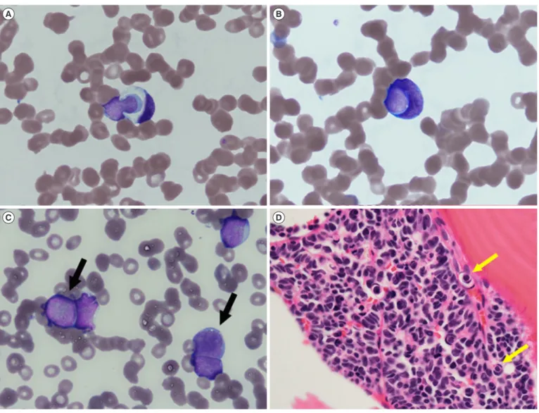
9/L. A PB smear showed a few suspicious tumor cells (5% of total nucleated cells) exhibiting cellular cannibalism, which is characterized by a typically large cell with a crescentic nucleus engulfing another smaller cell with a round-to-oval nucleus rimmed by a halo of phagocytic vacuoles (Fig. 1A-C). Some flattening or
“molding” of adjacent cells was observed (Fig. 1C), which is a feature of small-cell carcinoma [8]. Subsequent bone marrow (BM) aspiration and biopsy revealed a markedly increa sed number of suspicious tumor cells exhibiting cellular cannibalism (Fig. 1D). The cells were large with a high nucleus-to-cytoplasm ratio, fine chromatin pattern, and scant cytoplasm. Myeloperoxi- dase and periodic acid-Schiff staining were negative. Flow cyto- metric analysis of the suspicious tumor cells in BM aspirate
Received: July 26, 2018
Revision received: January 2, 2019 Accepted: February 5, 2019
Corresponding author: Do-Hoon Kim, M.D., Ph.D.
https://orcid.org/0000-0002-9854-7850
Department of Laboratory Medicine, Keimyung University School of Medicine, 56 Dalseong-ro, Jung-gu, Daegu 41931, Korea
Tel: +82-53-250-7085, Fax: +82-53-250-7275, E-mail: [email protected]
© Korean Society for Laboratory Medicine
This is an Open Access article distributed under the terms of the Creative Commons Attribution Non-Commercial License (http://creativecommons.org/licenses/by-nc/4.0) which permits unrestricted non-commercial use, distribution, and reproduction in any medium, provided the original work is properly cited.
1 / 1 CROSSMARK_logo_3_Test
2017-03-16 https://crossmark-cdn.crossref.org/widget/v2.0/logos/CROSSMARK_Color_square.svg