뇌동맥류에서 혈관형성 인자와 혈관벽 기질 단백에 대한 면역조직화학적 연구
계명대학교 의과대학 신경외과학교실, 병리학교실*
김재홍·임만빈·이창영·김상표*
= Abstract =
Immunohistochemical Study for the Angiogenesis Factors and Vascular Wall Matrix Proteins in Intracranial Aneurysms
Jae Hong Kim, M.D., Man Bin Yim, M.D., Chang Young Lee, M.D., Sang Pyo Kim, M.D.*
Department of Neurosurgery and Pathology,* Keimyung University School of Medicine, Taegu, Korea
bjective:Until now, it has been little known about the biological mechanisms associated with the genesis, growth, and rupture of intracranial aneurysm. This study was performed to investigate and understand a part of these mechanisms.
Materials and Methods:Immunohistochemical stains for angiogenesis growth factors(basic fibroblast growth factor (bFGF) and vascular endothelial growth factor(VEGF)) and selected vascular wall matrix proteins(alpha smooth muscle actin(αSMA) and collagen Type IV) were performed in fixed sections from a normal circle of Willis artery which was taken from the autopsy specimen as a control vessel and 17 aneurysmal wall specimens which was taken during surgical clipping of aneurysms. The staining intensity and distribution of immunoreactivity to angiogenesis growth factors and selected wall matrix proteins in control vessel and aneurysmal wall were examined and compared with each other. The difference of staining intensity according to the size of aneurysm was also investigated.
Results:There was no immunoreactivity to bFGF and VEGF in the control vessel. bFGF immunoreactivity was exhibited in 15 of 17 aneurysm specimens around smooth muscle cells within the media of aneurysm. VEGF immu- noreactivity was also exhibited in all aneurysm specimens in patches or diffusely affecting all layers of the aneurysmal wall. The degrees of intensity of bFGF and VEGF immunoexpression were proportionate roughly to the size of aneurysm. Strong immunoexpression of both factors were noticed in large aneurysm. A regularly arranged and defined band of immunoreactivity of αSMA was noticed in the media of the control vessel, whereas diffuse, faint, irregularly arranged αSMA was noticed in the aneurysmal wall. A regularly defined band of collagen Type IV immunoreactivity was also noticed in the subendothelium of the control vessel, whereas diffuse disorganized immu- noreactivity of collagen Type IV was noticed in the entire wall of the aneurysm.
Conclusion:These results indicate substantial evidences of abnormal expression of angiogenesis factors and changes of selected vascular wall matrix proteins in the wall of intracranial aneurysm. The unbalanced changes of angiogenesis factors and vascular wall matrix proteins in the wall of aneurysm may be one of the biological mec- hanisms for the growth and rupture of aneurysm.
KEY WORDS:Aneurysm・Basic fibroblast growth factor・Vascular endothelial growth factor・Alpha smooth muscle actin・Collagen type IV.
OOOO
서 론
뇌동맥류의 발생과 성장 및 파열에 대한 생물학적 기전에 대하여서는 아직까지도 잘 밝혀져 있지 않다2)22)23).
뇌동맥류의 발생기전은 크게 2가지로 현재 제시되고 있 다23). 하나는 선천적으로 뇌동맥의 구조적인 결손(struc- ural defect)이 존재하여 발생한다는 설이고, 다른 하나는 후천적으로 뇌동맥에 퇴행성 변화가 진행되어 발생한다는 설 이다23). 선천적인 뇌혈관의 구조적인 결손은 과거에는 뇌혈관 분지부의 중막 결손(media defect)이라고 알려져 왔으나23), 근래에는 뇌혈관 중막의 망상섬유 결손2)10-12)20)21)
또는 혈 관벽 교원질의 결핍이 중요 원인으로 제시되고 있다16)19). 후 천적으로는 혈류역동학적 부하(stress)가 뇌혈관에 영향을 주어 뇌혈관의 퇴행성 변화를 촉진시키므로써 동맥류가 형 성된다는 설로23), 고혈압, 고령 및 흡연은 이러한 퇴행성 변 화를 촉진시켜 뇌동맥류의 발생에 관여하는 위험인자로 보 고되어 있다3)18).
한편 뇌동맥류의 성장 및 파열에 대한 연구는 드물다22). 특히 어떤 뇌동맥류는 크기가 작은 경우에도 파열되나, 어떤 뇌동맥류는 거대 뇌동맥류가 될 때까지 파열 없이 성장하는 예들도 있다. 또한 어떤 뇌동맥류는 파열없이 환자가 사망 한 후 부검에서 발견되는 예들도 있다3). 이와같은 생물학적 특성의 차이는 동맥류 벽을 구성하는 물질중에서, 동맥류 벽 을 용해시키는 물질과 강화시키는 물질간의 불균형 혹은 동 맥류 벽의 손상시 이 손상된 부위를 복구하는 기전상의 차 이가 하나의 원인으로도 추정할 수 있다. 따라서 저자들은 수술 중 얻은 뇌동맥류의 조직에서 혈관벽을 약화시키는 물 질로 알려진 혈관형성 인자와 혈관벽의 강도를 유지시키는 데 중요한 역할을 한다고 알려진 혈관벽 기질 단백에 대해 면역조직화학적 염색을 시행한 후 그 발현여부를 파악하고, 이들의 발현정도와 대조혈관에서의 발현정도를 상호 비교하 여, 뇌동맥류의 발생, 성장 및 파열의 기전에 대하여 알아보 고자 하였다.
재료 및 방법
1996년 12월부터 1997년 6월까지 본원에서 수술한 뇌 동맥류 환자들 중에서 뇌동맥류 결찰후 동맥류 조직의 채취 가 가능하였던 17례를 조사대상으로 하였고, 대조혈관은 뇌 혈관질환 없이 사망한 부검된 1례에서 채취한 Willis환의 혈 관을 사용하였다. 대상 환자의 특성은 남자가 8례, 여자가 9례였고, 연령은 28세에서 78세사이였다. 17례중 16례가
파열 동맥류였고 1례가 비파열 동맥류였다. 동맥류 크기는 3~5mm의 소동맥류가 5례, 8~10mm의 중간 크기의 동맥 류가 11례, 20mm인 대동맥류가 1례였다. 동맥류 발생 부 위는 후교통동맥 동맥류 2례, 전맥락동맥 동맥류 1례, 내경 동맥 분기부 동맥류 1례, 전대뇌동맥 수평분절 동맥류 1례, 전교통동맥 동맥류 6례, 중대뇌동맥 동맥류 5례 및 후하소 뇌동맥 동맥류 1례였고 이는 말초성 동맥류였다. 동맥류 벽 은 비후한 예가 12례, 얇은 예가 5례였다(Table 1).
동맥류 조직의 채취 방법은 수술시 동맥류 경부를 결찰한 다음 동맥류 기저부(fundus)를 뇌조직에서 박리 노출시킨 후, 미세 수술가위로 이 부위의 동맥류 조직을 채취하였다. 채취 한 동맥류 조직은 즉시 포르말린(formalin)용액으로 고정후 파라핀 포매 조직을 만든 다음 면역조직화학 염색을 시행하 였다.
대조혈관과 뇌동맥류 조직에서의 혈관형성 인자로써는 basic fibroblast growth factor(bFGF)와 vascular endo- thelial growth factor(VEGF)를, 혈관벽 기질 단백으로는 alpha smooth muscle actin(αSMA)과 교원질(collagen) Type IV를 조사하였다. 면역조직화학 염색을 시행하여 이 들의 발현상태를 관찰하고, 대조혈관과 뇌동맥류 조직간 발현 부위 및 발현정도를 비교하였다. 발현정도는 발현이 없는 경 우을 0, 경미한 경우 1, 중등도 2 및 고도인 경우 3으로 평가 하였다.
혈관형성 인자 및 혈관벽 기질 단백에 대한 면역조직화학 염색 방법은 일차 항체로 bFGF(polyclonal antibody, Cal- biochem, U.S.A.), VEGF(polyclonal antibody, Neomar- kers, U.S.A.), αSMA(monoclonal antibody, DAKO, U.S.A.) 및 교원질 type IV(monoclonal antibody, DA- KO, U.S.A.)를 각각 20~100배로 희석하여 사용하였다.
대표되는 조직절편을 탈파라핀과 탈수를 시행한 후 계열 에 탄올을 거쳐 함수하고, 에탄올과 30% H2O2를 9:1로 혼합 한 용액에 30분간 실온에서 방치한 후 0.01M citrate buffer (pH 6.0) 용액에 담근 뒤 microwave로 5분씩 2회 가열하 였다. 그 후 1% 정상 마혈청(normal horse serum, Vec- tastain kit, U.S.A.)으로 37℃에서 30분간 둔 후 30μl의 일 차 항체를 조직절편위에 놓고 coverslip을 덮은 다음 37℃
에서 2시간 두었다. 세척후 이차 항체(biotinylated anti- mouse immunoglobulin, Vectastain Elite Kit, U.S.A.)와 avidinbiotin peroxidase complex(Vector, U.S.A.)를 1:
200으로 조직절편위에 적당량 놓은 후 각각 37℃에서 30 분간 두었다. 각 과정의 세척은 phosphatebuffered saline (PBS)을 사용하였다. Diaminobenzidine tetrahydrochloride (DAB)-H2O2 용액으로 발색하고 Meyer’s hematoxylin
으로 대조 염색후에 관찰하였다.
결 과
1. 혈관형성 인자의 발현
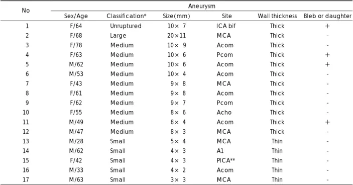
bFGF는 대조혈관에서는 발현이 없었다(Fig. 1A). 동맥류 벽에서는 17례중 15례에서 발현되었고, 모두 동맥류 벽의 중막 근육세포 주위에서 발현되었다(Fig. 1B). 발현이 없었 던 2례중 1례는 크기가 작은 후하소뇌동맥 말초성 동맥류였 고, 1례는 중등도 크기의 후교통동맥 동맥류였다. bFGF가 발현된 예중 발현의 정도가 경미한 예는 5례, 중등도 예는 9례, 고도인 예는 1례였다. bFGF의 발현된 정도와 동맥류 크 기와의 관계를 조사하였다. 경미하게 발현된 5례중 소동맥 류는 2례였고, 중등도 크기의 동맥류는 3례였다. 중등도로 발현된 9례중 소동맥류는 2례였고, 6례는 중등도 크기의 동 맥류였으며 1례는 비파열 동맥류였다. 고도로 발현된 1례는 대동맥류였다(Table 2).
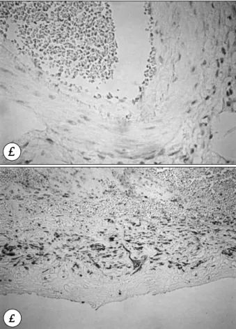
VEGF도 대조혈관에서는 발현이 없었다(Fig. 2A). 동맥 류 조직에서는 17례 모두에서 발현되었고, 대부분 내피하층 (subendothelium), 중막 및 외막(adventitia)에 국한하여 혹은 광범위 하게 발현되었다(Fig. 2B). VEGF의 발현정도
는 경도인 예가 7례, 중등도 8례, 고도인 예가 2례였다. 소 동맥류 5례에서는 3례가 경도로, 2례가 중등도로 발현되었 고, 중등도 크기의 동맥류 10례에서는 경도로 발현된 예가 3례, 중등도 6례, 고도로 발현된 예가 1례였다. 대동맥류 1례 는 고도로 발현되었고 비파열 동맥류 1례는 경도로 발현되 었다.
전체적으로 크기가 큰 동맥류는 bFGF와 VEGF 모두 고 도로 발현되었고, 비파열 동맥류는 bFGF는 중등도, VEGF 는 경도로 발현되었다. 크기가 작은 동맥류들은 bFGF와 VEGF 모두 경도로 발현되는 예들이 많았다. 따라서 bFGF 와 VEGF는 공히 동맥류가 크고 벽이 두터운 예들에서 강하 게 발현되고, 크기가 작고 벽이 얇은 예들에서는 경도로 발현 되는 경향을 보였다(Table 2).
2. 혈관벽 기질단백의 발현
대조혈관에서 αSMA은 중막에서 규칙적으로 정열되어 하 나의 띠로 강하게 발현되었다(Fig. 3A). 동맥류 조직에서도 1례를 제외한 16례에서 발현되었고 모두 중막에서 발현되 었다(Fig. 3B). 소동맥류 5례에서는 말초성 동맥류인 후하 소뇌동맥 동맥류 1례에서는 발현이 없었고, 1례에서는 경도, 2례는 중등도, 1례는 고도로 발현되었다. 크기가 중등도인
Table 1. Summary of characteristics of intracranial aneurysms
Aneurysm
No Sex/Age Classification* Size(mm) Site Wall thickness Bleb or daughter
1 F/64 Unruptured 10× 7 ICA bif Thick +
2 F/68 Large 20×11 MCA Thick -
3 F/78 Medium 10× 9 Acom Thick -
4 F/63 Medium 10× 6 Pcom Thick +
5 M/62 Medium 10× 6 Acom Thick +
6 M/53 Medium 10× 4 Acom Thick -
7 F/43 Medium 9× 8 MCA Thick -
8 F/61 Medium 9× 8 Acom Thick -
9 F/62 Medium 9× 7 Pcom Thick -
10 F/55 Medium 8× 6 Acho Thick -
11 M/49 Medium 8× 4 Acom Thick +
12 M/47 Medium 8× 3 MCA Thick -
13 M/28 Small 5× 4 MCA Thin -
14 M/62 Small 4× 3 A1 Thin -
15 F/42 Small 4× 3 PICA** Thin -
16 M/33 Small 4× 2 Acom Thin -
17 M/63 Small 3× 3 MCA Thin -
*:Small:2----6mm, medium:6----15mm, large:15----25mm, giant:>25mm
**:Peripheral aneurysm
ICA bif:internal carotid artery bifurcation, MCA:middle cerebral artery, Acom:anterior communicating artery, Pcom:
posterior communicating artery, Acho:anterior choroidal artery, A1:horizontal segment of the anterior cerebral artery, PICA:posterior inferior cerebellar artery
10례에서는 경도로 발현된 예가 2례, 중등도 3례, 고도 5 례였으며, 비파열 동맥류는 중등도, 대동맥류는 고도로 발현 되었다.
대조혈관에서의 교원질 Type IV는 내피하층에서 규칙적 으로 중등도로 발현되었다(Fig. 4A). 그리고 동맥류 조직에 서는 말초성 동맥류인 후하소뇌동맥 동맥류를 제외한 전례에 서 불규칙적으로 내피하층, 중막 혹은 외막에서 국한하여 또 는 광범위하게 발현되었고 특히 동맥류의 중막에서 발현되는 예들이 많았다(Fig. 4B). 발현되는 정도는 동맥류의 크기와 관계없이 모두 중등도 또는 고도로 발현되었으나, 규칙적으 로 배열되어 발현되는 대조혈관에서의 소견과는 달리 동맥류 조직에서는 모두 불규칙적으로 배열되어 발현되었다. 따라 서 전체적으로 혈관벽 기질단백도 동맥류가 크고 벽이 두터 운 예들에서는 강하게 발현되었으나, αSMA은 크기가 작고 벽이 얇은 예들에서는 약하게 발현되는 경향을 보였고, 교 원질 Type IV는 모든 예들에서 강하게 발현되었으나 불규 칙적으로 배열된 소견을 보였다(Table 3).
고 찰
선천적이나 혹은 후천적으로 야기된 뇌혈관벽의 기질적 결 손이나 손상은 뇌동맥류의 발생과 성장 및 파열의 기전에서 절대적인 역할을 한다1)2)10-12)16)19)20)21)23)
. 뇌혈관은 두개강 외의 혈관과 달리 구조적으로 외탄력층(external elastic lamina)이 존재하지 않기 때문에 내탄력층(internal elastic lamina)과 근육세포를 싸고 있는 세망섬유(reticular fiber) 가 뇌혈관을 기질적으로 정상으로 유지하는데 매우 중요한 역할을 하며20), 만약 이들이 결손되거나 손상을 받으면 뇌 동맥류가 발생한다. 발생된 뇌동맥류는 혈류역동학적 부하에 의해 점차적으로 커지다가 파열되거나, 거대 동맥류로 성장 하거나, 또는 환자가 사망후 부검에서 비파열 동맥류로 발견 되기도 한다3). 이와같이 뇌동맥류가 성장하고 파열하는 생 물학적 기전은 다양한데 이에 대해서 연구한 문헌은 많지 않 다22). Skirgaudas 등22)은 동맥류가 파열하는 기전의 하나로 혈관형성 인자가 동맥류 벽의 기질단백을 파괴함으로써 동 맥류가 파열한다는 설을 제시하였다.
Table 2. Intensity and distribution of the angiogenesis factors in control and intracranial aneurysms
bFGF VEGF No Classification
Intensity* Distribution Intensity* Distribution
Control 0 0
1 Unruptured 2 med 1 sub 2 Large 3 med 3 med, adv 3 Medium 2 med 1 diffuse 4 Medium 1 med 3 sub, med 5 Medium 1 med 1 sub, med 6 Medium 2 med 2 med 7 Medium 2 med 1 sub, med 8 Medium 2 med 2 diffuse
9 Medium 0 2 sub, med
10 Medium 2 med 2 diffuse 11 Medium 2 med 2 sub, med
12 Medium 1 med 2 med
13 Small 2 med 1 adv
14 Small 2 med 2 med, adv
15 Small 0 1 sub
16 Small 1 med 2 sub, med, adv
17 Small 1 med 1 med
*:Grading of immunostaining intensity, 0:no immunore- activity, 1:faint staining, 2:moderate staining, 3:strong staining
bFGF:basic fibroblast growth factor, VEGF:vascular en- dothelial growth factor, sub:subendothelium, med:media, adv:adventitia
Fig. 1. Immunohistochemical findings for basic fibroblast growth factor(bFGF) in control and intracranial aneurysm. Ne- gative immunoreactivity in control(A, ×200). bFGF im- munoreactivity around smooth muscle cells within the media of aneurysmal wall(B, ×200).
AA AA
B BB B
혈관형성 인자는 신생혈관의 형성, 성장 및 분화의 과정에 중요한 역할을 하며 창상의 치유나 종양의 성장과 같은 병적 과정에도 관여한다6)7). 많은 혈관형성 인자가 알려져 있으나 대표적인 것으로 bFGF와 VEGF가 있다6). bFGF는 섬유세 포(fibrocyte), 근세포(myocyte) 및 내피세포(endothelial cell)를 표적으로 하는 강력한 혈관형성 유도체이다17)22). 이 인자는 세포간질의 분해와 세포손상을 활성화시키기도 하 고22), 내피세포와 평활근 세포의 유사분열물질로써 작용하여 창상치유에 관여하기도 한다8). VEGF는 처음에는 혈관 투 과 인자(vascular permeability factor)로 인식되었던 인자 로서4) 혈관내피세포를 표적으로 하여 이들의 성장을 유도
하고5)13)14) 혈관형성의 시작에 중요역할을 한다7). 또한 이
인자는 내피세포간의 연결에서 누출을 야기하고 세포간질 단 백의 효소분해를 야기하는 과정을 활성화시키는 작용도 있 다22). 한편 동맥류 벽을 구성하는 대표적인 기질단백은 교 원질 Type IV, fibronectin 및 근육세포들이다9)15)22). 교원 질은 13개의 아형(subtype)들이 있으나 이중 Type I, III 및 IV가 동맥혈관의 구성성분으로서 중요하다1)9). 교원질
Type I과 III가 동맥혈관의 전체 교원질중 80~90%를 차 지하고, 혈관의 내막(intima), 중막 및 외막의 세포외기질 (extracellular matrix)의 주요 구성물이 되며, Type IV는 기저막(basement membrane)의 구성물이 된다9). Fibron- ectin은 정상 혈관에서 내층하(subintima), 평활근 세포 주 위 및 외막에 존재하는 혈관 구성 물질이며9), αSMA은 혈 관 중막에서의 평활근의 밀도와 조직화의 정도를 나타낸다22). 따라서 뇌동맥류 벽에서 혈관형성 인자인 bFGF와 VEGF가 발현되고 혈관벽 기질 단백인 교원질 Type IV와 평활근의 밀도를 나타내는 αSMA이 감소하는 것이 확인되면 이는 뇌 동맥류의 발생, 성장 및 파열에 관여할 것으로 추정된다. 본 연구에서 대조혈관에서는 혈관형성 인자인 bFGF와 VEGF 가 발현되지 않았으나 동맥류 조직에서는 bFGF는 17례중 15례에서 중막에, VEGF는 전례에서 내피하층, 중막 및 외막 에 국한하여 혹은 광범위하게 발현되었다. 이는 정상혈관에 서는 존재하지 않는 혈관형성 인자가 동맥류 벽에서는 존재 하는 것으로 뇌동맥류의 발생과 관계가 있을 것으로 생각된
Fig. 2. Immunohistochemical findings for vascular endothelial growth factor(VEGF) in control and intracranial ane- urysm. Negative immunoreactivity in control(A, ×200).
VEGF immunoreactivity in the entire wall of aneurysm (B, ×200).
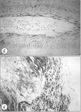
Fig. 3. Immunohistochemical findings for alpha smooth muscle actin(αSMA) in control and intracranial aneurysm. Re- gularly arranged, well-defined band of immunoreactivity of αSMA in the media of the control vessel(A, ×100).
Diffuse, faint, irregularly arranged αSMA within disrupted media of the aneurysmal wall(B, ×200).
AA AA
B BB B
AA AA
B B B B
다. 즉 뇌혈관의 분지부에 혈류역동학적 부하와 퇴행성 병 변이 야기될 때 혈관형성 인자가 발현되어 뇌혈관의 기질적 손상을 진행시켜 동맥류가 발생되고 진행하는데 관여할 것 으로 추정한다. 발현되는 정도는 bFGF와 VEGF 공히 전체 적으로 동맥류가 크고 벽이 두터운 예들에서 강하게 발현되 고 크기가 작고 벽이 얇은 예들에서는 경도로 발현되는 경향 을 보였다. 이의 결과는 아마도 뇌동맥류의 성장과 관계있 을 것으로 추정된다. 혈관형성 인자와 기질단백의 불균형으 로 일부의 예들에서는 조기에 파열되나, 일부의 예들에서는 손상된 동맥류 벽이 치유되면서 두터워지고 이러한 예들에서 는 혈관형성 인자의 발현이 증가하지 않나 추측된다. 특히 본 연구에서 bFGF 중막에서만 발현되는 것은 이 인자가 근 육세포의 분열을 촉진시켜 동맥류 벽의 손상치유에 관여하 여 동맥류가 대동맥류나 거대 동맥류로 진행하는데 관여할 것으로 역시 추정된다. 한편 Skirgaudas 등22)의 연구에서는 VEGF가 2가지의 형태로 발현되었다. 하나는 구조가 파괴된 전체 동맥류 벽에서 반점형태로 발현되는 예들로서 이는 동 맥류의 파열과 관계가 있고, 다른 하나는 거대 동맥류 벽의 신생혈관 부위에서 발현되는 형으로서 거대 동맥류로 성장하 고 진행하는데 관여하는 것으로 추정하였다. 본 연구에 서는 거대 동맥류의 예가 없어 신생혈관에서 발현되는 예는 관찰
할 수 없었고, 모든 예들이 동맥류 벽에서 부분적으로 혹은 광범위하게 발현되는 예들이었다.
본 연구에서 대조혈관에서의 αSMA은 혈관의 중간층에서 규칙적으로 정열되어 하나의 띠로 강하게 발현되었다. 동맥 류 조직에서는 17례중 16례에서 발현되었으나 대조혈관과 비교하여 전체적으로 불규칙적으로 약하게 발현되었다. 또한 대조혈관에서 교원질 Type IV는 내피하층에서 규칙적으로 중등도로 발현되었으나, 동맥류 벽에서는 대조혈관과 비교하 여 모두 불규칙적으로 배열된 양상을 보였다. 이와같은 소견 은 동맥류 조직에서 혈관벽 기질 단백의 감소를 의미하고 이 러한 감소가 역시 동맥류 파열에 관여될 것으로 생각된다.
마지막으로 언급할 것은 말초성 동맥류인 후하소뇌동맥 동 맥류 1례에서는 혈관형성 인자도 혈관벽 기질 단백도 모두 발현되지 않았다. 이 동맥류는 4mm 크기의 동맥류 벽이 얇 은 예였다. 이는 아마도 혈관을 구성하는 성분을 가지지 않은 가성 동맥류(pseudoaneurysm)로 생각된다.
Skirgaudas 등22)의 진균성(mycotic) 동맥류에서는 혈관
Table 3. Intensity and distribution of the vascular wall ma- trix proteins in control and intracranial aneurysms αSMA Collagen type IV No Classification
Intensity* Distribution Intensity* Distribution
Control 3 med 2 sub
1 Unruptured 2 med 3 med 2 Large 3 med 3 sub, med
3 Medium 1 med 3 med
4 Medium 3 med 3 med, adv 5 Medium 1 med 2 med, adv
6 Medium 3 med 3 med
7 Medium 2 med 2 med
8 Medium 3 med 3 sub, med 9 Medium 2 med 2 sub, diffuse 10 Medium 2 med 3 diffuse 11 Medium 3 med 2 med, adv
12 Medium 3 med 3 med
13 Small 2 med 3 med
14 Small 3 med 3 sub, med
15 Small 0 0
16 Small 2 med 3 diffuse
17 Small 1 med 3 med
*:Grading of immunostaining intensity, 0:no immuno- reactivity, 1:faint staining, 2:moderate staining, 3:strong staining αSMA:alpha smooth muscle actin, sub:suben- dothelium med:media, adv:adventitia
Fig. 4. Immunohistochemical findings for collagen Type IV in control and intracranial aneurysm. Regularly defined band of collagen Type IV immunoreactivity in the sub- endothelium of control vessel(A, ×200). Diffuse irregularly arranged collagen Type IV immunoreactivity in the entire wall of aneurysm(B, ×200).
AA AA
BB BB
형성 인자와 혈관벽 기질 단백이 모두 발현되었다.
결 론
저자들은 대조혈관과 수술 중 얻은 뇌동맥류의 조직에서 혈관벽을 약화시키는 물질로 알려진 혈관형성 인자와 혈관 벽의 강도를 유지시키는데 중요한 역할을 한다고 알려진 혈 관벽 기질 단백에 대해 면역조직화학적 염색을 시행하여 그 발현여부를 알아보고, 이들의 발현정도와 대조혈관에서의 발 현정도를 상호 비교하여, 뇌동맥류의 발생, 성장 및 파열의 기전에 대하여 알아보았다.
혈관형성 인자는 대조혈관에서 발현되지 않았으나 동맥류 조직에서는 거의 모든 예들에서 발현되었고, 동맥류가 크고 벽이 두터운 예들에서 강하게 발현되었다. 혈관벽 기질 단백 은 대조혈관에서는 규칙적으로 정열되어 중등도 또는 강하 게 발현되었으나, 동맥류 조직에서는 불규칙적으로 배열된 양 상을 보이거나 약하게 발현되었다.
결론적으로 뇌동맥류의 성장과 파열에 혈관형성 인자들과 혈관벽 기질 단백의 변화가 관여 될 것으로 생각한다. 혈관형 성 인자들이 뇌동맥류 벽을 약화시키고, 혈관벽 기질 단백 이 동맥류 조직에서 감소하는 것은, 동맥류의 성장과 파열의 생물학적 기전중의 하나가 될 것으로 추정된다.
•논문접수일:2000년 2월 7일
•심사완료일:2000년 9월 25일
•책임저자:임 만 빈
700-712 대구광역시 중구 동산동 194 계명대학교 의과대학 신경외과학교실
전화:053) 250-7332, 전송:053) 250-7356 E-mail:[email protected]
References
1) Austin G, Fisher S, Dickson D, Anderson D, Richardson S:
The significance of the extracellular matrix in intracranial aneurysms. Ann Clin Lab Sci 23:97-105, 1993
2) Chyatte D, Reilly J, Tilson MD:Morphometric analysis of reticular and elastin fibers in the cerebral arteries of patients with intracranial aneurysms. Neurosurgery 26:939-943, 1990 3) de la Monte SM, Moore GW, Monk MA, Hutchins GM:Risk factors for the development and rupture of intracranial berry aneurysms. Am J Med 78:957-964, 1985
4) Dvorak HF, Brown LF, Detmar M, Dvorak AM:Vascular permeability factor/vascular endothelial growth factor, micro- vascular hyperpermeability, and angiogenesis. Am J Pathol 146:1029-1039, 1995
5) Ferrara N, Houck KA, Jakeman LB, Winer J, Leung DW:
The vascular endothelial growth factor family of polypeptides.
J Cell Biochem 47:211-218, 1991
6) Folkman J, Klagsbrun M:Angiogenic factors. Science 235: 442-447, 1987
7) Fong GH, Rossant J, Gertsenstein M, Breitman ML:Role of the Flt-1 receptor tyrosine kinase in regulating the assembly of vascular endothelium. Nature 376:66-70, 1995
8) Futami K, Yamashita J, Tachibana O, Kida S, Higashi S, Ik- eda K, et al:Basic fibroblast growth factor may repair experimental cerebral aneurysms in rats. Stroke 26:1649- 1654, 1995
9) Futami K, Yamashita J, Tachibana O, Higashi S, Ikeda K, Ya- mashima T:Immunohistochemical alterlations of fibronectin during the formation and proliferative repair of experimental cerebral aneurysms in rats. Stroke 26:1659-1664, 1995 10) Hegeds K:Some observations on reticular fibers in the me-
dia of the major cerebral arteries:A comparative study of patients without vascular diseases and those with ruptured berry aneurysms. Surg Neurol 22:301-307, 1984
11) Hegeds K:Reticular fiber deficiency in the intracranial art- eries of patients with dissecting aneurysm and review of the possible of the pathogenesis of previously reported cases. Eur Arch Psychiatry Neurol Sci 235:102-106, 1985
12) Hegeds K:Pattern of reticular fibers of the major cerebral arteries in cases of unexplained subarachnoid hemorrhage. J Neurol 233:44-47, 1986
13) Jakeman LB, Armanini M, Phillips HS, Perrara N:Develo- pmental expression of binding sites and messenger ribonucleic acid for vascular endothelial growth factor suggests a role for this protein in vasculogenesis and angiogenesis. Endocrin- ology 133:848-859, 1993
14) Leung DW, Cachianes G, Kuang WJ, Goeddel DV, Ferrara N:
Vascular endothelial growth factor is a secreted angiogenic mitogen. Science 246:1306-1309, 1989
15) Madri JA, Bell L, Marx M, Merwin JR, Basson C, Prinz C:
Effect of soluble factors and extracellular matrix compon- ents on vascular cell behavior in vitro and in vivo:Models of de-endothelialization and repair. J Cell Biochem 45: 123-130, 1991
16) Majamaa K, Myllyla VV:A disorder of collagen biosynth- esis in patients with cerebral artery aneurysm. Biochim Biophys Acta 1225:48-52, 1993
17) Mignatti P, Rifkin DB:Release of basic fibroblast growth factor, an angiogenic factor devoid of secretory signal sequ- ence:A trivial phenomenon or a novel secretion mechanism?
J Cell Biochem 47:201-207, 1991
18) Morris KM, Shaw DM, Foy PM:Smoking and subarachnoid haemorrhage:A case control study. Br J Neurosurg 6:429- 432, 1992
19) Neil-Dwyer G, Bartlett JR, Nicholls AC, Narcisi P, Pope FM:
Collagen deficiency and ruptured cerebral aneurysms:A cli- nical and biochemical study. J Neurosurg 59:16-20, 1983
20) Østergaard JR, Reske-Nielsen E, Oxlund H:Histological and morphometric observations on the reticular fibers in the arterial beds of patients with ruptured intracranial saccular aneurysms. Neurosurgery 20:554-558, 1987
21) Østergaard JR, Reske-Nielsen E, Buhl J:Deficiency of reti- cular fibers in cerebral arteries. On the etiology of saccular aneurysms in childhood. Br J Neurosurg 3:113-115, 1989
22) Skirgaudas M, Awad IA, Kim J, Rothbart D, Criscuolo G:
Expression of angiogenesis factors and selected vascular wall matrix proteins in intracranial saccular aneurysms. Neuro- surgery 39:537-547, 1996
23) Stehbens WE:Etiology of intracranial berry aneurysms. J Neurosurg 70:823-831, 1989