door laminoplasty has been considered as an effective and safe method for decompressing the multi-segmental cervical lesions caused by CSM, OPLL, or intervertebral disc herniation with de- velopmental canal stenosis, as well as for achieving the immedi- ate stability of these lesions1,3,8,9,14,19). For spinal canal decompres- sion and stability of the spine, various materials were used in this procedure, which include : autologous grafts, hydroxyapa- tite (HA) spacers, and allogeneic bone (AB) spacers. However, the pros and cons of individual implants have not been thor- oughly investigated. In addition, many studies have compared heterogeneous groups or small individual case series, but there is still a lack in direct comparisons among these options. In this study, we compared double-door laminoplasty using HA spac- ers (Apacerum®; Asahi Optical Co., Ltd., Tokyo, Japan) and AB spacers (Allo-Spine®; CG Bio, Seoul, Korea) to analyze the clini- cal and radiological outcomes in patients.
INTRODUCTION
Cervical laminoplasty was introduced in the early 1980s as a successful means of addressing multilevel cervical spinal steno- sis secondary to either cervical spondylotic myelopathy (CSM) or ossification of posterior longitudinal ligament (OPLL)23). The goal of laminoplasty is to indirectly decompress the spinal canal by opening one or both sides of the lamina. In this procedure, the “open-door” technique, which was popularized by Hirabayas- hi et al.2) can be used. Later on, other adaptations of this surgical technique including the double-door approach where the spi- nous process is split open were developed. Although both tech- niques are effective in improving neurologic outcomes, double- door laminoplasty is better than open-door laminoplasty when it comes to clinical outcomes such as axial neck pain, periopera- tive complication, and anatomical alignment13,15,18). Double-
A Comparison of Implants Used in Double Door Laminoplasty : Allogeneic Bone Spacer versus Hydroxyapatite Spacer
Dong Yoon Lee, M.D., Chang Kyu Lee, M.D., In-Soo Kim, M.D., Ph.D.
Department of Neurosurgery, Keimyung University, School of Medicine, Dongsan Medical Center, Daegu, Korea
Objective : The aim of this study was to compare the clinical and radiological outcomes associated with the use of hydroxyapatite (HA) spacer and allogeneic bone (AB) spacer in laminoplasty.
Methods : From January 2006 to July 2014, 79 patients with cervical spondylotic myelopathy or ossification of the posterior longitudinal ligament underwent cervical laminoplasty. The radiologic parameters were obtained from plain radiography and three-dimensional computed tomography. All images were taken before and after surgery. Cervical lordosis, spinal canal dimension, fusion between lamina and spacer, and resorption of spacer were checked. Clinical outcomes were assessed using visual analog scale and Japanese Orthopedic Association.
Results : Double-door laminoplasty was performed on 280 levels : 182 in the HA group and 98 in the AB group. The mean follow-up was 23.1 months (range : 4–69 months). Similar fusion rates were found in these groups (p=0.3). The resorption rate between lamina and spacer was lower in the HA group (p<0.001). During the immediate postoperative period, the canal dimension of both groups increased compared with the results in the preoperative period. However, the canal dimension of the AB group decreased over time compared with that of the HA group (p<0.001).
Conclusion : Double-door laminoplasty improved the clinical outcomes of both groups. However, the spinal canal dimension in the AB group showed a greater degree of reduction than in the HA group at the final postoperative follow-up. Therefore, we suggest that surgeons consider the use of larger-sized AB spacers in double-door laminoplasties.
Key Words : Allograft · Hydroxyapatites · Cervical myelopathy · Ossification of posterior longitudinal ligament · Spinal fusion.
Clinical Article
•Received : May 25, 2016 •Revised : June 2, 2016 •Accepted : September 22, 2016
•Address for reprints : In-Soo Kim, M.D., Ph.D.
Department of Neurosurgery, Keimyung University, School of Medicine, Dongsan Medical Center, 56 Dalseong-ro, Jung-gu, Daegu 41931, Korea Tel : +82-53-250-7730, Fax : +82-53-250-7356, E-mail : [email protected]
•This is an Open Access article distributed under the terms of the Creative Commons Attribution Non-Commercial License (http://creativecommons.org/licenses/by-nc/3.0) which permits unrestricted non-commercial use, distribution, and reproduction in any medium, provided the original work is properly cited.
MATERIALS AND METHODS Patient selection
We retrospectively identified 79 patients (59 men, 20 women) with CSM or OPLL who underwent double-door laminoplasty between January 2006 and July 2014. These patients were divid- ed into two groups : laminoplasty using HA spacer (HA group) and AB spacer (AB group). Patients with cervical kyphosis, evi- dence of cervical instability, serious medical problems, as well as those who underwent prior anterior cervical surgery were excluded. Patient characteristics including gender, age, bone mineral density, alcohol, and smoking histories were obtained (Table 1).
Radiological evaluation
Plain radiography and three-dimensional computed tomog- raphy (3-D CT) were performed pre- and postoperatively. Plain radiography was performed in all 79 patients during the follow-
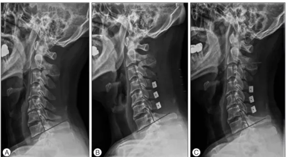
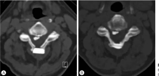
up period. However, only 39 of them underwent CT during the follow-up due to death and personal reasons such as financial concerns. Those who were followed up only underwent plain radiography. Cervical lordosis, spinal canal dimension, fusion between lamina and spacer, and resorption of spacer were ex- amined. Cervical lordosis was defined as the angle between C2 and C7 inferior endplates. Change in overall cervical lordosis was assessed using the radiographic images (Fig. 1). The trans- verse area of the cervical canal was measured at the level of the vertebral pedicles using a computer software (Marosis, M-View Ver. 5.4). As visualized on CT images, bone fusion of the spacer was determined as the disappearance of the gap at the interface between the grafted spacer and host bone with new bone forma- tion. On the other hand, spacer resorption was defined as the change in the size and shape of the spacer on CT images (Fig. 2).
Surgical procedure
All laminoplasties were performed by a single surgeon (Dr.
Table 1. Characteristic of the two patient groups
Characteristic HA group (n=51) AB group (n=28) p value
Mean age, years (range) 63.1 (37–87) 62.8 (42–83) 0.931
Mean follow up duration, months (range, months) 33.75 (4–69) 17.1 (7–35)
Sex (male : female) 35 : 16 24 : 4 0.095
OPLL (%) 34 (66.7) 23 (82.1) 0.142
Osteoporosis (%) 22 (43.1) 13 (46.4) 0.778
Smoking (%) 18 (35.3) 11 (39.3) 0.725
Alcohol (%) 26 (51.0) 13 (46.4) 0.699
Surgical levels (total) 182 98
C2 2 2
C3 40 24
C4 48 28
C5 50 26
C6 40 18
C7 2 0
Osteoporosis defined BMC <-2.5. HA : hydroxyapatite, AB : allogeneic bone
Fig. 1. Cervical lordosis, defined as the angle between C2 and C7 inferior end plates. A : Pre-op. B : Immediately post
op. C : Final follow up. A B C
I.S. Kim). For surgical positioning, the Mayfield headrest was used wherein the neck was fixed from the neutral to the flexion position. After a vertical incision from the C2 to the C7 areas was made, the paravertebral muscle was carefully denuded. The researchers used double-door laminoplasty using an HA spacer or an AB spacer and the shallowest 1.7 mm Match Head Tool (Midas Rex® Legend EHS Stylus® High-Speed Surgical Drill;
Medtronic, Inc., Fridley, MN, USA) to maximize the distance between split spinous processes and to enlarge the spinal canal area after applying a spacer between these areas. The outer cor- tex of the hinge site was minimally drilled to allow the reattach- ment of each site after splitting the spinous processes and to help with rapid bone fusion. Gentle care was taken while de- nuding the paravertebral muscle to minimize bleeding and pain after the operation. As much as possible, the researchers did not use electrocautery during dissection to prevent muscle atrophy at the follow-up period.
Clinical evaluation
Neurological status of the patient was preoperatively and postoperatively assessed using the Japanese Orthopedic Associ- ation (JOA) scores, while the pain intensity of the neck was as- sessed from 0–10 using the neck visual analog scale (VAS; 0=no pain; 10=worst pain).
Statistical analysis
Parameters were analyzed using the Repeated Measures ANONA. The differences in the results were considered signifi- cant when p values were <0.05. All results are expressed as mean±standard deviation. Analyses were performed using the IBM Statistical Package for the Social Sciences Version 18.0 (SPSS Inc., Chicago, IL, USA).
RESULTS
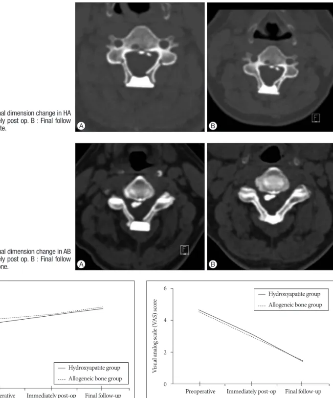
The mean follow-up duration was 23.1 months (range : 4–69 months). Of the 79 included patients, 22 were diagnosed with CSM and 57 with OPLL. Surgeries were performed on 280 lev- els : 182 for the HA group and 98 for the AB group. Mean cervi- cal lordosis changed from 13.54°±9.21° to 12.16°±8.68° in the HA group and from 16.93°±9.07° to 11.13°±7.08° in the AB group (p=0.131) (Table 2). Immediately after the surgery, the spinal canal dimension increased from 189.31 mm2 to 275.25 mm2 in the HA group and from 185.48 mm2 to 307.22 mm2 in the AB group, but it decreased to 243.71 mm2 and 254.50 mm2, respectively, at the last follow-up (p<0.001) (Table 3) (Fig. 3, 4).
The dimension decreased during the last follow-up in both groups, but the degree of decrease in dimension was greater in the AB group than in the HA group. JOA scores changed from 12.35±4.67 to 15.02±3.72 in the HA group and from 13.04±4.19
A B
Fig. 2. Spacer resorption was defined that the shape of spacer is smaller or changed on CT im- ages. A : Immediately post op. B : Final follow up.
Table 2. Cervical lordosis change
Groups Pre-op
mean±SD (degrees) Immediately post OP
mean±SD (degrees) Final follow up
mean±SD (degrees) p value
HA group (n=28) 13.16±9.00 11.61±8.04 12.17±8.60 0.131*
AB group (n=11) 16.93±9.07 11.25±8.42 11.13±7.08
*Repeated measures ANOVA. HA : hydroxyapatite, AB : allogeneic bone
Table 3. Spinal canal dimension change
Groups Pre-op
mean±SD (mm2) Immediately post OP
mean±SD (mm2) Final follow up
mean±SD (mm2) p value
HA group (n=28) 189.31±26.05 275.25±34.21 243.71±31.12 <0.001*
AB group (n=11) 185.48±32.62 307.22±45.47 254.50±54.70
*Repeated measures ANOVA. HA : hydroxyapatite, AB : allogeneic bone
to 15.29±3.13 in the AB group (p=0.674) (Fig. 5). Neck VAS scores changed from 4.65±3.10 to 1.41±1.51 in the HA group and from 4.54±1.71 to 1.46±0.88 in the AB group (p=0.598) (Fig. 6). To com- pare both fusion and resorption rates, we chose the group with a similar follow-up term of 18 months. In the HA group, a total of 74 implants were used from which 55 achieved union. Mean- while, in the AB group, a total of 37 implants were used from which 24 achieved union. However, there was no statistically significant difference found between the two groups (p=0.3) (Table 4). In terms of resorption rate, a total of 74 spacers in the HA group were used from which resorption occurred in 6. On
the other hand, a total of 37 spacers in the AB group were used from which resorption occurred in 19 (p<0.001) (Table 5).
DISCUSSION
Numerous modifications and supplementary instruments were utilized to retain hinge patency and provide secure fixa- tion in open-door or double-door laminoplasties4,17). Some ex- amples of these are as follows : HA spacer, AB spacer, ceramic lamina, miniplate osteosynthesis, and autogenous bone graft-
ing4,24). To be specific, AB spacer, HA spacer, autogenous bone
graft, and ceramic block were used for the maintenance of an
A B
Fig. 4. The spinal canal dimension change in AB group. A : Immediately post op. B : Final follow up. AB : allogeneic bone.
A B
Fig. 3. The spinal canal dimension change in HA group. A : Immediately post op. B : Final follow up. HA : hydroxyapatite.
Fig. 5. Clinical outcome in JOA scores compared between preoperative, immediately post operative and final follow up.
18 16 14 12 10 8 6 4 2
0 Preoperative Immediately post-op Final follow-up
Japanese orthopaedic association (JOA) score
Hydroxyapatite group Allogeneic bone group
6
4
2
0 Preoperative Immediately post-op Final follow-up
Visual analog scale (VAS) score Hydroxyapatite group
Allogeneic bone group
Fig. 6. Clinical outcome in VAS scores compared between preoperative, immediately post operative and final follow up.
opened lamina. Although these promote the bony healing of the grafted side and the maintenance of the laminar arch when spaces were used, these posed the risk of root or cord compres- sion because of a graft kick-out4,5,22). In laminoplasty, HA and AB spacers have several benefits compared to the autologous bone graft. These include good biomechanical stability and re- ductions in operation time, blood loss, and donor site morbidi- ty7,10-12,16). It was reported that the fusion rates after the use of an HA spacer and AB spacer were 48.8% and 51%, respectively16). However, no study has actually directly compared these two groups. In this study, the fusion between the lamina and the spac- er, as well as the resorption, was checked through the fine-sec- tion reconstruction of the CT. Since an HA spacer only has an osteoconduction activity compared with an AB spacer with both osteoconduction and osteoinduction activities, the fusion rate was expected to be higher in the AB group. In the current study, 55 of the 74 changes in the HA group and 24 of the 37 changes in the AB group were fused. While the result was not statistical- ly significant, the HA group showed a similar fusion rate with AB group. Although the clinical outcomes improved in both groups, there were no statistically significant differences be- tween them. This is because the shape and size of the spacers are similar, which allowed the canal dimension to increase in a similar proportion with the canal expansion postoperatively. In agreement with previous findings, this study observed the di- mensional reduction of the spinal canal after an immediate postoperative widening7,16). However, the degree of dimensional reduction was greater in the AB group on the final follow-up CT imaging. This may be taken into consideration in conjunction with the fusion rate. In addition, it may be related to the fusion of the newly formed bone, which narrowed the canal dimension.
Moreover, spacer resorption may be important in this change in dimension. Originally, the spacer can widen the width of the canal by occupying the space between the spinous processes.
However, with the increase in spacer resorption, its shape is changed, and the space between the spinous processes is nar- rowed. It is believed to result in a decrease because of its canal- widening effect. Aside from having been observed in many other studies, kyphotic deformities were also observed after
laminoplasty in this study. However, additional follow-up is needed3,6,18,20,21).
Limitations of the study
This study had several limitations. It was a retrospective anal- ysis involving a relatively small number of patients. In particu- lar, limited CT data were available because of the retrospective nature of this study. In addition, the fusion rates between the two groups were not accurately compared because the mean follow-up periods of each group was different. Thus, it is neces- sary to measure them again after sufficient follow-up periods.
Prospective controlled studies using patient-based outcomes are required to compare outcomes and complications associated with these two implants. As supplements to this study, a broader scope and a longer follow-up duration are thus needed.
CONCLUSION
After double-door laminoplasty was performed with an AB spacer on one group and an HA spacer on another group, the clinical outcomes of both groups improved. In addition, similar fusion rates were found in these groups. However, the AB group had a greater degree of reduction in the spinal canal dimension than the HA group at the final postoperative follow-up. There- fore, we suggest that surgeons should consider the use of larger- sized AB spacers in double-door laminoplasties.
References
1. Chiba K, Ogawa Y, Ishii K, Takaishi H, Nakamura M, Maruiwa H, et al. : Long-term results of expansive open-door laminoplasty for cervical my- elopathy--average 14-year follow-up study. Spine (Phila Pa 1976) 31 : 2998-3005, 2006
2. Hirabayashi K, Watanabe K, Wakano K, Suzuki N, Satomi K, Ishii Y : Ex- pansive open-door laminoplasty for cervical spinal stenotic myelopathy.
Spine (Phila Pa 1976) 8 : 693-699, 1983
3. Iwasaki M, Kawaguchi Y, Kimura T, Yonenobu K : Long-term results of expansive laminoplasty for ossification of the posterior longitudinal lig- ament of the cervical spine : more than 10 years follow up. J Neurosurg 96(2 Suppl) : 180-189, 2002
4. Jin SW, Kim SH, Kim BJ, Choi JI, Ha SK, Kim SD, et al. : Modified open- Table 4. Fusion between lamina and spacer
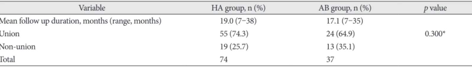
Variable HA group, n (%) AB group, n (%) p value
Mean follow up duration, months (range, months) 19.0 (7–38) 17.1 (7–35)
Union 55 (74.3) 24 (64.9) 0.300*
Non-union 19 (25.7) 13 (35.1)
Total 74 37
*chi-square test. HA : hydroxyapatite, AB : allogeneic bone
Table 5. Spacer resorption rate
Variable HA group (%), (n=74) AB group (%), (n=37) p value
Mean follow up duration, months (range, months) 19.0 (7–38) 17.1 (7–35)
Resorption (%) 06 (8.10) 19 (51.4) <0.001*
*chi-square test. HA : hydroxyapatite, AB : allogeneic bone
door laminoplasty using hydroxyapatite spacers and miniplates. Korean J Spine 11 : 188-194, 2014
5. Kanemura A, Doita M, Iguchi T, Kasahara K, Kurosaka M, Sumi M : Delayed dural laceration by hydroxyapatite spacer causing tetraparesis following double-door laminoplasty. J Neurosurg Spine 8 : 121-128, 2008 6. Kawaguchi Y, Kanamori M, Ishihara H, Ohmori K, Nakamura H, Kimu- ra T : Minimum 10-year followup after en bloc cervical laminoplasty.
Clin Orthop Relat Res (411) : 129-139, 2003
7. Kimura A, Seichi A, Inoue H, Hoshino Y : Long-term results of double- door laminoplasty using hydroxyapatite spacers in patients with com- pressive cervical myelopathy. Eur Spine J 20 : 1560-1566, 2011 8. Kimura I, Shingu H, Nasu Y : Long-term follow-up of cervical spondy-
lotic myelopathy treated by canal-expansive laminoplasty. J Bone Joint Surg Br 77 : 956-961, 1995
9. Kong Q, Zhang L, Liu L, Li T, Gong Q, Zeng J, et al. : Effect of the decom- pressive extent on the magnitude of the spinal cord shift after expansive open-door laminoplasty. Spine (Phila Pa 1976) 36 : 1030-1036, 2011 10. Kubo S, Goel VK, Yang SJ, Tajima N : Biomechanical evaluation of cervi-
cal double-door laminoplasty using hydroxyapatite spacer. Spine (Phila Pa 1976) 28 : 227-234, 2003
11. Martin-Benlloch JA, Maruenda-Paulino JI, Barra-Pla A, Laguia-Gar- zaran M : Expansive laminoplasty as a method for managing cervical multilevel spondylotic myelopathy. Spine (Phila Pa 1976) 28 : 680-684, 12. Nakano K, Harata S, Suetsuna F, Araki T, Itoh J : Spinous process-split-2003
ting laminoplasty using hydroxyapatite spinous process spacer. Spine (Phila Pa 1976) 17(3 Suppl) : S41-S43, 1992
13. Nakashima H, Kato F, Yukawa Y, Imagama S, Ito K, Machino M, et al. : Comparative effectiveness of open-door laminoplasty versus French- door laminoplasty in cervical compressive myelopathy. Spine (Phila Pa 1976) 39 : 642-647, 2014
14. Ogawa Y, Chiba K, Matsumoto M, Nakamura M, Takaishi H, Hirabayas- hi H, et al. : Long-term results after expansive open-door laminoplasty for the segmental-type of ossification of the posterior longitudinal liga-
ment of the cervical spine : a comparison with nonsegmental-type lesions.
J Neurosurg Spine 3 : 198-204, 2005
15. Okada M, Minamide A, Endo T, Yoshida M, Kawakami M, Ando M, et al. : A prospective randomized study of clinical outcomes in patients with cervical compressive myelopathy treated with open-door or French- door laminoplasty. Spine (Phila Pa 1976) 34 : 1119-1126, 2009 16. Park JH, Jeon SR : Midline-splitting open door laminoplasty using hy-
droxyapatite spacers : comparison between two different shaped spac- ers. J Korean Neurosurg Soc 52 : 27-31, 2012
17. Patel CK, Cunningham BJ, Herkowitz HN : Techniques in cervical lam- inoplasty. Spine J 2 : 450-455, 2002
18. Ratliff JK, Cooper PR : Cervical laminoplasty : a critical review. J Neuro- surg 98(3 Suppl) : 230-238, 2003
19. Sakaura H, Hosono N, Mukai Y, Ishii T, Iwasaki M, Yoshikawa H : Long- term outcome of laminoplasty for cervical myelopathy due to disc her- niation : a comparative study of laminoplasty and anterior spinal fusion.
Spine (Phila Pa 1976) 30 : 756-759, 2005
20. Shimamura T, Kato S, Toba T, Yamazaki K, Ehara S : Sagittal splitting laminoplasty for spinal canal enlargement for ossification of the spinal ligaments (OPLL and OLF). Semin Musculoskelet Radiol 5 : 203-206, 2001
21. Suk KS, Kim KT, Lee JH, Lee SH, Lim YJ, Kim JS : Sagittal alignment of the cervical spine after the laminoplasty. Spine (Phila Pa 1976) 32 : E656- E660, 2007
22. Tanaka N, Nakanishi K, Fujimoto Y, Sasaki H, Kamei N, Hamasaki T, et al. : Expansive laminoplasty for cervical myelopathy with interconnect- ed porous calcium hydroxyapatite ceramic spacers : comparison with autogenous bone spacers. J Spinal Disord Tech 21 : 547-552, 2008 23. Tsuji H : Laminoplasty for patients with compressive myelopathy due to
so-called spinal canal stenosis in cervical and thoracic regions. Spine (Phila Pa 1976) 7 : 28-34, 1982
24. Yang SC, Niu CC, Chen WJ, Wu CH, Yu SW : Open-door laminoplasty for multilevel cervical spondylotic myelopathy : good outcome in 12 pa- tients using suture anchor fixation. Acta Orthop 79 : 62-66, 2008