∙ Received: October 15, 2007. Accepted: October 30, 2007.
∙ Corresponding author: Hoon Hee Park
Dept. of Nuclear Medicine, Yonsei University Health System 134 Shinchon-Dong, Seodaemun-Gu, Seoul, 120-749, Korea Tel: +82-2-2228-6064 Fax: +82-2-2227-7062
E-mail: [email protected]
Original Article Gated Cardiac Blood Pool scan에서의 심장 주위
배후방사능 관심영역 설정시 산란선의 영향을 감소시키기 위한 연구
연세의료원 세브란스병원 핵의학과
김정열⋅박훈희⋅남궁혁⋅조석원⋅김재삼⋅이창호
Study a Technique for Reducing the Influence of Scattered Rays from Surrounding Organs to the Heart during Gated Cardiac Blood Pool scan
Jung Yul Kim, Hoon Hee Park, Hyuk Nam Koong, Suk Won Cho, Jae Sam Kim, Chang Ho Lee Department of Nuclear Medicine, Severance Hospital, Yonsei University Health System
Purpose: The Gated cardiac blood pool scan is non-invasive method that a quantitative evaluation of left ventricular function. Also this scan have shown the value of radionuclide ejection fraction measurements during the course of chemotherapy as a predictor of cardiac toxicity. Therefore a reliable method of monitoring its cardiotoxic effects is necessary. the purpose of this study is to minimize the overestimate of left ventricular ejection fraction (LVEF) by modified body position to reduce the influence of scattered rays from surrounding organs of the heart in the background region of interest.
Materials and Methods: Gated cardiac blood pool scan using in vivo
99mTc-red blood cell (RBC) was carried out in 20 patients (mean 44.8±8.6 yr) with chemotherapy for a breast carcinoma.
Data acquisition requires about 600 seconds and 24 frames of one heart cycle by the multigated acquisition mode, Synchronization deteriorates toward the end of the cycle and with the distance from the trigger signal (R-wave) by ECG gating. Gated cardiac blood pool scan was studied with conventional method (supine position and the detector head in 30-45° left anterior oblique position and caudal 10-20° tilt) and compared with modified method (left lateral flexion position with 360 mL of drinking water). LVEF analysis was performed by using the automatically computer mode. Results: The ROI counts of modified scan method were lower than LV conventional method (1429±251 versus 1853±243, <0.01). And LVEF of modified method was also decrease compared with conventional method (58.3±5.6% versus 65.3±6.1%, <0.01). Imaging analysis indicated that stomach was expanded because of water and spleen position was changed to lateral inferior compared with conventional method. Conclusion: This study shows that the modified method in MUGA reduce the influence of scattered rays from surrounding organs. Because after change the body position to left lateral flexion and drinking water, the location of spleen, left lobe of liver and stomach had changed and they could escaped from background ROI. Therefore, modified method could help to minimize the overestimate LVEF (%). (Korean J Nucl Med Technol 2008;12(1):33-38)
Key Words : Gated cardiac blood pool, RBC, LV EF, ROI
서 론
게이트 심장 혈액 풀 스캔(Gated Cardiac Blood Pool scan) 은 심전도를 지표로 사용하여 심박주기 동안 심장내의 혈액 풀 변화를 영상화하는 비침습적인 방법이다.13) 급성심근경색
핵의학기술 제12권 제1호 2008
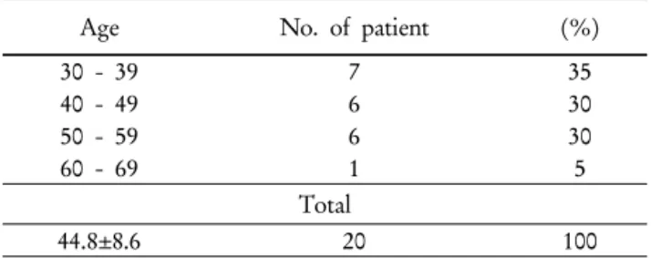
Table 1. Distribution of age in the patients with breast cancer
Age No. of patient (%)
30 - 39 7 35
40 - 49 6 30
50 - 59 6 30
60 - 69 1 5
Total
44.8±8.6 20 100
증, 관상동맥질환, 심근증, 심장판막질환, 호흡곤란환자의 심 장기능 평가 등 여러 심질환에서의 심장기능 특히 좌심실 기 능을 평가하는데 유용하며 심박출계수가 가장 유용한 지표로 사용되고 있다.2) 항암 화학요법 치료 시 안스라사이클린계열 이나 독소루비신과 에피루비신 같은 항암제가 심근증, 울혈성 심부전의 심장 손상 증상을8,10-12) 미리 예측 할 수 있는 검사 법 중 하나이다.6,7) 이외 검사법으로는 환자에게 고통을 주지 않고 방사능의 피폭이 없는 간편한 검사 방법으로 심초음파 가 있지만 광범위하게 사용되는 방법임에도 불구하고 좌심실 용적과 심박출계수 측정의 재현성은 떨어진다고 알려져 있다. 3차원 자기공명영상은 좌심실 용적 측정의 금과옥조검사이 지만 비싼 경비와 제한된 기술 때문에 많이 사용되지 않는다.
항암 화학요법 치료로 인한 심근증의 증상은 약제를 중지 하면 회복이 가능하며 그 위험도가 항암제의 용량과 비례한
다.11,12) 재현성이 좋은 게이트 심장 혈액 풀 스캔으로 추적 검
사를 통하여 항암제의 계속적인 투여와 용량을 결정하게 되 며8) 심박출계수의 정확도와 높은 재현성이 요구된다. 심박출 계수는 심실 내 시간 방사능 곡선으로부터 구할 수 있으며, 이를 위하여 좌심실과 배후 방사능 관심영역을 정해야 하는 데 올바른 관심영역의 설정은 심박출계수의 정확도와 밀접한 관련이 있다. 특히 배후 방사능영역 설정시 간의 좌엽, 비장, 위, 대동맥 등이 배후 방사능 영역에 포함되어 배후 방사능치 가 높게 측정될 경우 심박출계수가 과대평가될 수 있다. 본 연구에서는 배후 방사능 관심영역에 심장주위 장기의 산란선 영향으로 인해 심박출계수가 과대평가 될 수 있는 요인들을 최소화할 수 있도록 영상의 획득 방법을 변형하여 정확성에 초점을 두어 기술을 하고자 한다.
대상 및 방법
1. 대상
2007년 9월부터 10월까지 세브란스 병원에서 유방암 절제 술이나 생검으로 확진된 원발성 유방암 환자로 각 병기에 따 라 수술 전 혹은 수술 후에 항암화학요법 치료를 병행하고자 하는 환자 20명를 대상으로 하였다. 대상 환자의 연령은 33세 에서 63세 사이에 분포하였고 평균 연령은 44.8±8.6세였다.
모든 대상 환자는 과거력상 심장병 또는 고혈압이 없고 정상 이학적 소견 및 심전도 소견이었다(Table 1).
2. 검사방법
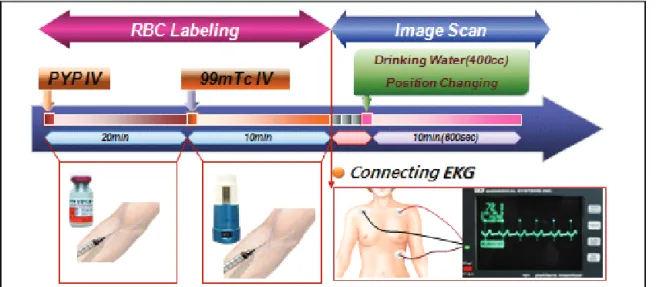
대상군에 in vivo method로 표지된 99mTc-RBC 370 MBq (10 mCi)를 정맥 주사한 뒤 10분 후 안정상태에서 심전도를 부착한 후에 앙와위에서 스캔을 시행했다. 스캔은 저 에너지 고해상력 조준기(collimator)을 사용하였으며, 심실중격이 분명히 보이고, 좌심실과 우심실의 분리가 잘되는 최적 중격 상이 될 수 있도록 35~45도의 좌전사위상에서 single-head gamma camera (Argus ADAC/Philips; Milpitas, CA)를 이용 하여 영상을 얻었고, 심전도를 연결시켜 각 심박주기의 R파 를 기준으로 얻은 영상자료를 PegasysTM ultra computer에 수 록 분석하였다.
자료획득방식은 심전도상의 R파를 시작신호로 사용하여 R-R간격 내에 24개의 단위영상을 얻어 서로 상응하는 단위 영상을 정해진 시간이나 계수치를 얻을 때까지 더하여 정적 영상을 얻는 다중게이트 획득방식(multiple gated acquisition mode)을 사용했으며 영상은 가로, 세로 64×64 행렬방식 및 4×4 mm 크기의 화소(pixel)가 설정되었다. 한 주기를 24화면 으로 나눈 단위영상을 얻어 총 600초 동안 촬영하였고 자료 획득 전에 20개의 심박주기 동안의 심전도를 분석하여 적절 한 심박주기와 이에 대한 10%의 식별영역(window)을 정하 여 이에 해당되는 심박주기의 영상만을 획득하였다. 기존의 방법으로 영상을 획득한 후 360 ml의 물을 먹이고 오른쪽 팔을 머리 위로 거상시키고 좌측 굴곡자세(left flexion po- sition)로 앙와위에서 이와 같이 환자 자세를 변형시키고 이 외 다른 조건들은 동일하게 하여 영상을 다시 한 번 획득하였 다(Fig 1, 2).
3. 분석방법
PegasysTM ultra computer에서 Gated analysis, Ver. 3.40를 이용하여 수록된 두 가지의 방법으로 영상을 획득한 각 단위
Fig. 1. Red blood cells were labeled in vivo by intravenously injecting unlabeled stannous pyrophosphate, followed 20 minutes later
by 10-25 mCi of technetium-99m as pertechnetate. After 10 minutes later by ECG gating and with conventional method and acquired with modified method (left lateral flexion position, 360 mL of drinking water).Fig. 2. (A) Right arm uprising and left lateral flexion position. This position is
characterized by the slight curving of the body to the left. (B and C) Camera angle is obtained at an angle ~45° less than the “best septal” view and used for 10~15° caudal-cephalic tilt to help separate the ventricles from the atria.BKG EDV
ESV EF EDV
−
= −
count background BKG
volume systolic end
ESV
volume diastolic end
EDV
fraction ejection EF
: : : :
−
−
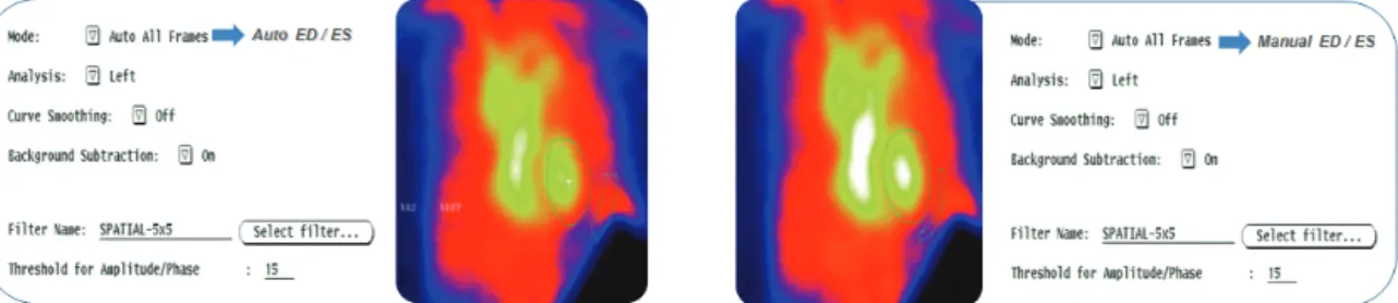
영상 모두에서 컴퓨터 자동 관심영역 설정 모드로3) 좌심실 관심영역과 배후방사능 관심영역이 설정되었다. 배후방사능 치를 보정한 좌심실 영역 내의 방사능치를 이용하여 좌심실 의 시간-방사선곡선 즉 좌심실 용적곡선을 그려 좌심실의 심 박출계수를 측정하게 되는데, 다음 공식에 의해 산출하였다.1)
이렇게 하여 자동관심영역 설정 모드에서 산출한 심박출 계수와 배후방사능 관심영역의 계수치는 객관화를 위해 1년 이상의 경험을 가진 방사선사가 각각 5회 반복 측정하여 서로 비교 평가하였다(Fig. 3). 모든 통계적 숫자는 평균±표 준편차로 표시하였다. 각 군간의 통계적 처리는 SPSS, Ver.
12를 이용하여 paired t-test를 시행하였으며 p-value는 0.05 이하를 통계적으로 의미있는 것으로 하였다.
결 과
영상 획득 후 컴퓨터 자동 관심영역 설정 모드를 이용하여 분석한 결과로 기존의 검사방법에서 좌심실 배후방사능 관심
핵의학기술 제12권 제1호 2008
Fig. 3. ROIs analysis was compared using auto ED/ES and manual ED/ES mode.
Fig. 4. Computer Tomography (CT) was compared as variation of patient position (A) and dinking water (360 ml) or not (B) with
Gated Cardiac Blood Pool scan (GCBP) (C, D). (A-1, A-2) CT images showed anatomical variation by changing patient position;A-1: coronal plane, A-2: axial plane. (C) GCBP images showed variation of spleen (arrow). (B-1, B-2) CT images showed anatomical variation by dinking water (360 ml) or not; B-1: coronal plane, B-2: axial plane. Circle (yellow) showed expansion of stomach. These variation showed in GCBP (D; blue arrow: stomach, pink arrow: spleen).
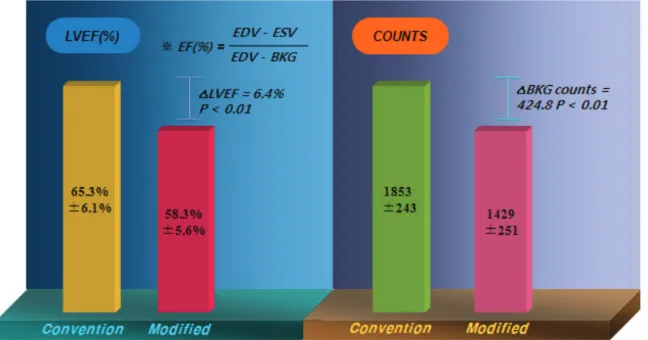
영역 계수가 1853±243이였고, 변형방법에서는 1429±251로 배후방사능 관심영역 계수가 유의하게 감소하였다(p<0.01).
또한 좌심실의 심박출계수는 기존 앙와위(supine)자세로 검 사한 방법에서 65.3%±6.1%이였고, 변형 방법인 소량의 물과 오른쪽 팔을 거상시키고 좌측굴곡시킨 자세로 검사한 방법에 서는 58.3%±5.6%으로 유의하게 감소하였다(p<0.01). 기존의 방법에서 획득한 영상과 변형 방법에서의 획득된 영상을 비교하였을 때 변형방법에서 위(stomach)에 모여 있는 free technetium이 물에 의해 감소되었고, 물에 의해 팽창된 위와
환자 자세의 변형으로 인해 비장의 위치가 외측하부로 변화 되어 배후 방사능 관심영역에서 제외됨을 확인하였다(Fig.
4,5).
고 찰
심장질환 환자에서 좌심실 기능의 평가는 그 환자의 진단, 치료 및 예후판정에 중요하다. 이러한 좌심실 기능분석의 지 표로는 심박출량, 1회 심박동량, 박출압, 좌심실의 확장기말
Fig. 5. Results of GCBP: LVEF (%) was statistically decrease in all patients modified with patient position and some dinking water
compared with the control group (65.3%±6.1% Vs 58.3±5.6%, p<0.01). Also, counts was decrease too (1853±243 Vs 1429±251,p<0.01).
Table 2. LVEF(%) and BKG countsin conventional and modified method
No. Conventional method Modifid method Defference
p-value
LVEF (%) 65.3±6.1 58.3±5.6 6.4±1.9 <0.01
BKG 1853.4±243.7 1429.8±251.0 424.1±201.6 <0.01
압 또는 확장기말 용적 및 심박출계수 등이 있으며 이중 심박 출계수가 가장 흔히 이용되는 지표이다.6) 이러한 검사법으로 는 X-선 심장영화조영술과 심초음파가 있으나 앞서 지적한 바와 같이 여러 가지 시행상의 어려운 점과 불완전성 등의 단 점이 있다. 이에 반해 gated cardiac blood pool scan은 1971년 Strauss등의 보고이래 좌심실의 기능을 비침습적으로 검사할 수 있는 간편한 방법으로 알려져 이용되고 있으며 또한 여러 가지 좌심실의 박출능력의 계측뿐 아니라 computer를 이용 하여 심실벽의 운동상을 얻음으로 좌심실벽의 국소적인 또는 전체적인 운동상태를 관찰할 수 있다.4,5)
이러한 gated cardiac blood pool scan은 심근증에 의한 심 실기능장애를 조기진단할 수 있으며 심근증의 치료에 대한 평가나 예후 판정에 유용하다. 안스라사이클린계열이나 독소 루비신과 에피루비신은 효과적인 항암요법제이지만 심근 증을 유발시켜 치명적인 심부전이 생길 수 있어서 사용에 제한이 있다.7,8,10) 특히 독소루비신 치료 1년 내에 종격동 (mediastinum)에 대한 방사선치료를 했거나, 누적용량이 550 mg/m2 이상일 때, 고혈압, 협심증 등의 심장질환이 있을 때
등이 심근증 유발의 위험인자로 생각되고 있다. 이러한 독소 루비신의 부작용은 예측하기 어려워서 550 mg/m2 이하의 누적용량에서도 생길 수 있으며 1000 mg/m2 이상에도 없을 수 있다. 그러므로 임상증상이나 심부전이 오기 전에 심독 성(cardiotoxicity)을 예측할 수 있는 예민한 비침습적인 검사 가 필요하다. 약물투여 전에 좌심박출계수의 기저치를 얻어 야 하며 누적용량이 300 mg/m2와 450 mg/m2가 될 때 반복검 사를 하여야 한다. 좌심실박출계수가 15% 이상 감소하거나 45% 이하일 때는 투여를 중단하여야 한다.6,10) 본 연구에서의 결과를 보면(Table 2), 두 방법 사이의 심박출계수 차이는 최저 4%, 최고 10.5%, 평균 7%±1.9%였고, 좌심실 배후방사 능 관심영역에서의 계수의 차이는 최저 88, 최고 738, 평균 423±201이였다. 심박출계수와 배후방사능 관심영역 계수는 통계적으로 유의하게 감소하게 나타났으며 이는 좌심실의 배 후방사능 관심영역 설정시 위, 비장, 대동맥등이 배후방사능 영역에 포함되어 배후방사능치가 높게 측정되었고 심박출 계수가 과대평가되었음을 나타낸다 하겠다. 변형방법으로 인 해 이러한 과대평가를 감소시킬 수 있었다. 본 연구의 결과를
핵의학기술 제12권 제1호 2008
분석하자면 사용한 방사성의약품은 in vivo method로 표지 된 99mTc-RBC이다. 체내표지법은 표지효율이 약 85% 정도 이며 표지되지 않은 과산화테크네슘이 위, 갑상선 및 세포 간질(interstitium)로 들어가서 표적/배후 비(target to back- ground ratio)가 다른 방법에 비하여 낮은 것이 단점이다.5) 그 러나 방법이 간편하여 반복검사에 적합하다는 장점이 있다.
위에 방사능 섭취가 증가된 것은 적혈구에 표지되지 않은 유리 테크네슘이 많음을 의미한다. 360 ml의 물을 섭취하여 위에 있는 유리 테크네슘을 세척하여 방사능치를 줄일 수 있 었다. 또한 팽창된 위로 인하여 비장의 위치 변화를 관찰 할 수 있었다. 오른쪽 팔을 거상시키고 좌측 굴곡 자세로 간의 좌엽, 비장, 대동맥이 배후방사능영역과 중첩을 피할 수 있었 다. 컴퓨터 단층 촬영의 영상을 보면 기존 자세와 변형 자세 에서의 간의 좌엽 부분이 변형자세에서 하부쪽으로 변화되어 짐을 알 수 있었다. 또한 비장 및 대동맥 또한 외측하부 그리 고 내측으로 각각 위치변동되어 나타났다.
심박출계수 외에 게이트 심장 혈액 풀 스캔으로 측정 가능 한 심수축기 및 심이완기지표의 정량적 분석은 심질환의 특 성을 이해하는데 아주 중요한 지표가 되어주지만 본 연구에 서는 유방암 환자들에게 있어 항암제의 심장독성에 따른 심 박출계수들의 변화를 본 것이므로 이를 계측하여 평가되지 않았다. 또한 수축률이나 이완율을 정확히 측정하기 위해서 는 최소한 30개 이상의 단위영상을 얻어야 한다고 알려 있지 만 검사 시간이 많이 소요되는 점 때문에 부득이 24개의 단위 영상으로 시행하였으나 이 점은 앞으로 더 많은 영상을 얻도 록 고려되어야 할 것이다. 또한 X-선 심장영화촬영술과 심초 음파 그리고 SPECT와의 심박출계수9)와 다른 심지표간의 연 관관계를 비교 평가하지 못하였으며 앞으로의 논의에서 본 연구 방법에 따른 일치도의 평가가 필요할 것으로 생각된다.
이런 모든 결과들이 중요한 의미를 가지기에는 스캔의 정 확성이나 재현성, 각 심기능지표들에 대한 정확한 생리학적 이해 등의 여러 가지 문제들이 남아 있긴 하지만 심박출계수 측정시 심장주위 장기인 간의 좌엽, 위, 비장 등으로부터 나 오는 산란선의 영향으로 인해 배후 방사능치가 증가하여 과 대평가되는 것을 변형방법을 통하여 산란선의 영향을 최소화 하고 좀 더 정확한 검사가 이루어질 수 있음을 보여 주었다.
결 론
항암화학요법시 심장독성의 부작용을 가지고 있는 항암제
를 사용할 때 gated cardiac blood pool scan에 의한 좌심실계 수 평가의 심장주위 장기 산란선의 영향을 최소화하여 과대 평가를 방지하고자 항암화학요법을 시행받고 있는 유방암 환자를 대상으로 gated cardiac blood pool scan을 시행하고 다름과 같은 결과를 얻었다.
기존의 방법보다 자세 변형과 소량의 물을 투여한 변형 방법에서 심장 주위 관심영역 설정시 간의 좌엽이나 비장 및 위의 일부분이 제외됨으로써 배후 방사능 계수치가 감소하였 고, 그 결과로 심박출계수 또한 감소하였다. 요약하면 이 연 구는 심장 기능의 지표인 심박출 계수의 과대평가되는 것을 최소화는 데에 도움이 될 수 있을 것으로 사료된다.
REFERENCES
1. Wackers, et. al. Multiple Gated Cardiac Blood Pool Imaging for Left Ventricular Ejection Fraction: Validation of the technique and assessment of variability. Am J Cardiol 1979;43:1159-1166.
2. Michael V. Green, et al. Ejection Fraction by count rate from gated images. J Nucl Med 1978;19:880-883.
3. Johan H. C. Reiver, et al. Clinical Validation of Fully Automated Computation of Ejection Fraction from Gated Equilibrium Blood- Pool Scintigrams. J Nucl Med 1983;24:1099-1107.
4. Chang Woon Choi, et al. Analysis of Left Ventricular Diastolic Function in coronary artery disease with gated blood pool scan.
Korean J Nucl Med 1986;20(2):39-45.
5. Jae Hwang Kang, et al. Analysis of Left Ventricular Functional parameters in normal korean subjects by ECG gated blood pool scan. Korean J Nucl Med 1994;28(1):52-61.
6. Seok Gun Park. Assessment of Ventricular Function using Gated Blood Pool scan and Gated Blood Pool SPECT. Korean J Nucl
Med 2005;39(2):100-106.
7. Sonja Parmentier, et al. Assessment of Left Ventricular diastolic function in patients receiving anthracycline therapy. Eur J Nucl
Med 1988:13:563-567.
8. Charles L. shapiro, Abram recht. side effects of adjuvant treat- ment of breast cancer. N Engl J Med 2001;344(26):1997-2008.
9. Bartlett ML, Srinivasan G, Barker WC, Kitsiou MN, Dilsizian V, Bacharach SL. Left ventricular ejection fraction: Comparison of results from planar and SPECT gated blood-pool studies. J Nucl
Med 1996;37:1795-9.
10. Jones LW, Haykowsky MJ, Swartz JJ, et al. Early breast cancer therapy and cardiovascular injury. J Am Coll Cardiol 50:1435- 1441, 2007.
11. Cardinale D, Sandri M, Colombo A, et al. Prognostic value of troponin I in cardiac risk stratification of cancer patients under- going high-dose chemotherapy. Circulation 109:2749-2754, 2004.
12. Kalay N, Basar E, Ozdogru I, et al. Protective effects of carvedilol against anthracycline-induced cardiomyopathy. J Am
Coll Cardiol 48:2258-2262, 2006.
13. A Konstam, et al. Use of equilibrium (gated) radionuclide ventriculography to quantitate left ventricular output in patients with and without left-sided valvular regurgitation, Circulation 64;
578-585, 1981.