2
조선대학교 치의학전문대학원 구강악안면외과학교실
이 성 석, 김 수 관, 오 지 수, 유 재 식, 김 원 기, 양 정 은, 임 형 섭
Comparative inorganic analysis of the mesiodens and the 3rd molar as the
autogenous tooth graft materials
Comparative inorganic analysis of the mesiodens and the 3rd molar as the autogenous tooth graft materials
Department of Oral and Maxillofacial Surgery, School of Dentistry, Chosun University Sung-Suk Lee, Su-Gwan Kim, Ji-Su Oh, Jae-Seek You, Won-Gi Kim, Jung-Eun Yang, Hyoung-Sup Lim Objective: The aim is to evaluate the potential of the mesiodens through the comparative inorganic analysis of 3rd molar teeth prior to clinical study.
Material and methods: The extracted mesiodens and the 3rd molar teeth were prepared. The teeth are prepared as in the process of the autogenous tooth bone graft. Scanning electron microscopy (SEM), energy dispersive X-ray spectroscopy (EDS), X-ray diffraction (XRD) analysis was performed for inorganic analysis.
Results: Rough and porous surfaces were observed in all materials in SEM analysis. Ca/P ratio of mesiodens was 1.55 and the 3rd molar was 1.22 in EDS analysis. XRD analysis shows that the 3 main peaks position were similar. This means that the graft materials are very similar to that of the crystallinity.
Conclusions: The mesiodens and the 3rd molar teeth are very similar to the inorganic component. These results provide the reasonable rationale that mesiodens can be used as autogenous tooth bone graft in a clinic.
Key words : Bone graft, Dental implant, Teeth ABSTRACT
Corresponding Author Su-Gwan Kim, DDS, PhD
Department of Oral and Maxillofacial Surgery, School of Dentistry, Chosun University, 375, SeoSukDong, DongGu, GwangJu City, Republic of Korea.
Phone : 82-62-220-3819, Fax : 82-62-228-7316, E-mail: sgckim@chosun.ac.kr
Acknowledgment
This study was supported by research fund from Chosun University, 2016.
ar as the autogenous tooth graft materials
Ⅰ. Introduction
The autogenous bone graft had been used for the reconstruction of maxillofacial hard tissue defect, but the disadvantage such as morbidity of donor site1). The development of xenogeneic bone and synthetic bone have been made in order to overcome this limitation, but the problem such as the delay of the bone remodeling, immune rejection was raised1. Thus, recently, the research about the autogenous teeth graft material using extracted tooth was actively performed and the clinically good result was reported in many researches1, 2). Also, the concern with the autogenous teeth graft material has been growing, patients try to utilize extracted tooth as the autogenous teeth graft material. The autogenous teeth graft material is made by mainly using the 3rd molar, sound tooth which is extracted with the periodontal problem, extracted premolar for the orthodontic treatment. Kim3)reported the good bone revision by using the autogenous teeth graft material using the premolar in the bone graft for implant surgery. Thus, the author paid attention to the possibility as the autogenous teeth graft material of the supernumerary tooth, not the 3rd molar or other teeth.
The generation of the supernumerary tooth was not clearly clarified but the insistences that extra tooth bud was formed by growth of extra dental lamina or superactivity of dental lamina are widely supported4). The supernumerary teeth have the form which is similar to the neighboring natural teeth. Mesiodens are the most common supernumerary teeth. It can occur in 0.15% to 1.9% of the population and show up individually or numerously5). In addition, mesiodens can change both position and eruption path of the
permanent incisor clinically then cause malocclusion6). Once a mesiodens has been diagnosed, the result that it is clinically better to extract a tooth during the early mixed dentition stage was reported7).
Thus, this study evaluate possibility as the autogenous teeth graft material of mesiodens by comparing and analyzing the surface structure, the Ca/P ingredient and crystallinity.
Ⅱ. Materials and Methods
We got approval(CUDHIRB-1609-042) of Chosun Dental Hospital Clinical Trial Center Institutional Review Board(IRB). Teeth of the patients before the age of 30 regardless of sex were selected considering teeth extraction period. First, each 5 samples of the mesiodens and the 3rd molars which are extracted from the patients was put into the saline solution and stored in the refrigerator. We did not consider the position of the 3rd molars. Then, the following process8)was gone through to make the autogenous teeth graft material by using extracted teeth(Table 1).
The scanning electron microscope(Scanning Electronic Microscopy, SEM: JSM 840-A, JEOL co., Japan) was used. The sample was examined at 100, 500, 1,500, and 5,000 magnification according to the surface analysis method specified by the Food &
Drug Association safety office “physicochemical characteristic evaluation guideline of the dental graft material”.
Energy Dispersive X-ray Spectrometer EDS(ISIS 310, Oxford, UK) was used to analyze qualitatively, quantitively the components including the calcium and phosphorus of the mesiodens and the 3rd molars. Then
we analyze the average and standard deviation of each 5 pieces.
X-ray Diffraction (X’Pert PRO MRD, PANalytical co., Netherlands) analysis was performed separately to compare and analyze the crystallinity of mesiodens and the 3rd molar. Each graft materials were made with the powdered form for the XRD analysis. XRD analysis was performed with each graft material put into the glass sample case in the Celsius 10-90 degree.
Ⅲ. Results
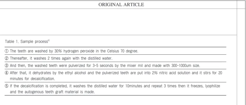
Surface structureThe morphology such as the particle size, surface roughness, pore size could be analyzed by observing the surface of the mesiodens and the 3rd molars via the scanning electron microscope. The particle size and form of graft materials are similar and the 300-1000um uniform-size could be confirmed in the low resolution ( 100). Also, the high density, and the various forms were observed in mesiodens and the 3rd molars. In 500, 1,500, the surface roughness of each graft materials can be observed. There are many micropore, which is dentinal tubule. In 5,000, the irregular micropores of about 10um size can be observed(Fig. 1).
Surface component
In the results of EDS of the mesiodens, the carbon occupied about 31.0% and the oxygen was the most abundant about 51.1%. The calcium and phosphorus atomic ratio (Ca/P) was about 1.55. In the results of EDS of the 3rd molar, carbons were the most abundant about 61.7%, oxygen occupied about 32.6% and the Ca/P ratio was about 1.22. The carbon(C), oxygen(O), phosphorus(P) and calcium(Ca) only existed in all graft materials (Table 2).
In our research, the Ca/P ratio of the mesiodens was high more than the 3rd molar. There is a difference between the organic matter and collagen in the tooth.
The ratio of the carbon of the mesiodens and the 3rd molar shows the big difference in this research. It is considered that the ratio of the organic component of the mesiodens such as the dentin or pulp is smaller than that of the 3rd molar9).
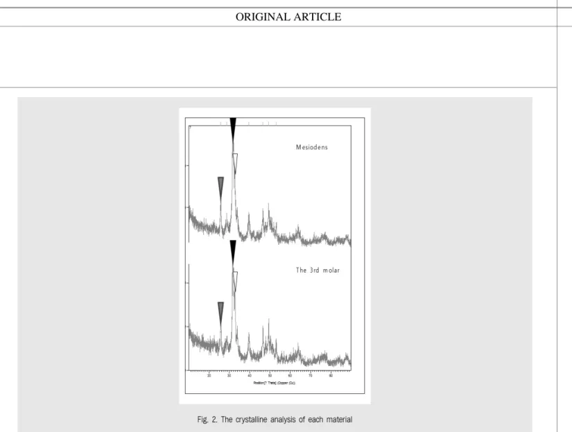
Crystallinity analysis
X-ray diffraction analysis(XRD) was performed separately on each graft materials to analyze crystallinity of the mineral components. The analysis
① The teeth are washed by 30% hydrogen peroxide in the Celsius 70 degree.
② Thereafter, it washes 2 times again with the distilled water.
③ And then, the washed teeth were pulverized for 3-5 seconds by the mixer mil and made with 300-1000um size.
④ After that, it dehydrates by the ethyl alcohol and the pulverized teeth are put into 2% nitric acid solution and it stirs for 20 minutes for decalcification.
⑤ If the decalcification is completed, it washes the distilled water for 10minutes and repeat 3 times then it freezes, lyophilize and the autogenous teeth graft material is made.
Table 1. Sample process8)
ar as the autogenous tooth graft materials
Fig. 1. The SEM analysis of surface of the mesiodens(A) and the 3rd molar(B)
C 31.03 ± 2.00 61.71 ± 2.79
O 51.06 ± 2.30 32.59 ± 1.13
P 6.96 ± 0.60 2.49 ± 0.75
Ca 10.68 ± 1.64 3.15 ± 1.41
Ca/P 1.55 ± 0.14 1.22 ± 0.21
Table 2. EDS Analysis of the components of each group
Element Mesiodens The 3rd molar
At%(mean ± SD) At%(mean ± SD)
was performed between the Celsius 10-90 degrees and displayed the phenomenon that 3 major peaks consistency. This shows that the inorganic component crystallinity of each 2 graft materials is very similar (Fig. 2).
Ⅳ. Discussions
The healing mechanism of the bone graft material is classified into the osteogenesis, osteoinduction, osteoconduction. Autogenous bone graft material has limited harvest volume and cannot avoid the re- absorption and have the second defect and can cause postoperative inconvenience1). Allogeneic bone involves the risk of the transition of the infectious
disease10~12).
The xenogenic bone graft material uses the animal bone of pig, cow and it is easy to purchase. However, there is the risk of infectious disease of the other animals. Also, in the disinfection process, bone reproduction property may be reduced by the deproteinized process13~15). The synthetic bone graft is the artificial material for comprising bone and mainly contains the inorganic component like the calcium phosphate including the hydroxyapatite16). It has excellent biocompatibility and stability but it takes a long time to absorb and it delays bone regeneration speed. Also, it has no osteoinductive properties17, 18).
These products are at risk of immune rejection, infection, etc. and lack the characteristic including the osteogenesis, osteoinduction, and the cost is expensive.
Fig. 2. The crystalline analysis of each material
ar as the autogenous tooth graft materials Thus, Kim1, 19, 20)had been researching on the graft
material that uses the autogenous teeth since 1993.
Extracted tooth in the person is classified as the shipment so it should be discarded. Moreover, the allogenous bone graft is contraindicated in the clinical procedure due to a legal problem. But, its own tooth is processed and can be used in the implant surgery with the consent of the patient. The technique intentionally making the tooth root remain to the alveolar bone for alveolar bone conservation in the past been introduced.
The root rest which is not taken with the infection doesn't cause the special problem in the alveolar bone because the ingredient of the tooth is similar to the that of jaw bone21, 22). If the 3rd molar comes close to the inferior alveolar nerve canal or its extraction is difficult, the long-term safety was proved by performing intentional coronectomy as alternative of extraction23, 24). Based on this kind of the clinical basis, the autogenous tooth is processed for the bone graft material. Recently, the Kim25)reported that autogenous teeth graft material is the most similar to the autogenic bone in comparison with the other bone graft material by analyzing the surface structure and physico- chemical property of the autogenous teeth bone graft material by using the scanning electron microscope, X- ray diffraction analysis, and Ca/P solubility inspection.
Also, there was no immune reaction or foreign body reaction and the bio-compatibility was excellent and there was no risk of infection, the psychological rejection of the patients can be reduced since the bone graft material using the autogenous tooth1).
Mesiodens is a supernumerary tooth located in the maxillary central incisor region and usually do not erupt5). According to the research of Orhan26), one supernumerary tooth showed in the case of 76-86%, 2
supernumerary teeth showed up in 12-23%, and additional supernumerary tooth was reported as less than 1%. In the research of Ray27), mesiodens in the male is twice as frequently as that in the females. The sex difference was reported in the other research. Rajab
& Hamdan28)reported supernumerary tooth affect more male than female with the ratio of 2.2:1. Mesiodens can be classified into cornical shape, incisor shape, and tubercle shape on the basis of their morphology. The conical shape is the commonest among these29, 30). In the several patients, the mesiodens sometimes erupts normally. However, it is usually impacted or conversely positioned. This supernumerary tooth has the abnormal eruption path or it is considered to ectopic eruption31). If the tooth is unerupted, the eruption of the neighboring permanent tooth can be obstructed or the malocclusion such as the diastema, change of the angle of adjacent permanent tooth, crowding can be caused32). Also, it was reported that the mesiodens is rarely related to dentigerous cyst33). For this reason, most dentists and oral, maxillofacial surgery dentist suggests to extract mesiodens in the appropriate time.
We performed SEM, XRD and EDS analysis in order to confirm the possibility as the autogenous tooth graft material. SEM analysis is the method of the morphology figure of the surface structure. SEM can analyze sample material size, a degree of crystallization, micropore and macropore, pore size, surface roughness and porosity. These are closely linked to bioactivity in implanting the graft material.
The degree of crystallinity is defined as the ratio of the calcium phosphate and it plays an important role in the protein absorption, cell adhesion and dissolution of the biomaterial34). Micropore plays a significant role in the penetration and attachment between cell and body fluid
and means the pore structure less than 10um35). Through the uneven surface and micropore observed above research, it is considered that autogenous tooth graft material of the mesiodens forms the surface of the ideal bone graft material.
EDS equipment evaluates the components by analyzing the emitted X-ray from graft material by collision of electron. The EDS equipment has a relatively high energy and penetrates deep into the graft material36). Generally, the inorganic component of the tooth is comprised of the calcium phosphate (the hydroxyapatite(HA), tricalcium phosphate(TCP), octacalcium phosphate(OCP), amorphous calcium phosphate(ACP), and dicalcium phosphate dehydrate(DCPD))37). It is important to understand the relationship between the Ca/P ratio, acidity and solubility due to the various molecular structure of the calcium phosphate. If the Ca/P ratio is smaller than 1, the acidity and solubility is climbing sharply. If the ratio is very low, the absorption becomes fast.
Consequently, graft material doesn't function as scaffold normally. On the contrary, if the Ca/P ratio is high and it becomes close to 1.67 which is HA with the chemical formula, the acidity and solubility reduce.
There is a disadvantage that the water-absorption is so low38). HA is equal to the crystallographic structure of the bone mineral. Bone graft material based on HA generally is considered nonabsorbable material. On the contrary, TCP displays 1.5 of the Ca/P ratio and is the precursor of the bone and is closer to the amorphous structure and it has high osteogenic property in the bone defect. Also, TCP is observed in the high ratio in the newly formed bone25). Saffar39)inserted TCP to the intrabony cystic lesion of 4 patients. They took a biopsy after 10~16 month and observed histologically.
Moreover, they reported that TCP have the property promoting the new bone formation and when the osteogenesis is initiated, TCP simultaneously begins to be actively absorbed. TCP was completely absorbed when the osteogenesis is completed. And, Kim40) reported that the Ca/P ratio of the crown was close to HA in 1.75, and that of root was close to ACP in 1.32.
That is, crown has high rates of HA, root have high rates of ACP. Our research indicates that Ca/P ratio of the mesiodens was close to TCP in 1.55 and the 3rd molar displayed 1.22. Since the ratio of TCP is high, it suggests the possibility of osteogenic property of autogenous tooth graft material.
In this research, the ratio of the carbon oxygen of the mesiodens and the 3rd molar showed the big difference. We suppose that it's because of the organic component of the mesiodens, the ratio which the dentin or pulp occupies, is smaller than that of the 3rd molar.
The X-ray Diffraction(XRD) is the method to investigate the solid crystal structure by analyzing the radiological diffraction pattern of the powdery specimen. Even if the chemical formula of the materials is equal, the crystal structure can be different.
The crystal structure and size can be estimated by X- ray diffraction. The more width of XRD peak is wide, the less the crystallinity is low and the more narrow and steep, the high crystallinity is shown41~44). It is previously reported that main component of tooth ash processed at high temperature is comprised of HA and -TCP19). Meanwhile, autogenous tooth bone graft material contains all the calcium phosphates in 4 stage.
XRD pattern displays the other aspect on the crown and root. That is, the root mainly consisting of the dentin and cementum was the low crystallinity and crown which has high rates of enamel was high
ar as the autogenous tooth graft materials crystallinity. If the crystallinity is high and the particle
size is big, the disintegration is impossible and the osteoconduction property is very low and the depolymerization by the osteoclast cannot be done.
Also the low crystallinity carbonic apatite shows the most excellent bone conduction property45). In the other research including the Kim37), the XRD patterns of the autogenous bone, autogenous tooth graft material and allogenous bone was similar and show low crystallinity. However, the XRD patterns of the xenogeneic bone and synthetic bone displayed narrow and steep similarly. This showed that 2 materials are high crystallinity. In above research, it is difficult to identify whether or not it is high crystallinity. We could know that their crystallinity is similar because the value
of the theta which the peak shows up about the form of the mesiodens, the 3rd molar was consistently observed.
Ⅴ. Conclusion
The possibility of the osteogenic property as the autogenous tooth graft material was confirmed through EDS analysis. crystallinity of these was very similar through the XRD analysis. This means that mesiodens is very similar to the inorganic component of the 3rd molar. The morphology and size of mesiodens is very various, further studies are needed to investigate the clinical application.
1. Kim YK, Kim SG, Byeon JH, et al. Development of a novel bone grafting material using autogenous teeth. Oral Surg Oral Med Oral Pathol Oral Radiol Endod. 2010;109:496-503.
2. Kim YK. The development of new biomaterial for restoration of hard tissue defects. J Korean Dent Assoc. 1998;36:289-295.
3. Kim YK, Lee HJ, Kim KW, Kim SG, Um IW. Guide bone regeneration using autogenous teeth: case reports. J Kor Assoc Oral Maxillofac Surg.
2011;37:142-147.
4. Black G. Supernumerary teeth. Dent Summ.
1909;29:83-110.
5. Mukhopadhyay S. Mesiodens: a clinical and radiographic study in children. J Indian Soc Pedod Prev Dent. 2011;29:34-38.
6. Luten Jr JR. The prevalence of supernumerary teeth in primary and mixed dentitions. J Dent Child (Chic). 1967;34:346-353.
7. Russell KA, Folwarczna MA. Mesiodens-diagnosis and management of a common supernumerary tooth. J Can Dent Assoc. 2003;69:362-367.
8. Tazaki J, Murata M, Yuasa T, et al. Autograft of human tooth and demineralized dentin matrices for bone augmentation. J Ceram Soc JPN.
2010;118:442-445.
9. Fuller BT, Richards MP, Mays S. Stable carbon and nitrogen isotope variations in tooth dentine serial sections from Wharram Percy. J Archaeol Sci.
2003;30:1673-1684.
10. Mulliken JB, Glowacki J, Kaban LB, Folkman J, Murray JE. Use of demineralized allogeneic bone implants for the correction of maxillocraniofacial deformities. Ann Surg. 1981;194:366-372.
11. Goldberg VM, Stevenson S. Natural history of autografts and allografts. Clin Orthop Relat Res.
1987:7-16.
12. Urist M. Bone morphogenetic protein induced bone formation in experimental animals and patients with large bone defects. Cell and Molecular Biology of Vertebrate Hard Tissues, Symposium (ed 18),
136CIBA Foundation, London, England; 1988. p.281.
13. Carmagnola D, Adriaens P, Berglundh T. Healing of human extraction sockets filled with Bio-Oss. Clin Oral Implants Res. 2003;14:137-143.
14. Wenz B, Oesch B, Horst M. Analysis of the risk of transmitting bovine spongiform encephalopathy through bone grafts derived from bovine bone.
Biomaterials. 2001;22:1599-1606.
15. Yeo SI, Park SH, Noh WC, Park JW, Lee JM, Suh JY. A comparative analysis of basic characteristics of several deproteinized bovine bone substitutes. J Korean Acad Periodontol. 2009;39:149-156.
16. Lewandrowski KU, Gresser JD, Wise DL, Trantol DJ.
Bioresorbable bone graft substitutes of different osteoconductivities: a histologic evaluation of osteointegration of poly(propylene glycol-co-fumaric acid)-based cement implants in rats. Biomaterials.
2000;21:757-764.
17. Laurencin CT. Bone graft substitutes: ASTM International; 2003.
18. Sun JS, Lin FH, Hung TY, suang YH, Chang WH, Liu HC. The influence of hydroxyapatite particles on osteoclast cell activities. J Biomed Mater Res.
1999;45:311-321.
19. Kim YK. An experimental study on the tissue reaction of toothash implanted in mandible body of the mature dog. J Korean Assoc Maxillofac Plast Reconstr Surg. 1993;15:129-136.
20. Kim Y, Lee H, Kim S, Um I, Lim S, Kim S.
Analysis of inorganic component and SEM analysis of autogenous teeth bone graft material and histomorphometric analysis after graft. J Korean Acad Implant Dent. 2009;28:1-9.
21. Gongloff RK. Vital root retention. Int J Oral Maxillofac Surg. 1974;3:97-99.
22. Fareed K, Khayat R, Salins P. Vital root retention:
a clinical procedure. J Prosthet Dent. 1989;62:430- 434.
23. Freedman GL. Intentional partial odontectomy:
report of case. J Oral Maxillofac Surg.
1992;50:419-421.
참 고 문 헌
ar as the autogenous tooth graft materials 24. Kim YK. Development of autogenous teeth bone
graft material and clinical evaluation. J Korean Dent Assoc. 2011;49:159-169.
25. Jensen SS, Bornstein MM, Dard M, Bosshardt DD, Buser D. Comparative study of biphasic calcium phosphates with different HA/TCP ratios in mandibular bone defects. A long-term histomorphometric study in minipigs. J Biomed Mater Res B Appl Biomater. 2009;90:171-181.
26. Orhan AI, özer L, Orhan K. Familial occurrence of nonsyndromal multiple supernumerary teeth: a rare condition. Angle Orthod. 2006;76:891-897.
27. Ray D, Bhattacharya B, Sarkar S, Das G. Erupted maxillary conical mesiodens in deciduous dentition in a Bengali girl--a case report. J Indian Soc Pedod Prev Dent. 2005;23:153-155.
28. Rajab LD, Hamdan MA. Supernumerary teeth:
review of the literature and a survey of 152 cases.
Int J Paediatr Dent. 2002;12:244-254.
29. Gallas MM, Garcia A. Retention of permanent incisors by mesiodens: a family affair. Br Dent J.
2000;188:63-64.
30. Fernández Montenegro P, Valmaseda Castell?n E, Berini Ayt?s L, Gay Escoda C. Retrospective study of 145 supernumerary teeth. Med Oral Patol Oral Cir Bucal. 2006;11:339-344.
31. Pakdaman A, Meighani G. Diagnosis and management of supernumerary (mesiodens): a review of the literature. J Dent (Tehran).
2010;7:41-49.
32. Asaumi J, Shibata Y, Yanagi Y, et al. Radiographic examination of mesiodens and their associated complications. Dentomaxillofac Radiol.
2004;33(2):125-127.
33. Dinkar AD, Dawasaz AA, Shenoy S. Dentigerous cyst associated with multiple mesiodens: a case report. J Indian Soc Pedod Prev Dent. 2007;25:56- 59.
34. Berube P, Yang Y, Carnes DL, Stover RE, Boland EJ, Ong JL. The effect of sputtered calcium phosphate coatings of different crystallinity on osteoblast differentiation. J Periodontol.
2005;76:1697-1709.
35. Rohanizadeh R, Padrines M, Bouler JM, Couchourel D, Fortun Y, Daculsi G. Apatite precipitation after incubation of biphasic calcium-phosphate ceramic in various solutions: influence of seed species and proteins. J Biomed Mater Res. 1998;42:530-539.
36. Shibli JA, Marcantonio E, d'Avila S, Guastaldi AC, Marcantonio E, Jr. Analysis of failed commercially pure titanium dental implants: a scanning electron microscopy and energy-dispersive spectrometer x- ray study. J Periodontol. 2005;76:1092-1099.
37. Kim YK, Kim SG, Yun PY, et al. Autogenous teeth used for bone grafting: a comparison with traditional grafting materials. Oral Surg Oral Med Oral Pathol Oral Radiol. 2014;117:e39-45.
38. Vallet-Regi M, Gonz?lez-Calbet JM. Calcium phosphates as substitution of bone tissues.
Progress in Solid State Chemistry. 2004;32:1-31.
39. Saffar JL, Colombier ML, Detienville R. Bone formation in tricalcium phosphate-filled periodontal intrabony lesions. Histological observations in humans. J Periodontol. 1990;61:209-216.
40. Kim YK, Kim SG, Oh JS, et al. Analysis of the inorganic component of autogenous tooth bone graft material. J Nanosci Nanotechnol.
2011;11:7442-7445.
41. Klug HP, Alexander LE. X-ray diffraction procedures: for polycrystalline and amorphous materials. X-Ray Diffraction Procedures: For Polycrystalline and Amorphous Materials, 2nd Edition, by Harold P. Klug, Leroy E. Alexander, Wiley-VCH, 1974;1:992.
42. Tadic D, Epple M. A thorough physicochemical characterisation of 14 calcium phosphate-based bone substitution materials in comparison to natural bone. Biomaterials. 2004;25:987-994.
43. Balasundaram G, Sato M, Webster TJ. Using hydroxyapatite nanoparticles and decreased crystallinity to promote osteoblast adhesion similar to functionalizing with RGD. Biomaterials.
2006;27:2798-2805.
44. Kirik S, Solovyov L, Blokhin A, Yakimov I.
참 고 문 헌
Structures of [Pd (NH3) 2X2] and its chemical transformation in the solid state. Acta Crystallographica Section B: Structural Science.
2000;56:419-425.
45. Lee S. Low crystalline hydroxyl carbonate apatite. J Korean Dental Assoc. 2006;44:524-533.
참 고 문 헌