Res. Plant Dis. 17(3) : 376−380 (2011) © The Korean Society of Plant Pathology
토마토 종자로부터 PCR을 이용한 Pseudomonas syringae pv. tomato의 검출
조정희·임규옥1·이혁인1·예미지1·차재순*
*충북대학교 식물의학과, 1농림수산검역검사본부 식물검역부
Development and Evaluation of PCR-Based Detection for Pseudomonas syrinage pv. tomato in Tomato Seeds
Jung-Hee Cho, Kyu-Ock Yim1, Hyok-In Lee1, Mi-Chi Yea1 and Jae-Soon Cha* Department of Plant Medicine, Chungbuk National University, Cheongju 361-763, Korea
1Animal, Plant and Fisheries Quarantine and Inspection Agency, 433-1 Anyang, 430-016, Korea (Received on November 4, 2011; Revised on November 28, 2011; Accepted on November 28, 2011)
The bacterial speck of tomato caused by Pseudomonas syringae pv. tomato leads to serious economic losses especially on fruits of susceptible genotype. Thus, Pseudomonas syringae pv. tomato is a plant quarantine bacterium in many countries including Korea. In this study, we developed specific PCR assays for detection of the bacterium from tomato seeds. A specific primer set is designed from the hrpZ gene for specific detection of Pseudomonas syringae pv. tomato. A 501 bp PCR product corresponding to hrpZ gene was amplified only form Pseudomonas syringae pv. tomato strains, but no PCR product was amplified from other tomato bacterial pathogens, such as Pseudomonas syringae pv. glycinea, P. syringae pv. maculicola, P. syringae pv.
atropurpurea, P. syringae pv. morsprunorum, and from other P. syringae pathovar strains. The nested-PCR primer set corresponding to an internal fragment of the 501 bp sequence (hrpZ) gine was used to specific detection of Pseudomonas syringae pv. tomato in tomato seed. A 119 bp PCR product using nested PCR primer was highly specific and sensitive to detect low level of Pseudomonas syrigae pv. tomato in tomato seeds.
We believe that the PCR assays developed in this study is very useful to detect Pseudomonas syringae pv.
tomato from the tomato seeds.
Keywords : Bacterial speck, Plant quarantine, Seed-borne disease
Pseudomonas syringae pv. tomato(Pst)는 종자 전염성 병 원세균으로 다양한 토마토 품종에서 bacterial speck병을 일으키고, 토마토의 재배지역에서 중요한 병원균 중 하나 로 알려져 왔다(Cuppels와 Elmgirst, 1999). Pst에 의한 병 징은 과실과 잎에서 다각형의 점무늬가 나타나고, 점무늬 주위에 점차 노란색의 달무리가 생긴다(Zaccardilli 등, 2005).
Pst는 발병 시 토마토의 과실에 피해를 입혀 큰 경제적 손실을 가져오기 때문에 중요한 식물 세균병원균으로 알 려져 왔고, 국내에서도 식물검역 관리병으로 지정되어 관 리되고 있다(Zaccardilli 등, 2002).
지금까지 Pst의 검출을 위해 다양한 방법들이 개발되어
사용되고 있으나, 현재 사용 중인 방법들은 무병종자 및 무병종묘의 인증 프로그램과 식물검역 현장에서 사용했 을 때, 여러 가지 문제점이 있다. Vogel-Bonner-tartate (VBTar)와 KBC 선택배지(Ovod 등, 1997; Schaad 등, 2001)의 경우 특별한 기기를 필요로 하지 않고 간단하게 검출할 수 있는 장점이 있지만, 병원균의 콜로니를 확인 할 때까지 많은 시간이 필요하며, 부생균과 Pst의 콜로니 를 구별하기 어려운 문제점들이 있다(Cuppels 등, 1999, 2006). IFAS(Indirect fluorescent antibody staining)과 ELISA(enzyme linked immunsorbent assay)등과 같이 항혈 청을 이용한 방법은 현재까지도 식물검역과 무병종자 및 무병종묘 인증 프로그램에서 많이 사용하고 있지만, 이 방법들은 항체의 교차반응에 의한 거짓 양성반응과 종자 에서 병원세균을 검출하기에는 민감도가 낮은 문제가 있 다(Zaccardelli 등, 2005). Bereswill 등(1994)에 의해 개발
*Corresponding author
Phone)+82-43-261-2554, Fax) +82-43-271-4414 Email) [email protected]
http://dx.doi.org/10.5423/RPD.2011.17.3.376 Note Open Access
된 세균독소인 coronatine 생합성에 관련 유전자인 cf1 염 기서열에서 제작한 프라이머를 이용한 PCR 방법의 경우 coronatine은 Pst뿐만 아니라 Pseudomonas syringae pv.
glycinea, P. syringae pv. maculicola, P. syringae pv.
atropurpurea, P. syringae pv. morsprunorum 등과 같이 토 마토에 병을 일으키는 다른 병원세균도 합성하는 독소이 므로 Pst만을 특이적으로 검출하는 방법으로는 적합하지 않다(Palmer와 Bender, 1993). Manceau와 Horvais(1997)에 의해 개발된 Pst의 16S-23S rDNA 영역에서 디자인한 프 라이머를 이용하는 PCR방법의 경우 Pst의 16S-23S rDNA 영역이 다른 P. syringae 균주들과 거의 차이가 나지 않 기 때문에 Pst 검출만을 위한 방법으로 사용되기는 어렵 다. 본 연구에서는 새로운 PCR 프라이머를 이용하여 기 존의 방법보다 더 빠르고, 특이성이 높으며, 민감한 검출 방법을 개발하고자 하였다. PCR 프라이머는 Pst hrp pathogenicity island의 내부에서 hrpZ 유전자 부분을 이용 하여 디자인 하였으며, PCR의 검출 민감도를 높이기 위 해 1st PCR 산물의 내부 영역에서 nested PCR 프라이머 를 개발하였다. 또한 검출방법을 단순화하기 위해 토마토 종자 추출물을 직접 PCR에 사용하였다.
PCR 프라이머의 특이성 및 민감도. 본 연구에 사용한 균주는 Table 1과 같으며, 각각 한국농업미생물자원센타 (Korean Agricultural Culture Collection, Korea), 한국미생 물자원센타(Korean Collection for Type Cultures, Korea),
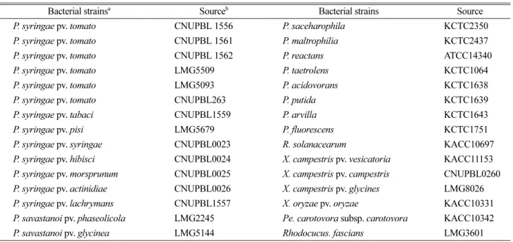
벨기에 BCCM-LMG(Belgian Co-ordinated Collections of Micro-organisms, Belgium)에서 분양 받았다. 또한 일부는 충북대학교 세균병학실험실 수집균주도 사용되었다. 본 연구에서 Pst의 검출을 위해 개발된 프라이머는 NCBI에 서 보고된 Pst DC3000균주의 hrp pathogenicity island (AF232004.3)의 염기서열 내부에서 1113 bp 크기의 hrpZ 유 전자 부분을 blast를 통해 확인 후 Primer3 프로그램을 이 용하여 증폭산물이 501 bp 크기를 가지는 Pst-JH-F(5'-RGY TRA TYY RYG RAA AGY TC-3')와 Pst-JH-R(5'-CRT CGR YYT CGR GRT TTC YY-3') 1st PCR 프라이머를 선 발하였다. PCR검출의 민감도를 높이기 위해서 1st PCR의 증폭 산물의 내부의 염기서열로부터 Pst-JH-F-ne(5'-RCA AGR CCC RGY YCC CYA CY-3')와 Pst-JH-R-ne(5'-TTG RYY AAY GRC RYC RAR AG-3')의 nested PCR용 프라 이머를 디자인하였다. PCR 프라이머들의 특이성을 확인 하기 위해서 Cho 등(2011)의 방법에 의해서 Pst균주 6개 와 Pseudomonas spp., 그리고 주요 식물세균병원균들의 DNA와 프라이머 Pst-JH-F/R을 이용하여 PCR을 수행하 였다. PCR 실험결과, 오직 Pst 균주들에서만 501 bp 크기 의 밴드가 증폭되는 것을 확인하였고, 다른 균주들에서는 어떤 DNA밴드도 검출되지 않았다(Fig. 1A). 종자에 낮은 농도로 감염되어 있는 Pst의 경우 일반 PCR 검출방법으 로 검출이 불가능하기 때문에 민감도를 높이기 위해서 1st PCR 증폭산물의 내부 영역에서 nested PCR 프라이머인
Table 1. Bacterial strains used in this study
Bacterial strainsa Sourceb Bacterial strains Source
P. syringae pv. tomato CNUPBL 1556 P. saceharophila KCTC2350
P. syringae pv. tomato CNUPBL 1561 P. maltrophilia KCTC2437
P. syringae pv. tomato CNUPBL 1562 P. reactans ATCC14340
P. syringae pv. tomato LMG5509 P. taetrolens KCTC1064
P. syringae pv. tomato LMG5093 P. acidovorans KCTC1638
P. syringae pv. tomato CNUPBL263 P. putida KCTC1639
P. syringae pv. tabaci CNUPBL1559 P. arvilla KCTC1643
P. syringae pv. pisi LMG5679 P. fluorescens KCTC1751
P. syringae pv. syringae CNUPBL0023 R. solanacearum KACC10697 P. syringae pv. hibisci CNUPBL0024 X. campestris pv. vesicatoria KACC11153 P. syringae pv. morsprunum CNUPBL0025 X. campestris pv. campestris CNUPBL0260 P. syringae pv. actinidiae CNUPBL0026 X. campestris pv. glycines LMG8026 P. syringae pv. lachrymans CNUPBL1557 X. oryzae pv. oryzae KACC10331 P. savastanoi pv. phaseolicola LMG2245 Pe. carotovora subsp. carotovora KACC10342 P. savastanoi pv. glycinea LMG5144 Rhodocucus. fascians LMG3601
a P: Psuedomonas, R: Ralstonia, X: Xanthomonas, Pe: Pectobacterium
b LMG: Belgian Co-ordinated Collections of Micro-organisms, KACC: Korean Agricultural Culture Collection, KCTC, Korean Collection for Type Cultures: CNUPBL, Chungbuk National University Plant Bacteriology Lab.
Pst-JH-F/R-ne을 선발하였다. 선발된 nested PCR 프라이 머의 특이성을 확인하기 위한 PCR에서 Pst 균주들에서 만 119 bp 크기의 밴드가 증폭되는 것을 확인하였고, 다 른 균주에서는 어떤 DNA도 증폭되지 않았다(Fig. 1B).
개발된 프라이머가 토마토 DNA를 주형으로 DNA를 증 폭할 수 있는지 여부를 확인하기 위해서 2종류의 토마토 종자, 2종류의 고추 종자, 1종류의 알팔파 종자에서 DNA 를 추출하여 동일한 PCR을 수행하였다. 실험결과, 모든 토마토 종자의 DNA에서 어떤 특이적인 DNA도 증폭되 지 않았다(자료 미제공).
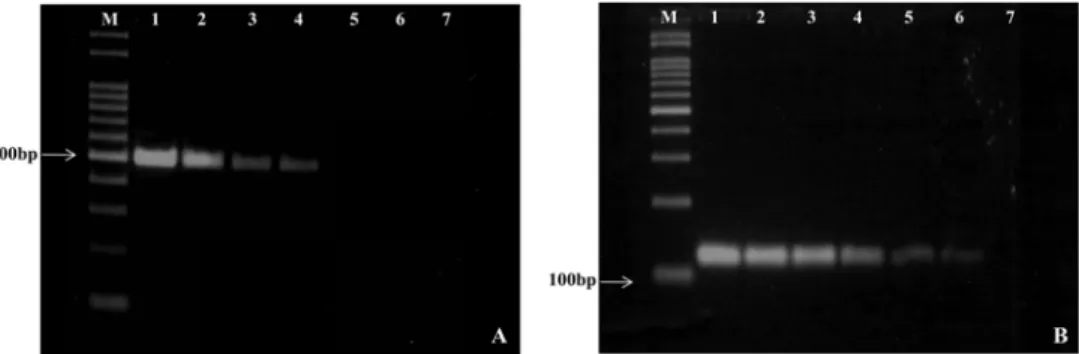
본 연구에서 개발한 PCR방법에 의해 증폭되는 주형 DNA의 농도 한계를 측정하기 위해 DNA를 10 ng 기준으 로 10배씩 희석하여 PCR을 수행하였다. 실험결과, 10 ng의 1000배 희석농도인 0.01 ng에서 501 bp 크기의 특이적 DNA 가 증폭되었다(Fig. 2A). 또한 1st PCR의 증폭산물들을 10 배 희석 후 1 µl을 사용하여 nested PCR을 수행했을 때 1st PCR의 증폭 최종농도보다 100배 더 희석한 시료에서도 특이적 DNA가 증폭되었다(Fig. 2B).
토마토 종자에서 병원균 검출. Pst에 농도 별로 인공오 염 시킨 토마토종자에서 DNA의 추출과정 없이 PCR을 수행하기 위해서 Cho 등(2010)의 방법을 이용하여 종자 추출물을 제조하였다. 토마토종자의 추출물을 이용하여 기존의 Pst의 검출방법으로 사용되었던 반선택배지(KBC) 의 경우 배양 72시간 후에 조사하였을 때, Pst의 전형적
인 콜로니가 배지상에서 나타났고, 104 CFU/ml의 접종농 도까지 검출이 가능하였다(Table 2). 접종농도 103 CFU/ml 미만에서는 반선택배지에 부생성 세균의 생장으로 인해 정확하게 Pst의 콜로니를 구별하기는 어려웠다. 혈청학적 검출방법인 ELISA의 경우 인공오염 된 토마토 종자에서 병원균 접종농도 103CFU/ml까지 양성반응을 확인하였다 (Table 2). 본 연구에서 개발된 PCR 프라이머를 이용하여 인공오염 시킨 토마토 종자에서 PCR을 수행한 결과, 1st PCR의 경우 103 CFU/ml까지 특이적 DNA가 증폭되었다.
1st PCR의 증폭산물을 10배 희석하여 사용한 nested PCR 에서 접종농도 10 CFU/ml까지 특이적 DNA가 증폭되었 다(Table 2). 이 결과는 nested PCR의 검출 민감도가 반 선택배지, ELISA보다는 100−1,000배 더 민감하고, 1st PCR 에 의한 검출보다 100배 더 검출 민감도가 높다는 것을 의미한다.
종자전염 세균병의 경우 102−103CFU/ml 정도의 낮은 농도로 종자에 존재하며, 적합한 환경조건하에서 매우 빠 르게 증식 및 전반을 통해 심각한 피해를 가져오는 것으 로 보고되었다(Cho 등, 2010). Bacterial speck병을 일으키 는 Pst는 전 세계적으로 토마토를 재배하는 지역에서 경 제적 심각한 피해주는 종자전염세균으로(Cuppels 등, 1999), 아직 국내에서는 발병이 보고되지 않았다(The Korean society of plant pathology, 2009). 오염된 종자에 의한 식 물병의 전파는 아직 병이 발생하지 않는 지역에 새로운 Fig. 1. PCRs were carried out with primers Pst-JH-F, Pst-JH-R and 10 ng of bacterial DNAs (A). Nested-PCRs were carried out with primers Pst-JH-F-ne, Pst-JH-R-ne and 1µl of 10 times dilution of 1st PCR products (B). Lane 1: P. syringae pv. tomato DC3000, 2: P.
syringae pv. tomato AV1, 3: P. syringae pv. tomato Y1, 4: P. syringae pv. tomato LMG5509, 5: P. syringae pv. tomato LMG5093, 6: P.
syringae pv. tomato CBNUPBL 263, 7: P. syringae pv. tabaci, 8: P. syringae pv. pisi, 9: P. syringae pv. syringae, 10: P. syringae pv.
hibisci, 11: P. syringae pv. maculicola, 12: P. syringae pv. actinidiae, 13: P. syringae pv. lachrymans, 14: P. savastanoi pv. phaseolicola, 15: P. savastanoi pv. glycinea, 16: P. saceharophila, 17: P. maltrophilia, 18: P. reactans, 19: P. taetrolens, 20: P. acidovorans, 21: P.
putida, 22: P. arvilla, 23: P. fluorescens, 24: R. solanacearum, 25: X. campestris pv. vesicatoria, 26: X. campestris pv. campestris, 27: X.
campestris pv. glycines, 28: X. oryzae pv. oryzae, 29: P. carotovora subsp. carotovora, 30: Rhodocucus fascians.
병원균이 도입되는 매우 중요한 수단으로 병의 피해를 최 소화하기 위해서는 종자로부터 빠르고 정확한 병원균의 검출방법의 개발이 필요하다(Cho 등, 2011; Kritzman, 1991). 현재까지 사용되는 Pst 검출방법 중 반선택배지의 경우 검출 시간이 최소 3일 이상 걸리고, KBC 배지의 경 우 Pst 뿐만 아니라, 다른 P. syringae 균주들의 검출이 가 능하다는 단점을 가지고 있다(Schaad 등, 2001). ELISA 의 경우 종자에서 검출을 할 때 교차반응에 의해 거짓반 성반응이 일어나고, 검출민감도가 낮아 종자에서의 Pst의 검출이 불가능하다는 단점을 가지고 있다(Cuppels 등, 2006).
본 연구에서 Pst의 특이적인 검출을 위해 Pst의 hrp pathogenicity island 영역에서 프라이머를 제작하여 PCR 에 사용되었다. Pst의 hrp island 영역은 다른 syringae 균 주들과 구별될 수 있는 특이적 염기 서열을 가지고 있다 (Zaccardelli 등, 2005). 또한 hrp-유전자를 이용한 PCR검 출방법은 P. syringae pv. papulans, X. campestris pv.
vesicatoria등 많은 병원세균들에서 신속한고 정확한 검출 을 위해 많이 사용되고 있다(Kerkoud 등, 2002; Leite 등, 1994).
본 연구에서 개발된 PCR방법과 nested PCR방법은 기존 의 PCR 검출방법들이 종자로부터 병원균을 검출할 수 있
을 만큼 민감도가 충분하지 않다는 단점을 개선하였고, 또 한 종자 추출액을 직접 PCR에 이용하는 방법은 기존의 Pst의 검출을 위해 DNA를 추출하여 PCR에 사용하던 방 법들(Berewill 등, 1994; Cuppels와 Anisworth, 1995;
Cuppels, 2006; Leite 등, 1994; Manceau와 Horvais, 1997;
Zaccardelli 등, 2005)에 비해 DNA의 추출로 소비되는 시 간을 절약할 수 있고, DNA의 오염 등에 의해 일어날 수 있는 거짓양성반응을 방지할 수 있다는 장점을 가졌다.
따라서 본 연구에서 개발된 PCR방법은 토마토 종자로부 터 빠르고 정확하게 Pst를 검출하는데 사용될 수 있을 것 으로 생각된다.
요 약
P. syringae pv. tomato는 토마토에서 bacterial speck병 을 일으키는 종자전염 세균으로, 감수성 품종에서 주로 발병하여 경제적으로 큰 손실을 입힌다. 따라서 P. syringae pv. tomato는 한국을 비롯한 많은 나라에서 식물 검역대 상 세균으로 지정하여 관리되고 있다. 본 연구에서 우리 는 토마토 종자로부터 PCR을 이용하여 Pst를 검출할 수 있는 방법을 개발하였다. P. syringae pv. tomato의 hrpZ Fig. 2. Detection sensitivity of PCRs and nested PCRs for P. syringae pv. tomato LMG 5509 PCRs were carried out with primers, Pst- JH-F and Pst-JH-R (A) Lane 1: 1 µl of DNA (10 ng), Lane 2-7: 10-fold serial dilution (2-7) (A). Nested-PCRs were carried out Pst-JH- F-ne and Pst-JH-R-ne (B).
Table 2. Detection of Pseudomonas syringae pv. tomato in the artificially inoculated tomato seed by semi-selective medium, ELISA and PCR assays
Assaya Bacterial concentration(CFU/ml) of inoculumb
108 107 106 105 104 103 102 10
KBCc + + + + + - - -
DAS-ELISA + + + + + + - -
1stPCR + + + + + + - -
Nested PCR + + + + + + + +
a Positive reaction (+), Negative reaction (-).
b The 20 ml of bacterial suspension containing each concentration was used to contaminate 1 g of tomato seeds artificially.
c KBC: King’s medium B cephalexin.
유전자에서 특이적인 프라이머를 개발하였다. 개발된 프 라이머는 P. syringae pv. tomato에서만 501 bp 크기의 특 이적 DNA를 증폭하였으며, P. syringae pv. glycinea, P.
syringae pv. maculicola, P. syringae pv. atropurpurea, P.
syringae pv. morsprunorum와 같은 다른 토마토 세균병원 균과 P. syringae pathovar 균주들에서는 증폭되지 않았다.
Nested PCR 프라이머를 이용한 PCR에서도 오직 P. syringae pv. tomato에서만 119 bp 크기의 특이적 DNA가 증폭되었 고, 토마토 종자에서 P. syringae pv. tomato을 정확하고 민감하게 검출하였다. 본 연구는 현재까지 사용되고 있는 Pst의 검출방법의 민감도를 비교한 최초의 보고로 본 연 구에서 개발된 PCR방법들은 토마토 종자에서 Pst을 검 출하는 매우 유용한 방법으로 생각된다.
Acknowledgement
This work was supported by Animal, Plant and Fisheries Quarantine and Inspection Agency, Republic of Korea.
References
Berewill, S., Bugert, P., Volksch, B., Ullrich, M., Bender, C. L.
and Geider, K. 1994. Identification and relatedness of coronatine-producing Pseudomonas syringae pathovars by PCR analysis and sequence determination of the amplification products. Appl. Environ. Microbiol. 60: 2924−2930.
Cho, J. H., Jeong, M. J., Song, M. J., Yim, K. O., Lee, H. I., Kim, J. H., Baeg, J. H. and Cha, J. S. 2010. Development of PCR primers to detect Pseudomonas savastanoi pv. phaseolicola from the bean seeds. Res. Plant Dis. 16: 129−135. (In Korean) Cho, J. H., Yim, K. O., Lee, H. I., Kim, J. H., Baeg, J. H. and Cha, J. S. 2011. Detection of the causal agent of bacterial wilt, Ralstonia solanacearum in the seeds of solanaceae by PCR.
Res. Plant Dis. 17: 184−190. (In Korean)
Cuppels, D. A. and Ainsworth, T. 1995. Molicular and physiological characterization of Pseudomonas syringae pv.
tomato and Pseudomonas syringae pv. maculicola strains that produce the phytotoxin coronatine. Appl. Environ. Microbiol.
61: 3530−3536.
Cuppels, D. A. and Elmhirst, J. 1999. Disease development and changes in the natural Pseudomonas syringae pv. tomato
populations on field tomato plants. Plant Dis. 83: 759−764 Cuppels, D. A., Louws, F. J. and Ainsworth, T. 2006.
Development and evaluation of PCR-based diagnostic assays for the bacterial speck and bacterial spot pathogens of tomato.
Plant Dis. 90: 451−458.
Kerkoud, M., Manceau, C. and Paulin, J. P. 2002. Rapid diagnosis of Pseudomonas syringae pv. papulans, the causal agent of blister spot of apple, by polymerase chain reaction using specifically designed hrpL gene primers. Phytopathology 92:
1077-1083.
Kritzman, G. 1991. A method for detection of seedborne bacterial diseases in tomato seeds. Phytoparasitica 19: 133−141.
Leite, R. P., Minsavage, G. V., Bonas, U. and Stall, R. E. 1994.
Detection and identification of phytopathogenic Xanthomonas strains by amplification of DNA sequences related to the hrp genes of Xanthmonas campestris pv. vesicatoria. Appl.
Environ. Microbiol. 60: 1068−1077.
Manceau, C. and Horvais, A. 1997. Assessment of genetic diversity among strains of Pseudomonas syringae by PCR- restriction fragment length polymorphism analysis of rRNA operons with special emphasis on P. syringae pv. tomato.
Appl. Environ. Microbiol. 63: 498−505.
Ovod, V., Rhudolph, K. and Krohn, K. 1997. Serological classification of Pseudomonas syringae pathovars based on monoclonal antibodies towards the lipopolysaccharide O- chains. pp. 526−531 Vol. 9: Pseudomonas syringae pathovars and related pathogens. Kluwer Academic Publishers, Boston, USA.
Palmer, D, A. and Bender, C. L. 1993. Effects of environmental and nutritional factors on production of the polyketide phytotoxin coronatine by Pseudomonas syringae pv. glycinea.
Appl. Environ. Microbiol. 59: 1619−1626.
Schaad, N. W., Jones, J. B. and Chun W. 2001. Lavoratory guide for identification of plant pathogenic bacteria, pp. 84−120.
APS press, Minnesota, USA.
The Korean society of plant pathology. 2009. List of plant disease in Korea. 5th ed. The Korean society of plant pathology, Suwon, Korea. pp 151−157. (In Korean)
Zaccardelli, M., Parisi, M., and Giordano, I. 2002. Susceptibility of tomato genotypes to Pseudomonas syringae pv. tomato in the field conditions. J. Plant Pathol. 84: 200
Zaccardelli, M., Spasiano, A., Bazzi, C. and Merighi, M. 2005.
Identificaion and in planta detection of Pseudomonas syringae pv. tomato using PCR amplification of hrpZPst. Eur. J.
Plant Pathol. 111: 85−90.