Original Article
Clinical Characteristics of Polypoidal Choroidal Vasculopathy Associated with Chronic Central Serous Chorioretionopathy
Han Sang Park, In Taek Kim
Department of Ophthalmology, Kyungpook National University Hospital, Daegu, Korea
Typical central serous chorioretinopathy (CSC) is char- acterized by acute-onset serous retinal detachment as well as a benign visual prognosis with the self-resolution of retinal detachment after several months. Chronic CSC, which is defined as a persistent serous retinal detachment or recurrent multifocal detachments, may lead to diffuse decompensation of the retinal pigment epithelium (RPE), gradual multifocal RPE leakage with fluorescein angiog- raphy (FAG), and finally a poor visual outcome due to the atrophic retina and RPE [1-10].
Chronic CSC, which is also known as diffuse retinal pigment epitheliopathy, shows specific retinal changes such as multiple RPE atrophies, RPE hyperpigmentations,
and gravitational atrophic RPE tracts. As the indocya- nine green angiography (ICGA) of the CSC demonstrated several choroidal findings such as choroidal hyperperme- ability, choroidal lobular ischemia and choroidal venous congestion, it is accepted that the pathogenesis of CSC is associated with the dysfunction of choroidal vessels [9-15].
Polypoidal choroidal vasculopathy (PCV) is characterized by a branching vascular network (BVN) with terminal polypoidal dilations. Many studies on PCV have been conducted in Asian countries since the incidence of PCV is relatively high in Asians [16-18]. PCV is reported to originate from inner choroidal vessel abnormalities similar to that of CSC, and PCV also has a specific ICGA finding of CSC, which is known as choroidal hyperpermeability.
It thus appears that the pathogeneses of these two diseases have some common features [19,20]. A history of CSC was actually found to be more prevalent in PCV in a study of the relationship between a past history of CSC and the onset incidences of PCV or typical age-related macular
© 2012 The Korean Ophthalmological Society
This is an Open Access article distributed under the terms of the Creative Commons Attribution Non-Commercial License (http://creativecommons.org/licenses /by-nc/3.0/) which permits unrestricted non-commercial use, distribution, and reproduction in any medium, provided the original work is properly cited.
Received: August 24, 2010 Accepted: March 7, 2011
Corresponding Author: In Taek Kim, MD, PhD. Department of Oph- thalmology, Kyungpook National University Hospital, #200 Dongdeok- ro, Jung-gu, Daegu 700-721, Korea. Tel: 82-53-420-5814, Fax: 82-53-426- 6552, E-mail: [email protected]
Purpose: To investigate the clinical characteristics of polypoidal choroidal vasculopathy (PCV) associated with chronic central serous chorioretinopathy (CSC).
Methods: We retrospectively reviewed the medical records of 246 PCV patients (283 eyes) between July 2004 and August 2009 and investigated the clinical characteristics of the PCV patients who had specific fundus findings of chronic CSC.
Results: Among PCV patients, 13 eyes (4.6%) of 13 PCV patients (5.3%) had fundus findings of chronic CSC. All of the PCV lesions had a solitary polyp located outside the atrophic retina, predominantly in the macular area (84.6%), most showed an exudative pattern (69.2%) and there were a few that showed a hemorrhagic pattern (30.8%). All of the lesions were smaller than 1 disc diameter. Most of the PCV lesions (76.9%) were cured with less than two treatments in a short period of 6.4 ± 1.9 months; however, visual acuity deteriorated (61.5%) or was not changed (30.8%) in most of the cases.
Conclusions: The PCV associated with chronic CSC had several clinical features such as a small exudative retinal lesion with a solitary polyp and frequent involvement of the macular area. Even though there was poor visual outcome due to the atrophic change, all of the PCV lesions were easily resolved in a short period with a simple treatment course and no recurrence.
Key Words: Chronic central serous chorioretinopathy, Indocyanine green angiography, Polypoidal choroidal
vasculopathy
degeneration (AMD). Moreover, specific funduscopic find- ings of CSC, like atrophic RPE tract or focal photocoagu- lation scars, were found more frequently in PCV [21].
We retrospectively review the medical records of pa- tients with PCV associated with chronic CSC and investi- gated the clinical characteristic of these PCV patients.
Materials and Methods
We retrospectively reviewed the medical records of 246 PCV patients, for a total of 283 eyes, who had visited our clinic (Kyungpook National University Hospital) between July 2004 and August 2009. We extracted the PCV patients that had specific fundus findings of chronic CSC and then investigated the clinical characteristics of these PCV pa- tients. All of the PCV patients underwent a comprehensive ophthalmic examination, that included Snellen visual acu- ity, biomicroscopy, fundus photography, FAG, ICGA (HRA system; Heidelberg Engineering, Heidelberg, Germany), and optical coherence tomography (OCT; Stratus OCT or Cirrus OCT, Carl Zeiss Meditec, Dublin, CA, USA).
Chronic CSC was objectively diagnosed based on the presence of specific fundus findings like an atrophic RPE tract, multiple RPE atrophies and an FAG finding showing hypofluorescence due to a window defect or partial hyper- fluorescence due to a leak from diffuse RPE decompensa- tion. We confirmed an atrophic lesion as chronic CSC with the exclusion of AMD or PCV with ICGA in cases with a small leakage area of FAG.
The clinical diagnosis of PCV in older patients was made on the basis of the fundus and ICGA findings. The hemorrhagic or exudative lesion was typically diagnosed as PCV based on the presence of the BVN with single or multiple polyp-like terminal dilations in ICGA and an elevated red-orange finding in the fundus. The diagnosis of PCV also included the presence of polyp-like terminal dilations, which was a distinctive finding of PCV, without identifiable BVN or an elevated red-orange lesion. All of the patients were interviewed on their histories of systemic disease such as hypertension, diabetes mellitus or other retinal diseases that were also evaluated in the other eye of the patient.
Fundus findings were classified as one of two patterns, either as an exudative pattern or a hemorrhagic pattern. In the exudative pattern, serous pigment epithelium detachment (PED) and/or retinal detachment was predominant with little or no hemorrhage. In the hemorrhagic pattern, the hemor- rhagic PED and/or submacular hemorrhage were predominant in the lesion. The polyp location and the polyp configuration were identified with ICGA. The polyp location was evaluated on the basis of the locations of the disc and fovea. There were two types of polyp configurations, the solitary type and the
of the atrophic retina of chronic CSC and whether the polyps were involved in a single area or in multiple areas of the fundus. The number and method of treatments as well as when the lesion state reached a stable condition after treat- ment were also investigated.
Results
Among 283 eyes of 246 PCV patients, 13 eyes (4.6%) of 13 patients (5.3%) (right eye, 9 [69.2%]; left eye, 4 [30.8%]) had specific fundus findings of chronic CSC (Table 1). The mean age of PCV onset was 69.8 ± 6.2 years (range, 55 to 79 years), and 11 (84.6%) of the patients were men. The overall duration of follow-up after PCV onset was 27.0 ± 18.4 months (range, 6 to 64 months). The resolution of the PCV lesion took 6.4 ± 1.9 months (range, 4 to 10 months) through various treatments after PCV onset, and there were no recurrences after the resolution of PCV for 20.0 ± 17.7 months (range, 1 to 55 months). Of these 13 PCV pa- tients, three had hypertension and none of them had diabe- tes. All of the patients were unilaterally affected with PCV, but 11 (84.6%) of the 13 patients also had specific fundus findings of chronic CSC in their fellow eyes.
Eleven polypoidal lesions (84.6%) in ICGA were located in the macular area, which were all within 0.5 disc diam- eters (DD) from the fovea (Fig. 1), and two of the lesions (15.4%) were in the peripapillary area, within 0.5 DD from the margin of the optic disc (Fig. 2). The exudative pattern was found in the fundus in nine of the patients (69.2%), and the hemorrhagic pattern was found in four of the pa- tients (30.8%). Among the four eyes with the hemorrhagic pattern, only two (15.4%) had a definite hemorrhagic PED.
Most of the lesions in the other two eyes were composed of exudative lesions, and there was only a small amount of retinal hemorrhage in the larger area of exudative lesion.
The sizes of all of the lesions, regardless of their pattern,
were less than 1 DD in area, and none of the lesions had
extensive hemorrhage or exudation. All 13 eyes had a soli-
tary polyp configuration. A prominent BVN could not be
found near any of the polyps, even though large choroidal
vessels were easily found in the atrophic area around the
polyp in all of the eyes. The ICGA findings showed that
three of 13 patients had polyps that were involved in mul-
tiple areas of the fundus, and the other 10 patients (76.9%)
had a single area of PCV involvement. In regard to the
location of a polyp compared to the atrophic retinal area,
there were no polyps that were identified within the atro-
phic retina of the chronic CSC lesion or distant from the
atrophic retina. All of the polyps were located around the
boundary of the atrophic retina and were identified within
0.5 DD of the normal retina from the boundary of the atro-
phic retina.
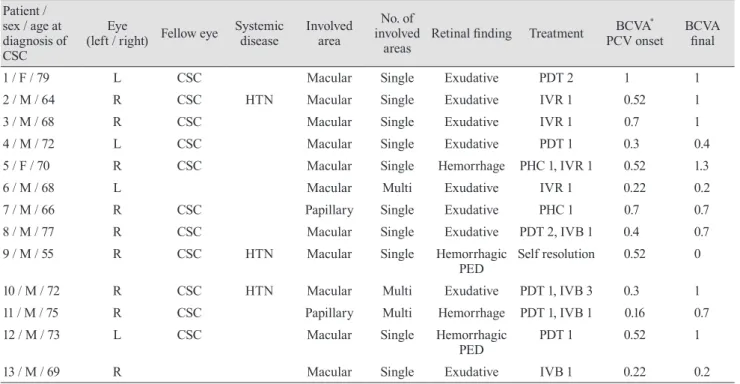
Table 1. Clinical data of patients with polypoidal choroidal vasculopathy Patient /
sex / age at diagnosis of CSC
(left / right) Fellow eye Eye Systemic
disease Involved area
No. of involved
areas Retinal finding Treatment BCVA
*PCV onset BCVA final
1 / F / 79 L CSC Macular Single Exudative PDT 2 1 1
2 / M / 64 R CSC HTN Macular Single Exudative IVR 1 0.52 1
3 / M / 68 R CSC Macular Single Exudative IVR 1 0.7 1
4 / M / 72 L CSC Macular Single Exudative PDT 1 0.3 0.4
5 / F / 70 R CSC Macular Single Hemorrhage PHC 1, IVR 1 0.52 1.3
6 / M / 68 L Macular Multi Exudative IVR 1 0.22 0.2
7 / M / 66 R CSC Papillary Single Exudative PHC 1 0.7 0.7
8 / M / 77 R CSC Macular Single Exudative PDT 2, IVB 1 0.4 0.7
9 / M / 55 R CSC HTN Macular Single Hemorrhagic
PED Self resolution 0.52 0
10 / M / 72 R CSC HTN Macular Multi Exudative PDT 1, IVB 3 0.3 1
11 / M / 75 R CSC Papillary Multi Hemorrhage PDT 1, IVB 1 0.16 0.7
12 / M / 73 L CSC Macular Single Hemorrhagic
PED PDT 1 0.52 1
13 / M / 69 R Macular Single Exudative IVB 1 0.22 0.2
CSC = central serous chorioretinopathy; BCVA = best-corrected visual acuity; PCV = polypoidal choroidal vasculopathy; HTN = hyper- tension; PDT = photodynamic therapy; IVR = intravitreal ranibizumab injection; IVB = intravitreal bevacizumab injection; PHC = con- ventional laser photocoagulation; PED = pigment epithelium detachment.
*