Glaucoma is an optic neuropathy characterized by pro- gressive injury to the optic nerve and retinal nerve fiber.
Since injury due to glaucoma is largely irreversible, early detection and prevention of glaucomatous damage is criti- cal. Examination of the optic nerve head and retinal nerve fiber layer (RNFL), as well as the visual field test, are considered essential for detecting glaucoma. Optical coher- ence tomography (OCT) allows for cross-sectional imag- ing and quantitative analysis of the peripapillary RNFL thickness, which may be useful in detecting patients with glaucoma. Within the last several years, OCT has become an important tool contributing to earlier and more accurate diagnosis of glaucoma. Currently, OCT is the most com-
mon monitoring modality, while spectral domain OCT (SD OCT) is an emerging technology. Considering the price and compatibility of OCT data with that of SD OCT, OCT is expected to be used commonly in the near future [1].
In East Asian countries, the prevalence of myopia is high, with increases in the number of new cases and the severity of the condition [2,3]. The association between myopia and glaucoma is well recognized, and there is an increased prevalence of myopia in patients with ocular hypertension, primary open-angle glaucoma, and normal- tension glaucoma [4-10]. However, optic disc appearances in myopic patients are often difficult to interpret and may mask early glaucomatous damage. Few studies have used RNFL photography in myopic eyes. This may be due to the fact that accurate RNFL measurements can be ob- scured by relatively small amounts of pigment in the reti- nal pigment epithelium in patients with myopia. The visual field test also has limitations in detecting glaucoma; it is less sensitive for early glaucoma, and abnormal results oc- cur frequently in patients with high myopia due to myopic
Original Article
The Effect of Axial Length on the Variability of Stratus Optical Coherence Tomography
Jeong Hun Bae
1, So Young Han
1, Hyunjoong Kim
2, Joon Mo Kim
1, Ki Ho Park
3, Jung Gon Cho
41
Department of Ophthalmology, Kangbuk Samsung Hospital, Sungkyunkwan University School of Medicine, Seoul, Korea
2
Department of Applied Statistics, Yonsei University, Seoul, Korea
3
Department of Ophthalmology, Seoul National University College of Medicine, Seoul, Korea
4
Yebon Eye Clinic, Seoul, Korea
© 2012 The Korean Ophthalmological Society
This is an Open Access article distributed under the terms of the Creative Commons Attribution Non-Commercial License (http://creativecommons.org/licenses /by-nc/3.0/) which permits unrestricted non-commercial use, distribution, and reproduction in any medium, provided the original work is properly cited.
Received: June 20, 2011 Accepted: September 2, 2011
Corresponding Author: Joon Mo Kim, MD. Department of Ophthalmol- ogy, Kangbuk Samsung Hospital, Sungkyunkwan University School of Medicine, #29 Saemunan-ro, Jongno-gu, Seoul 110-746, Korea. Tel: 82-2- 2001-2257, Fax: 82-2-2001-2262, E-mail: [email protected]
Purpose: To evaluate the effect of axial length on the variability of retinal nerve fiber layer (RNFL) thickness measurements using the Stratus optical coherence tomography (OCT) in normal and glaucomatous eyes.
Methods: We measured the RNFL thickness in 474 subjects using the Stratus OCT twice during the same day.
Axial length was measured with the IOLMaster, and refractive error was the absolute value of the spherical equivalent measured with an auto ref-keratometer. Standard deviation in overall mean RNFL thickness was used as the dependent variable to identify significant correlations.
Results: Long axial length affected the variability in the RNFL thickness value by stratus OCT at the temporal quadrant (p = 0.006) and clock-hour sector 9 (p = 0.001). Refractive error also affected the variability of the RNFL thickness value by stratus OCT at the temporal quadrant (p = 0.025) and clock-hour sector 9 (p = 0.024).
Conclusions: It is clinically significant that longer axial length demonstrates greater variability in temporal area as detected by OCT, a measurement which correlates with the preferably damaged position in the myopic glaucoma eye.
Key Words: Axial eye length, Glaucoma, Optical coherence tomography, Variability
degeneration. OCT is commonly used to measure RNFL thickness in a non-invasive manner; therefore, concern has been raised regarding the accuracy and reliability of OCT in myopic eyes. Despite this, OCT is rarely revised by measuring axial length in myopic patients, though it is not clear how this may influence clinical diagnosis. Previous studies in myopic patients have focused on peripapillary RNFL thickness distribution with OCT. The results have been conflicting; in some studies, the degree of myopia was correlated with the mean RNFL thickness measure- ments [11,12]. On the other hand, some studies have found that there is no significant correlation between level of myopia or axial length and RNFL thickness measurement [13]. In this present study, we investigated the effects of re- fractive error and axial length on the variability of stratus OCT measurements.
Materials and Methods
Subjects were recruited from the outpatient glaucoma service of the Department of Ophthalmology, Kangbuk Samsung Hospital. This prospective study looked at 668 eyes in 668 participants. Informed consent was obtained from each subject. This study was performed after ap- proval from the institutional review board and ethics com- mittee of the Kangbuk Samsung Hospital in Seoul, Korea.
All subjects underwent a full medical and ocular history and a detailed ocular examination including measurements of visual acuity, intraocular pressure using the Goldman applanation tonometer, and slit lamp and fundus examina- tions. Axial length was measured using the IOLMaster (Carl Zeiss Meditec, Dublin, CA, USA) as the mean of three measurements. Refractive error was defined as the absolute value of the spherical equivalent measured with an auto ref-keratometer (RK-F1; Canon, Tokyo, Japan).
Normal participants were included if they had a bilateral highest documented intraocular pressure of 20 mmHg, bilateral normal eye examination findings (including those from dilated fundus examinations), and bilateral normal visual field results, defined as pattern standard deviation within 95% of normal limits and a glaucoma hemi-field test result within 99% of normal limits. Subjects with glaucoma included those with diagnosis of open angle glaucoma, defined as optic disc abnormalities consistent with glaucomatous optic neuropathy with or without vi- sual field loss. Other inclusion criteria for both normal and glaucomatous subjects were age of 18 years or older, best- corrected visual acuity of 20 / 40 or better, no history of ocular or neurologic disease or surgery that may produce test results or visual changes that might confound recogni- tion of a test result due solely to glaucoma, and no history of amblyopia.
Optical coherence tomography technique
Subjects were scanned twice with a Stratus OCT (soft- ware ver. 4.0.1, Carl Zeiss Meditec) during the same day with short breaks between each measurement. The OCT scan was performed by a single technician through a dilat- ed pupil. Fast RNFL thickness protocols were performed with internal fixation. The operator applied artificial tears (Hyalein; Santen Pharmaceuticals, Osaka, Japan) before the subsequent scan.
The selected fundus image was sufficiently visible to distinguish the optic disc and the scanning circle. Images with poor scan quality, decentration, poor focus, low anal- ysis confidence, or low signal strength (less than 6) were excluded. For subjects who had both eyes scanned, one eye was randomly chosen for analysis. The analysis algorithm reported 17 RNFL thickness values: mean RNFL thickness around the entire circumference, average thickness within the four quadrants (temporal, superior, nasal, and inferior), and average thickness in each of 12 clock-hour sectors pro- vided, where clock-hours one to five represented the nasal clock hours and clock-hours seven to 12 represented the temporal ones. Left optic disc areas were considered to be mirror images of the right ones. In all tables, the measured areas of the nasal region were named clockwise from 12 to 6, and those of the temporal region from 6 to 12.
We used standard deviation (SD) to determine the repro- ducibility of RNFL thickness. The SD is a measure of the variability of a data set or a probability distribution. Each patient’s SD values were based on two measurements of RNFL thickness. A low SD indicated that the data points tended to be very close to the same value, while a high SD indicated that the data were spread out over a large range of values. We analyzed the reliability between measure- ments using the intraclass correlation coefficient (ICC), a ICC is commonly used statistic for assessing the reliability between two or more quantitative measures or ratings us- ing the ratio of the intrasubject components of the variance (the sum of intravisit variance components referred to as the intrasubject standard deviation) to the total variance.
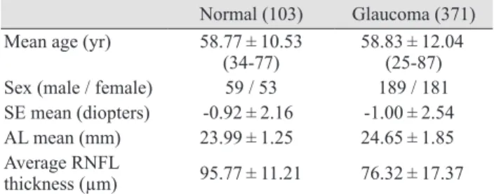
Table 1. Demographic and ophthalmic characteristics of the study participants
Normal (103) Glaucoma (371) Mean age (yr) 58.77 ± 10.53
(34-77) 58.83 ± 12.04 (25-87) Sex (male / female) 59 / 53 189 / 181 SE mean (diopters) -0.92 ± 2.16 -1.00 ± 2.54 AL mean (mm) 23.99 ± 1.25 24.65 ± 1.85 Average RNFL
thickness (µm) 95.77 ± 11.21 76.32 ± 17.37
SE = spherical equivalent; AL = axial length; RNFL = retinal
nerve fiber layer.
We analyzed the correlation between parameters using SPSS ver. 17.0 (SPSS Inc., Chicago, IL, USA).
Results
Six hundred sixty-eight subjects were initially enrolled in the study. We excluded 184 subjects because of poor
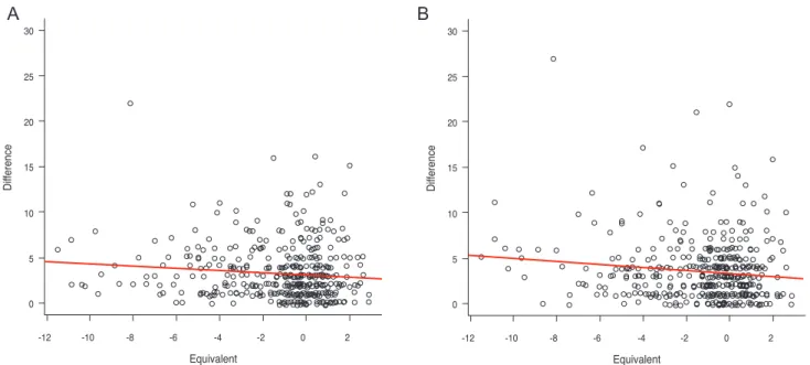
quality analysis (signal strength [SS] <6, low analysis con- fidence, or a difference between two SS figures) in one of the metrics. We included a group of 474 subjects com- prised of 103 normal subjects and 371 glaucoma patients in the final analysis. Participant demographic and ophthalmic characteristics are shown in Table 1. ICC and coefficients of variation of all 474 subjects are shown in Table 2. Cor- relations between variability (SD) in overall mean RNFL, quadrant RNFL, clock-hour RNFL, and spherical equiva- lent (SE) are shown in Table 3. Significant correlations were found between the SD in RNFL thickness in the tem- poral quadrant and SE. Significant correlations were also observed between the SD in RNFL thickness at clock-hour 9 and SE. The statistically significant variable portions (temporal area, clock-hour 9) were plotted against SE (Fig. 1).
Correlations between the variability (SD) in overall mean RNFL, quadrant RNFL, clock-hour RNFL, and axial length (AL) are shown in Table 3. Significant correlations were observed between the SD in RNFL thickness in the temporal quadrant and AL. Significant correlations were observed between the SD in RNFL thickness at clock-hour nine and AL. The statistically significant variable portions (temporal area, clock-hour 9) were plotted against AL (Fig.
2).
Discussion
Myopia is one of the greatest risk factors for glaucoma.
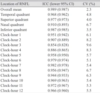
Myopic eyes have a 2- to 3-fold greater risk of developing glaucoma than emmetropic eyes. This increased risk is Table 2. ICC and CV for all 474 subjects
Location of RNFL ICC (lower 95% CI) CV (%)
Overall mean 0.989 (0.987) 2.3
Temporal quadrant 0.968 (0.962) 4.8
Superior quadrant 0.977 (0.973) 4.0
Nasal quadrant 0.910 (0.893) 6.7
Inferior quadrant 0.987 (0.985) 3.5
Clock-hour 1 0.951 (0.942) 6.1
Clock-hour 2 0.907 (0.889) 8.2
Clock-hour 3 0.854 (0.828) 9.6
Clock-hour 4 0.886 (0.865) 8.3
Clock-hour 5 0.958 (0.950) 5.7
Clock-hour 6 0.979 (0.974) 5.1
Clock-hour 7 0.982 (0.978) 5.4
Clock-hour 8 0.956 (0.947) 6.7
Clock-hour 9 0.944 (0.933) 6.3
Clock-hour 10 0.969 (0.963) 5.4
Clock-hour 11 0.972 (0.967) 5.3
Clock-hour 12 0.966 (0.960) 5.5
ICC = intraclass correlation co efficient; CV = coefficients of vari- ation; RNFL = retinal nerve fiber layer; CI = confidence interval.
Table 3. Reproducibility correlations with SE, AL and clock-hour measurements SE in RNFL
thickness AL in RNFL
thickness
Location of RNFL Regression r
2Coefficient Regression r
2Coefficient
Overall mean 0.025 0.011 -0.069 0.046 0.011 -0.122
Temporal quadrant 0.026 0.011 -0.125 0.006 0.022 -0.288
Superior quadrant 0.648 0 -0.029 0.581 0.001 -0.069
Nasal quadrant 0.823 0 -0.017 0.534 0.001 -0.093
Inferior quadrant 0.276 0.003 -0.065 0.184 0.005 -0.15
Clock-hour 1 0.876 0 -0.015 0.485 0.001 -0.125
Clock-hour 2 0.09 0.006 -0.181 0.451 0.002 -0.157
Clock-hour 3 0.819 0 -0.023 0.464 0.002 -0.137
Clock-hour 4 0.352 0.002 -0.09 0.236 0.004 -0.223
Clock-hour 5 0.942 0 -0.006 0.629 0.001 -0.08
Clock-hour 6 0.3 0.002 -0.098 0.384 0.002 -0.154
Clock-hour 7 0.623 0.001 -0.048 0.677 0 -0.078
Clock-hour 8 0.19 0.004 -0.102 0.077 0.009 -0.267
Clock-hour 9 0.011 0.014 -0.161 0.001 0.032 -0.405
Clock-hour 10 0.389 0.002 -0.059 0.837 0 -0.026
Clock-hour 11 0.912 0 -0.01 0.2 0.005 -0.223
Clock-hour 12 0.187 0.004 -0.119 0.194 0.005 -0.23
SE = spherical equivalent; AL = axial length; RNFL = retinal nerve fiber layer.
independent of other risk factors such as high intraocu- lar pressure [13]. Currently, the number of young myopic adults is increasing, with fundus examination for refractive surgery or routine check-up allowing for early detection of glaucoma. In high myopia, RNFL thinning appears to occur, and the higher visual field indicates deviation com- pared with emmetropia. The optic discs seen in myopic pa- tients may appear large, with greater cut-to-disc ratios than
emmetropic patients [11]. Also, in myopic patients, myopic visual field defects interfere with the diagnosis of glauco- ma by visual field tests. These similarities can complicate the diagnosis and monitoring of glaucoma in myopic eyes.
Imaging modalities such as OCT may be effective in these cases.
Our results may be an important finding as the correla- tion of axial length and refractive error has an influence on
Temporal
Equivalent
Di fferenc e
30 25 20 15 10 5
0
-12 -10 -8 -6 -4 -2 0 2
A Clock-hour 9
Equivalent
Di fferenc e
30 25 20 15 10 5
0
-12 -10 -8 -6 -4 -2 0 2
B
Fig. 1. Plot diagrams of temporal (A) and clock-hour 9 (B) with spherical equivalent. Positive correlations between the standard deviation in retinal nerve fiber layer thickness at clock-hour 9 and spherical equivalent.
Temporal
Length
Di fferenc e
30 25 20 15 10 5
0
22 24 26 28 30
A
Length
Di fferenc e
30 25 20 15 10 5
0
22 24 26 28 30