DOI 10.3349/ymj.2008.49.6.886
Operative laparoscopy was initially developed in the field of gynecology earlier on and the advent of laparoscopic surgery led to advances in general surgery as well. In the last few years, a number of articles have been published on the performance of surgical procedures using the robot-assisted laparoscopy.
The shortcomings of conventional laparoscopy have led to the development of robotic surgical system and future of telero- botic surgery is not far away, enabling a surgeon to operate at a distance from the operating table. The complete loss of tactile sensation is often quoted as a big disadvantage of working with robotic systems. Although the first generation da Vinci robotic surgical system provides improved imaging and instrumentation, the absence of tactile feedback and the high cost of the technology remain as limitations. New generations of the robotic surgical systems have been developed, allowing visualization of preoperative imaging during the operation.
Though the introduction of robotics is very recent, the potential for robotics in several specialties is significant. However, the benefit to patients must be carefully evaluated and proven before this technology can become widely accepted in the gynecologic surgery.
Key Words: Robotics, uterine cervical neoplasm, hysterectomy
INTRODUCTION
The term robot was first introduced in 1921 when the Czech writer Karel Capek described the notion in his play Rossum's Universal Robots (R.U.R.).1He depicted a plot in which people created a robot, which initially provides happiness but in the end produced despair in the form of social unrest and
unemployment. From then, robots have evolved from simple machines performing menial, re- petitive tasks to a highly sophisticated machine capable of performing specific tasks requiring precision. In 1950, a robot named Atom which pos- sessed both emotion and intelligence, was intro- duced in a Japanese cartoon. Robots became a popular concept in the 1970's, after the release and great success of the movie Star wars staring the robot R2D2.
Despite the advancement in technology, robotic systems in the medical field play a limited role and are still not applied to a variety of surgical opera- tions, especially in Asia. Therefore, it is meaningful and significant that we can discuss the current status and the future of the robotic surgery in gynecologic field.
ROBOT SURGERY
Operative laparoscopy was initially developed in the field of gynecology earlier on and the advent of the laparoscopic surgery led to advances in general surgery as well. In the last few years, a number of articles have been published on the performance of surgical procedures using the robot-assisted laparoscopy. For example, Computer Motion Inc. (Computer Motion, Inc., Santa Barbara, CA, USA) launched the first laparoscopic camera holder, AESOP (Automated Endoscopic System for Optimal Positioning).2 AESOP was designed in order to allow the surgeon greater control over visualization and to eliminate the need for a scope- holding assistant. The device holds the laparo- scope and the surgeon can command the laparo- scope by using voice-activated commands. Due to its convenience, AESOP has been used in over
Robotic Surgery in Gynecologic Field
Young Tae Kim, Sang Wun Kim, and Yong Wook Jung
Department of Obstetrics and Gynecology, Women's Cancer Clinic, Yonsei University College of Medicine, Seoul, Korea.
Received August 4, 2008
This study was supported by the Brain Korea (BK) 21 Project for Medical Science, Yonsei University and a grant of the Korean Health 21 R&D Project, Ministry of Health & Welfare, Republic of Korea (0412-CR01-0704-0001).
Reprint address: requests to Dr. Young Tae Kim, Division of Gynecologic Oncology, Department of Obstetrics and Gynecology, Yonsei University College of Medicine, 250 Seongsanno, Seodaemun- gu, Seoul 120-752, Korea. Tel: 82-2-2228-2230, Fax: 82-2-313- 8357, E-mail: [email protected]
10000 surgeries and some surgeons consider this as standard equipment for laparoscopic surgeries offering a cost advantage. Thanks to the rapid and continuing development of robotic technology, robotic surgery is being used not only in endoscopic surgery, but also in wide variety of surgical pro- cedures in the United States and other European countries.
In 1992, the first commercially available robotic system, ROBODOC, was described.3 This is a robotic arm designed and used in orthopedic hip prosthesis surgery. The ROBODOC makes precise cuts in the femur bone for the insertion of surgical implants based on the memorized three-dimen- sional CT image. The robot uses predetermined mapping to make a precise incision, while the surgeon controls the process by watching on a real time monitor. The program for total hip arthro- plasty using ROBODOC was developed by collabo- ration between Dr. Bargar and researchers at the University of California with funding from Inter- national Business Machines (IBM)'s Thomas J.
Watson Research Center. The ROBODOC system, which was approved by the US Food and Drug Administration (FDA), was used to perform suc- cessful total hip replacement in more than 10,000 patients. Similar products, upgraded, have been developed and currently being used throughout Europe and Asia. However, despite its wide distribution and usage, the limited role in surgical procedures makes it hard to call surgeries assisted by ROBODOC and AESOP a true robotic surgery.
In the 1990s, Computer Motion, Inc. (Computer Motion, Inc., Santa Barbara, CA, USA) developed several surgical robots called ZEUS and HERMES.
A computer engineer named Yulum Wang, a founder of the Computer Motion Inc., played a very important role in developing AESOP and revolutionizing the surgical practices by providing the baseline for integrated robotic surgery. After AESOP, Computer Motion unveiled the ZEUS sur- gical robotic system in 1998 which has a 2-dimen- sional imaging system similar to that of standard laparoscopy. On the other hand, the da Vinci surgical system was developed by Intuitive Sur- gical, Inc. (Intuitive Surgical, Mountain View, CA, USA) and the first successful surgery using the da Vinci surgical system was performed in Belgium in 1997.4
In contrast to ZEUS surgical system, the da Vinci surgical system is equipped with a 3-dimensional vision system in which double endoscopes generate two images resulting in the perception of a 3D image. In addition, with the development of endowrist, it reproduces the range of motion and dexterity of the surgeon hand, providing high precision, flexibility and ability to rotate instru- ments 360 degrees. Thus, the learning curve of achievement for the surgeons using the da Vinci surgical system was shortened. In 2001, a more advanced da Vinci surgical system with four robotic arms gained US FDA approval and is now being used in many surgical procedures through- out the world. The ongoing competition between the ZEUS and the da Vinci surgical system ended when Computer Motion Inc. was merged into Intuitive Surgical Inc. in 2003.
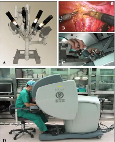
The da Vinci robotic system has three main components: the robotic cart, the operating console and the endoscopic stack (Fig. 1). The robotic cart is 2 meters high, approximately 1 meter long and 1 meter wide with a sliding system on the base which enables the cart to be placed freely according to patient's position. It is composed of four mechanical attached to a mobile base, which is connected to the operating console through a cable.
The robotic arms are mounted on a patient's side cart. The central arm contains the optic system consisting of an endoscope with two optical channels and two three-chip cameras. Three of these lateral arms hold surgical instrument. Each robotic arm has three or four joints enabling the arms to rotate freely. The surgeon, seated at the console, performs the procedure by manipulating specially designed joysticks. The movement is translated from the surgeon's fingers to the tip of the special instruments. There are six degrees of freedom at the instrument tip and a seventh degree of freedom is provided by the action of the instru- ment itself. Each instrument can be resterilized, but can be used only ten times. The computer is able to eliminate physiologic tremor and to down- scale the amplitude of motions enabling wide range of surgical procedures.
The operating console integrates the 3D viewing, the masters with two controllers (joysticks) and four foot pedals. The surgeon sits in an ergono- mically comfortable position at the console and
Fig. 1. Robotic cart with telerobotic arms (da Vinci surgical system); (A) da Vinci robot cart with 4 robotic arms (B) Surgical field; (C and D) Surgeon console. The motion of robotic instrument in the surgical field is operated by both hands.
his/her hands fit into the master instrument controllers. The movement is converted and trans-
lated from the surgeon's fingers to the tip of the instruments. The operating console has four foot pedals which can be manipulated to electrocau- terize for hemostatsis or to control the movement of the camera.
The da Vinci robotic surgical system replaces two-dimensional with three dimensional imaging with the optical channels and enhances the precision of anatomic dissection (Fig. 2). Further- more, the computer also provides motion scaling and tremor elimination, facilitating surgical pro- cedures that are typically more difficult.
One of the most significant advantages of this robotic system includes the three dimensional view that improves visualization of the surgical field, allowing greater precision and accuracy.
Another advantage is the wrist like motion of the robotic arm which provides finer and more dex- terous movements, enabling surgical procedures
Fig. 2. Inside vision system with endoscope.
A C
B
D
which were impossible with the conventional laparoscopy. In the United States, the da Vinci robotic surgical system is being used in the various fields of gynecology, urology, general surgery and thoracic surgery.
More than 645 da Vinci systems are in use around the world and about 41 da Vinci systems are currently in Asia. In Korea, the da Vinci system was first used at Yonsei University Medical Center in 2005. The Korean FDA first approved the da Vinci system in July 13th, 2005, and the system is currently being used at the departments of general surgery, urology, gynecology and thoracic sur- gery.5,6 The main disadvantages of the conventional laparoscopic procedures include two-dimensional imaging, lack of sensory feedback, the limited mobility of the instruments and the long learning curve. Therefore, much attention is now being paid to the promise of robotic surgery,
ROBOTIC HYSTERECTOMY IN GYNECO- LOGIC FIELD
Hysterectomy is the most common non-preg- nancy-associated surgical procedure in the United States.7 Laparoscopic hysterectomy was first re- ported in the literature over 15 years ago and since then, several surgical procedures including laparo- scopic assisted vaginal hysterectomy (LAVH), supracervical laparoscopic hysterectomy (SLH) and total laparoscopic hysterectomy (TLH) have been
introduced.8-17 However, the history of robotic laparoscopic hysterectomy is short and still developing. The first series which included eleven cases of successful robotic laparoscopic hysterec- tomy were performed by Diaz-Arrastia in 2002.18 The age of patients ranged from 22 to 77 years, and the indication for hysterectomy included cervical intraepithelial neoplasia III, endometrial cancer, myoma of uterus, postmenopausal bleeding and one case of ovarian cancer. In 2004, Advincular et al. reported 35 cases of robotic laparoscopic myomectomy, and 3 cases were converted into laparotomy (conversion rate of 8.6%)19: The weight of the uterus varied from 200 gram to 1200 gram, and the complication rate was fairly low. In 2006, Fiorentino et al. reported a pilot study assessing robotic laparoscopic hysterectomy and patient's outcomes.20 Twenty women with benign gyneco- logic conditions were included in this study, and the surgical procedure was converted to laparo- tomy in two patients (conversion rate 10%) because of poor visualization. The mean operating time was 3.2 hours and anesthesia time was 4 hours.
Mean estimated blood loss was 81 mL, and post- operative hospital day was 2 days. In addition, Reynolds et al. reported 16 cases of robotic laparo- scopic hysterectomy, and none was converted to laparotomy. The average weight of the uterus was 131.5 g and the mean operative time was 242 minutes. The average estimated blood loss was 96 mL, and the mean duration of hospital stay was 1.5 days. Four trocar ports were used for the
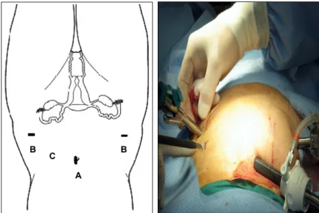
Fig. 3. Port placement: (A) The 12-mm camera port was placed in the umbilicus or above depending on the size of the uterus. (B) The 8-mm lateral ports for ro- botic instruments were mounted directly to the robotic arms and placed 2 to 3 cm medial and superior to the anterior supe- rior ileac spine with modification based on the size of the uterus. (C) The assis- tant port was placed between the camera port and the left lower quadrant port.
surgical procedures (Fig. 3). In Korea, the first robotic laparoscopic hysterectomy was done by Kim et al. in January 31st of 2006 after the approval by Korean FDA.21,22 In the gynecologic field, the da Vinci robotic surgical system is being used in a wide range of specialties, including surgeries for endometrial cancer, myoma of the uterus, adeno- myosis, endometrial hyperplasia and cervical in- traepithelial neoplasia.
CONCLUSION
The limitations of the conventional laparoscopy have led to the development of robotic surgical system, and future of telerobotic surgery is not far away enabling a surgeon to operate at a distance from the operating table.
The complete loss of tactile sensation is often quoted as a disadvantage of working with robotic systems. Although the first generation da Vinci robotic surgical system provides improved im- aging and instrumentation, the absence of tactile feedback and the high cost of the technology remain as limitations. New generations of the robotic surgical systems which allow visualization of the preoperative imaging during the operation have been developed. Although the robotics ex- perience is very early, the potential for robotics in several specialties is significant. However, the benefit to patients must be carefully evaluated and proven before this technology can become widely accepted in the gynecologic surgery.
REFERENCES
1. Capek K, Capek J. The Insect Play. In: Oxford MB, editor. 1st ed. New York: Oxford University Press;
1963.
2. Satava RM. Robotic surgery: from past to future-a personal journey. Surg Clin North Am 2003;83:1491- 500, xii.
3. Bann S, Khan M, Hernandez J, Munz Y, Moorthy K, Datta V, et al. Robotics in surgery. J Am Coll Surg 2003;
196:784-95.
4. Himpens J, Leman G, Cadiere GB. Telesurgical laparo- scopic cholecystectomy. Surg Endosc 1998;12:1091.
5. Kang CM, Chi HS, Hyeung WJ, Kim KS, Choi JS, Lee WJ, et al. The first korean experience of telemani-
pulative robot-assisted laparoscopic cholecystectomy using the da vinci system. Yonsei Med J 2007;48:540-5.
6. Lee YS, Han WK, Oh YT, Choi YD, Yang SC, Rha KH.
Robot-assisted laparoscopic radical prostatectomy: four cases. Yonsei Med J 2007;48:341-6.
7. Farquhar CM, Steiner CA. Hysterectomy rates in the United States 1990-1997. Obstet Gynecol 2002;99:229-34.
8. Wattiez A, Cohen SB, Selvaggi L. Laparoscopic hysterectomy. Curr Opin Obstet Gynecol 2002;14:417- 22.
9. Reich H. New techniques in advanced laparoscopic surgery. Baillieres Clin Obstet Gynaecol 1989;3:655-81.
10. Falcone T, Goldberg J, Garcia-Ruiz A, Margossian H, Stevens L. Full robotic assistance for laparoscopic tubal anastomosis: a case report. J Laparoendosc Adv Surg Tech A 1999;9:107-13.
11. Falcone T, Goldberg JM. Robotics in gynecology. Surg Clin North Am 2003;83:1483-9, xii.
12. Falcone T, Goldberg JM. Robotic surgery. Clin Obstet Gynecol 2003;46:37-43.
13. Falcone T, Goldberg JM, Margossian H, Stevens L.
Robotic-assisted laparoscopic microsurgical tubal an- astomosis: a human pilot study. Fertil Steril 2000;73:
1040-2.
14. Donnez J, Nisolle M. Laparoscopic supracervical (subtotal) hysterectomy (LASH). J Gynecol Surg 1993;9:
91-4.
15. Olive DL, Parker WH, Cooper JM, Levine RL. The AAGL classification system for laparoscopic hysterec- tomy. Classification committee of the American Asso- ciation of Gynecologic Laparoscopists. J Am Assoc Gynecol Laparosc 2000;7:9-15.
16. Semm K. [Hysterectomy via laparotomy or pelviscopy.
A new CASH method without colpotomy]. Geburt- shilfe und Frauenheilkunde 1991;51:996-1003.
17. Morrison JE Jr, Jacobs VR. 437 classic intrafascial supra- cervical hysterectomies in 8 years. J Am Assoc Gynecol Laparosc 2001;8:558-67.
18. Diaz-Arrastia C, Jurnalov C, Gomez G, Townsend C Jr.
Laparoscopic hysterectomy using a computer-enhanced surgical robot. Surg Endosc 2002;16:1271-3.
19. Advincula AP, Falcone T. Laparoscopic robotic gynecologic surgery. Obstet Gynecol Clin North Am 2004;31:599-609, ix-x.
20. Fiorentino RP, Zepeda MA, Goldstein BH, John CR, Rettenmaier MA. Pilot study assessing robotic laparo- scopic hysterectomy and patient outcomes. J Minim Invasive Gynecol 2006;13:60-3.
21. Kim YT, Kim SW, Yoon BS, Nahm EJ, Hur HW, Kim SH, et al. Robot-assisted total laparoscopic hysterec- tomy; initial experience in Korea. Korean J Obstet Gynecol 2006;49:2620-25.
22. Kim YT, Kim SW, Hyung WJ, Lee SJ, Nam EJ, Lee WJ.
Robotic radical hysterectomy with pelvic lympha- denectomy for cervical carcinoma: a pilot study.
Gynecol Oncol 2008;108:312-6.