INTRODUCTION
Despite the wide variety of phakic intraocular lenses (pIOLs) that yield good results, the correction of moderate-to-high
astigmatism remains a challenge.1 The foldable iris-fixated pIOL (hereafter, non-toric pIOL; Artiflex, Ophtec BV, Gronin- gen, the Netherlands) can be implanted through a small inci- sion,2 minimizing surgically induced astigmatism, but only corrects spherical error. The toric foldable iris-fixated pIOL (hereafter, toric pIOL; Toric Artiflex, Ophtec BV) is designed to correct cylindrical errors between -5.0 and -1.0 diopters (D) and spherical errors between -13.5 and -1.0 D. Several studies have shown that it effectively and safely corrects myopic astig- matism.2-4 However, accurate placement of the toric pIOL is critical to obtain satisfactory results concerning residual astig- matism, especially in eyes with high astigmatism. Moreover, toric pIOLs are quite expensive and the manufacturing time between placing the order and receiving the product is some-
Comparison of Toric Foldable Iris-Fixated Phakic Intraocular Lens Implantation and Limbal Relaxing Incisions for Moderate-to-High Myopic Astigmatism
Jeihoon Lee1, Hun Lee1,2, David Sung Yong Kang3, Jin Young Choi3, Eung Kweon Kim1,4, and Tae-im Kim1
1The Institute of Vision Research, Department of Ophthalmology, Yonsei University College of Medicine, Seoul;
2Department of Ophthalmology, International St. Mary’s Hospital, Catholic Kwandong University College of Medicine, Incheon;
3The Institute of Vision Research, Eyereum Eye Clinic, Seoul;
4Corneal Dystrophy Research Institute, Severance Biomedical Science Institute, and Brain Korea 21 Plus Project for Medical Science, Yonsei University College of Medicine, Seoul, Korea.
Purpose: To compare the effectiveness of toric foldable iris-fixated phakic intraocular lens (pIOL) implantation and non-toric fold- able iris-fixated pIOL implantation with limbal relaxing incisions (LRIs) for correcting moderate-to-high astigmatism in myopic eyes.
Materials and Methods: The medical records of 146 patients (195 eyes) with myopic astigmatism who underwent toric foldable iris-fixated pIOL implantation (toric group; 94 eyes) or non-toric foldable iris-fixated pIOL implantation with concurrent LRIs (LRI group; 101 eyes) were retrospectively reviewed. For subgroup analysis, the two groups were subdivided according to preoperative astigmatic severity [moderate, 2.00 to <3.00 diopters (D); high, 3.00–4.00 D]. Visual and astigmatic outcomes were compared 6 months postoperatively.
Results: The uncorrected distance visual acuity was at least 20/25 in 100% and 98% of the toric and LRI group eyes, respectively.
The toric group had lower mean residual cylindrical error (-0.67±0.39 D vs. -1.14±0.56 D; p<0.001) and greater mean cylindrical error change (2.17±0.56 D vs. 1.63±0.72 D; p<0.001) than the LRI group, regardless of the preoperative astigmatic severity. The mean correction index (1.10±0.16 vs. 0.72±0.24; p<0.001) and success index (0.24±0.14 vs. 0.42±0.21; p<0.001) also differed significantly between the groups.
Conclusion: Both surgical techniques considerably reduced astigmatism and had comparable visual outcomes. However, toric fold- able iris-fixated pIOL implantation was more reliable for correcting moderate-to-high astigmatism in myopic eyes.
Key Words: Lenses, intraocular, limbus corneae, astigmatism
pISSN: 0513-5796 · eISSN: 1976-2437
Received: December 3, 2015 Revised: April 15, 2016 Accepted: April 26, 2016
Corresponding author: Dr. Tae-im Kim, Department of Ophthalmology, Yonsei University College of Medicine, 50-1 Yonsei-ro, Seodaemun-gu, Seoul 03722, Korea.
Tel: 82-2-2228-3574, Fax: 82-2-312-0541, E-mail: [email protected]
•The authors have no financial conflicts of interest.
© Copyright: Yonsei University College of Medicine 2016
This is an Open Access article distributed under the terms of the Creative Com- mons Attribution Non-Commercial License (http://creativecommons.org/licenses/
by-nc/3.0) which permits unrestricted non-commercial use, distribution, and repro- duction in any medium, provided the original work is properly cited.
Yonsei Med J 2016 Nov;57(6):1475-1481 http://dx.doi.org/10.3349/ymj.2016.57.6.1475
what long.
Limbal relaxing incisions (LRIs) are another option for cor- recting astigmatism surgically. LRIs, also known as peripheral corneal relaxing incisions, flatten the steep meridian and cause coupling of the flat meridian.5 They are easy to perform and rel- atively inexpensive.6,7 In addition, LRIs can be combined with non-toric pIOL implantation to correct moderate-to-high astig- matism in myopic eyes. However, LRIs can weaken the cornea and/or decrease visual quality by increasing corneal aberra- tions and irregular astigmatism.8,9
If non-toric pIOLs combined with LRIs show comparable refractive and visual outcomes with those of toric pIOLs, non- toric pIOLs could be substituted to overcome the drawbacks of toric pIOLs, including their high cost, the long duration re- quired to receive the pIOL from the manufacturer, and chal- lenging elaborate implantation technique along an accurate axis of astigmatism. To the best of our knowledge, studies com- paring the astigmatism-reducing effects of these surgical pro- cedures are rare. Herein, we compared the effectiveness of toric pIOL implantation and non-toric pIOL implantation with LRIs for correcting moderate-to-high astigmatism in myopic eyes.
MATERIALS AND METHODS
Subjects
This retrospective comparative observational study was ap- proved by the Institutional Review Board of Yonsei University College of Medicine (Seoul, Korea). Its protocol adhered to the tenets of the Declaration of Helsinki and followed good clini- cal practice.
Patients were included if they were older than 20 years and had stable refraction and regular astigmatism between 2.00 and 4.00 D. The exclusion criteria were as follows: history of corneal refractive surgery or ocular disease that may affect vi- sual outcomes (e.g., color vision disturbance, chronic uveitis, glaucoma, and maculopathy); an anterior chamber depth
<3.0 mm from the endothelium, corneal endothelial cell den- sity (ECD) <2000 cells/mm2, white-to-white distance <11.0 mm, mesopic pupil diameter >7.0 mm, intraocular pressure (IOP)
>21 mm Hg; evidence of acute or chronic corneal infection, cor- neal inflammation, abnormal iris or pupil function, or cataract;
and the development of intraoperative or postoperative com- plications.
The same surgeon performed toric pIOL implantation (toric group) and non-toric pIOL implantation with concurrent LRIs (LRI group) in the standard fashion at the Eyereum Eye Clinic (Seoul, Korea) between November 2012 and October 2014.
The patient’s economic preference dictated the choice of surgi- cal method. The groups were further divided into the moderate (2.00 to <3.00 D) and high (3.00–4.00 D) astigmatic subgroups according to preoperative astigmatic severity.
All subjects received complete ophthalmic examinations, in-
cluding measurements of uncorrected distance visual acuity (UCDVA; Snellen) and IOP (noncontact tonometer NT-530, Nidek Co., Ltd., Aichi, Japan), manifest refraction, slit-lamp biomicroscopy (Haag-Streit AG, Köniz, Switzerland), and di- lated fundus examination. Central corneal thickness, kerato- metric values, and central ECD were measured by ultrasound pachymetry (UP-1000, Nidek Co.), autokeratometry (ARK- 530A, Nidek Co.), and specular microscopy (SP-3000P, Top- con Corporation, Tokyo, Japan), respectively. Manifest refrac- tion, UCDVA, and central ECD measurements were repeated 3 and 6 months postoperatively.
Surgical procedures
A small iridotomy was made with consecutive argon green and neodymium-doped yttrium aluminum garnet (Nd:YAG) lasers at least 1 week preoperatively. Immediately before surgery, the axis of astigmatism was marked along the negative axis at the slit-lamp to determine the flat meridian with the patient seated upright.
In the toric group, a 2.8-mm primary limbal incision was made at the marked meridians, and two sub-1.0-mm stab in- cisions were placed nasally and temporally to the primary in- cision, as seen fit by the surgeon. A miotic agent was injected intracamerally and 1.0% sodium hyaluronate was used to maintain the anterior chamber and coat the corneal endothe- lium. Then, the toric pIOL was inserted, fixated with special forceps, and aligned along the orientation marks on the cor- nea. Finally, the viscoelastic substance was removed by man- ual aspiration.
In the LRI group, the primary incision was placed on the steep meridian, with the length and location determined by using the Nichamin Age & Pachymetry-Adjusted Intralimbal Arcuate Astigmatic Nomogram.10 The non-toric pIOL was in- serted and fixated with special forceps. At the end of the sur- gery, an additional LRI was placed at the opposite meridian and the viscoelastic material was manually removed.
All surgeries were uneventful and no intraoperative compli- cation was noted. The subjects applied 0.5% moxifloxacin and 0.1% dexamethasone four times daily for the first postopera- tive week. Thereafter, dexamethasone was replaced with 0.1%
fluorometholone and the eye drops were continued four times daily for 1 month.
Statistical analyses
The results are expressed as means±standard deviations, where applicable. The Kolmogorov-Smirnov test was used to con- firm data normality. All data were analyzed by using SAS soft- ware (version 9.2, SAS Institute, Inc., Cary, NC, USA). Differ- ences between the groups were tested for statistical significance by using independent t tests. Paired t tests were used to test for differences between preoperative and residual astigmatism with- in each group. Statistical significance was defined as p<0.05.
Astigmatism was analyzed by the power vector method11
and Alpins method.12-14 The power vector method aids in visu- alizing complex changes in refractive error. The manifest re- fraction data, as written in conventional script notation, were used to calculate power vector coordinates and overall blur- ring strength (B) by the following formulas: B=(M2+J02+J452)1/2, where M=S+C/2, J0=(-C/2) cos(2α), J45=(-C/2) sin(2α), S=
sphere, C=cylinder, and α=axis. The calculated power vector length is a measure of the overall blurring strength of the refrac- tive error.
The Alpins method presents data in standard graphs for re- porting astigmatic outcomes of refractive surgery.12-15 The tar- get astigmatism is zero, because emmetropia is the goal. We calculated the target-induced astigmatic vector (TIAV), or the amount and direction of the dioptric force required to achieve emmetropia from the preoperative state, the surgically induced astigmatic vector (SIAV), which is the astigmatic change actu- ally induced by surgery, and the difference vector (DV), which represents the magnitude and axis of the difference in D be- tween the desired and achieved results. The correction index (CI) and success index (SI) were calculated as ratios of the SIAV to the TIAV and the DV to the TIAV, respectively. Free spread- sheets were used to present the data graphically (http://www.
standardgraphsforrefractivesurgery.com).15
RESULTS
The study included 195 eyes of 146 patients, with 94 eyes (65 subjects) in the toric group and 101 eyes (81 subjects) in the LRI group. There were no significant intergroup differences in the preoperative variables (Table 1).
The groups had comparable visual outcomes. In the toric
group, the UCDVA was 20/25 or better in all eyes (100%) and 20/20 or better in 91 eyes (96.8%). In the LRI group, it was 20/25 or better in 99 eyes (98%) and 20/20 or better in 83 eyes (82.2%).
The overall blurring strength significantly decreased from 8.69±2.31 D to 0.68±0.60 D in the toric group and from 8.62±
1.71 D to 0.72±0.25 D in the LRI group (both p<0.001). The to- ric group demonstrated a slightly lower blurring strength post- operatively, but the difference was not statistically significant (p=0.469).
Both groups had significantly reduced astigmatism after the surgery (Fig. 1). The residual cylindrical error was significantly lower (-0.67±0.39 D vs. -1.14±0.56 D; p<0.001) and the mean change in cylindrical error was significantly greater (2.17±0.56 D vs. 1.63±0.72 D; p<0.001) in the toric group than in the LRI group 6 months postoperatively. Furthermore, 47 (50.0%) and 85 (90.4%) of the 94 eyes in the toric group had residual cylin- drical errors within 0.50 and 1.00 D, respectively. On the con- trary, 19 (18.8%) and 43 (42.6%) of the 101 eyes in the LRI group demonstrated the same respective residual cylindrical error values (Fig. 2). The differences between the groups were sig- nificant (both p<0.001).
Subgroup analyses revealed that the mean residual cylin- drical errors were -0.64±0.37 and -1.09±0.58 D in toric and LRI group eyes with moderate astigmatism (p<0.001), respective- ly. For high astigmatism, the mean residual cylindrical errors were -0.72±0.41 and -1.16±0.66 D (p=0.001) in the respective groups. The mean changes in moderate (1.84±0.43 D vs. 1.33±
0.61 D; p<0.001) and high (2.55±0.44 D vs. 2.16±0.66 D; p=0.003) astigmatism were significantly greater in the toric group than in the LRI group.
In the analysis of astigmatism by the power vector method,11 most points were concentrated at the center of the graph, es- pecially in the toric group (Fig. 3). The toric group had J0 and J45 vector values between +0.50 and -0.50 D in 86 (91.5%) and 91 (96.8%) of the 94 eyes, respectively. In the LRI group, J0 and J45 vector values in the same range were noted in 76 (75.2%) and 69 (68.3%) of the 101 eyes, respectively. Both differences between the groups were significant (p=0.003 and p<0.001).
Table 1. Preoperative Demographic and Ocular Characteristics of the Subjects (n=146)
Parameter Toric group LRI group p value
Number of eyes 94 101
Age (yrs) 28.52±5.37 27.83±5.41 0.373
Female sex (%) 68.1 68.3 0.972
Right eye (%) 45.7 39.6 0.386
Refractive error (D)
Sphere -7.13±2.35 -7.12±1.63 0.964
Cylinder -2.85±0.48 -2.77±0.53 0.303
Spherical equivalent -8.56±2.34 -8.51±1.72 0.865 Keratometric value
Flat K 43.01±1.46 43.07±1.24 0.721
Steep K 45.66±1.48 45.48±1.32 0.368
ACD (mm) 3.34±0.22 3.39±0.22 0.173
ECD (cells/mm2) 3064.0±231.1 3075.6±256.2 0.739 Toric group, toric foldable iris-fixated phakic intraocular lens implantation; LRI group, non-toric foldable iris-fixated phakic intraocular lens implantation with limbal relaxing incisions; LRI, limbal relaxing incision; D, diopters; ACD, ante- rior chamber depth; ECD, endothelial cell density.
Data are expressed as means±standard deviations unless indicated otherwise.
Fig. 1. The mean profile graph of the toric and LRI groups. Toric group, to- ric foldable iris-fixated phakic intraocular lens implantation; LRI group, non-toric foldable iris-fixated phakic intraocular lens implantation with limbal relaxing incisions.
-3.50 -3.00 -2.50 -2.00 -1.50 -1.00 -0.50
0.00 3 mon
Toric group LRI group
Preop 6 mon
Refractive cylinder (D)
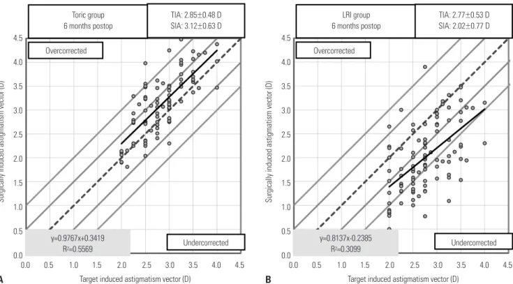
The subjects in the toric group had their astigmatism over- corrected, while those in the LRI group had undercorrected as- tigmatism (Fig. 4). However, the toric group had corrections much closer to the intended value (p<0.001). The mean CI and SI were significantly different between the groups (both p<
0.001) (Table 2).
The postoperative ECDs were 3034.2±236.0 and 3037.5±
265.4 cells/mm2 in the toric and LRI groups (p=0.926), respec- tively. The mean ECD changes were -29.8±120.9 (p=0.019) and -38.1±127.8 (p=0.003) cells/mm2 in the respective groups;
the intergroup difference was not significant (p=0.926). More-
over, the percentage change in ECD was -0.92±3.9% in the toric group and -1.19±4.2% in the LRI group (p=0.644).
DISCUSSION
Several articles have reported predictable and effective astig- matic correction with rigid toric iris-fixated pIOLs.16,17 Howev- er, they have some drawbacks, including a large amount of sur- gically induced astigmatism, mainly attributable to the length of the primary incision (up to 5.2 mm) required to implant the
Refractive astigmatism (D) Refractive astigmatism (D)
Postop Preoop
Postop Preoop Toric group
6 months postop
LRI group 6 months postop
≤0.50 D: 50%
≤1.00 D: 90%
≤0.50 D: 19%
≤1.00 D: 43%
Fig. 2. Comparison of astigmatism severity before and 6 months after surgery in the (A) toric and (B) LRI groups. Toric group, toric foldable iris-fixated pha- kic intraocular lens implantation; LRI group, non-toric foldable iris-fixated phakic intraocular lens implantation with limbal relaxing incisions; D, diopters.
80 70 60 50 40 30 20 10 0
80 70 60 50 40 30 20 10
≤0.25 0.26 0 ≤0.25
to 0.50
0.26 to 0.50 0.51
to 0.75
0.51 to 0.75 0.76
to 1.00
0.76 to 1.00 1.01
to 1.25
1.01 to 1.25 1.26
to 1.50
1.26 to 1.50 1.51
to 2.00
1.51 to 2.00 2.01
to 3.00
2.01 to 3.00
>3.00 >3.00
28%
11%
22%
8%
20%
13%
20.2%
10.9%
5.3%
27.7%
0% 0% 0% 0% 0% 0%3% 0% 0% 0% 0% 0% 0%
15%
5%
15%
0% 0% 4% 0%
27% 30%
68%
55%
1%
11%
% of eyes (%) % of eyes (%)
A B
A B
Fig. 3. Scatter plots of the J0 and J45 vectors [in diopters (D)], calculated by power vector analysis, in the (A) toric and (B) LRI groups. Black diamonds and grey squares indicate preoperative and 6-month postoperative values, respectively. Toric group, toric foldable iris-fixated phakic intraocular lens implan- tation; LRI group, non-toric foldable iris-fixated phakic intraocular lens implantation with limbal relaxing incisions.
2.5 2 1.5 1 0.5 0 -0.5 -1 -1.5 -2 -2.5
2.5 2 1.5 1 0.5 0 -0.5 -1 -1.5 -2 -2.5
-2.5 -2 -1.5 -1 -0.5 0 0.5 1 1.5 2 2.5 -2.5 -2 -1.5 -1 -0.5 0 0.5 1 1.5 2 2.5
J0 (D) J0 (D)
J45 (D) J45 (D)
Preop Postop
Preop Postop
lens. Although surgically induced astigmatism is accounted for in power calculations, it is not always accurately forecasted.
The toric pIOL can be inserted through a 3.2-mm incision, minimizing surgically induced and irregular astigmatism. In one study conducted by Ruckhofer, et al.,1 the mean residual as- tigmatism was -0.18±0.30 D after toric pIOL implantation. Oth- er studies have also shown relatively low residual cylindrical error with this method (multicenter study, 0.38±0.41 D2; two long-term follow-up studies, -0.39 D and -0.60 D3,4). In line with these results, we found residual astigmatism of -0.67±0.39 D at 6 months after toric pIOL implantation.
One study showed that toric IOL implantation and LRIs dur- ing cataract surgery yielded similar results for astigmatic cor-
rection.18 However, Hirnschall, et al.5 reported that toric IOL implantation reduced astigmatism more noticeably and pre- dictably than LRIs after cataract surgery. Further, Mingo-Botín, et al.6 showed that toric IOL implantation resulted in better refractive and visual outcomes in eyes with mild or moderate astigmatism. Consistent with these findings, toric pIOL im- plantation in our study, led to better astigmatic correction, re- sulting in lower residual astigmatism and a larger change in cylindrical error, regardless of the preoperative astigmatic se- verity, albeit the toric and LRI groups had comparable visual acuities and blurring strengths.
The CI is greater than 1.0 if overcorrection occurs, and less than 1.0 if there is undercorrection.12 In the present study, the mean CI was significantly larger in the toric group than in the LRI group (1.10 vs. 0.72). Despite the overcorrection, the re- fractive outcomes were less deviated from the ideal value (CI=
1.0) in the toric group (p<0.001). Furthermore, the mean SI was lower in this group (0.24 vs. 0.42). Considering that the SI is a relative measure of success and preferably zero,12 the re- duction in astigmatism was more successful in the toric group, implying that toric pIOL implantation corrects astigmatism more predictably and accurately than non-toric pIOL implan- tation with LRIs.
Several factors may explain why astigmatism was undercor- rected in the LRI group: improper identification of the steep meridian, incorrect calibration of the blade, oblique position- ing of the blade instead of apposition perpendicular to the
A B
Fig. 4. Relationship between the target-induced astigmatism vector and the surgically induced astigmatism vector in the (A) toric and (B) LRI groups. The dashed line represents the ideal situation, in which the intended and achieved cylindrical corrections are equal. Toric group, toric foldable iris-fixated phakic intraocu- lar lens implantation; LRI group, non-toric foldable iris-fixated phakic intraocular lens implantation with limbal relaxing incisions; D, diopters; TIA, target- induced astigmatism; SIA, surgically induced astigmatism.
4.5 4.0
3.5
3.0 2.5
2.0
1.5 1.0
0.5
0.0
4.5 4.0
3.5
3.0 2.5
2.0
1.5 1.0
0.5
0.0 0.5 1.0 1.5 2.0 2.5 3.0 3.5 4.0 4.5 0.00.0 0.5 1.0 1.5 2.0 2.5 3.0 3.5 4.0 4.5 Target induced astigmatism vector (D) Target induced astigmatism vector (D)
Toric group 6 months postop
LRI group 6 months postop
Overcorrected Overcorrected
Undercorrected Undercorrected
y=0.9767x+0.3419 R2=0.5569
y=0.8137x-0.2385 R2=0.3099 TIA: 2.85±0.48 D
SIA: 3.12±0.63 D TIA: 2.77±0.53 D
SIA: 2.02±0.77 D
Surgically induced astigmatism vector (D) Surgically induced astigmatism vector (D)
Table 2. Vector Analyses of Astigmatism at 6 Months Postoperatively
Group Toric group LRI group p value
TIAV 2.85±0.48 2.77±0.53 0.303
SIAV 3.12±0.63 2.02±0.77 <0.001
DV 0.67±0.39 1.14±0.56 <0.001
CI* 1.10±0.16 0.72±0.24 <0.001
SI 0.24±0.14 0.42±0.21 <0.001
Toric group, toric foldable iris-fixated phakic intraocular lens implantation; LRI group, non-toric foldable iris-fixated phakic intraocular lens implantation with limbal relaxing incisions; LRI, limbal releasing incision; TIAV, target-induced astigmatism vector; SIAV, surgically induced astigmatism vector; DV, differ- ence vector; CI, correction index (SIAV/TIAV); SI, success index (DV/TIAV).
Data are expressed as means±standard deviations.
*<1.0 indicates undercorrection and >1.0 indicates overcorrection.
limbus, and the far peripheral placement of incisions.10,19,20 LRIs are associated with other important problems. First, LRIs have a degree of uncertainty because equally relaxing incisions are difficult to create. We examined cases performed by only one surgeon to minimize the influence of variations in surgical style. Second, a relatively long period is needed after LRIs for the corrective effect to stabilize.9 The largest amount of refrac- tive regression occurs between 1 and 3 months after LRIs, and the refractive status stabilizes between 3 and 6 months post- operatively.21 Another study showed that surgically induced astigmatism continued to change up to 10 weeks after LRIs, but remained stable after 10 weeks and up to 3 years postop- eratively.22 We used 6-month follow-up data to exclude the possible effects of refractive regression.
The toric pIOL should be implanted along the correct cylin- drical axis, and especially in eyes with high astigmatism, be- cause small deviations can result in improper correction. Sev- eral studies have indicated that only 1.7–2.4% of eyes implanted with toric pIOLs had greater than 5° of misalignment.2,4
Endothelial cell loss is a possible complication of both toric and non-toric pIOL implantation. In our study, ECD signifi- cantly decreased in both groups, with comparable percent ch- anges from baseline to 6 months. These results are in accor- dance with previous results regarding iris-fixated pIOLs.1-3
Our study has some limitations, including its retrospective design and lack of results on visual quality. However, a previous study showed potential improvements in contrast vision and mean contrast sensitivity after toric pIOL implantation.23 In addition, this procedure does not alter or increase higher-order aberrations in myopic eyes.24 On the contrary, LRIs may in- crease corneal aberrations and decrease functional vision by degrading the optical quality of the cornea.25 Therefore, differ- ences in functional vision may exist between the study groups.
A prospective controlled study evaluating both the astigma- tism-reducing effects and visual quality of these methods is necessary.
In conclusion, both surgical methods considerably reduced astigmatism and had comparable visual outcomes. However, toric pIOL implantation was more reliable for moderate-to- high astigmatic correction than non-toric pIOL implantation with LRIs in myopic eyes.
ACKNOWLEDGEMENTS
This study was supported by a grant of the Korean Health Tech- nology R & D Project, Ministry of Health & Welfare, Republic of Korea (HI14C2044).
REFERENCES
1. Ruckhofer J, Seyeddain O, Dexl AK, Grabner G, Stoiber J. Correc- tion of myopic astigmatism with a foldable iris-claw toric phakic intraocular lens: short-term follow-up. J Cataract Refract Surg
2012;38:582-8.
2. Doors M, Budo CJ, Christiaans BJ, Luger M, Marinho AA, Dick HB, et al. Artiflex toric foldable phakic intraocular lens: short-term results of a prospective European multicenter study. Am J Ophthal- mol 2012;154:730-9.e2.
3. Guerin MB, Treacy MP, O’Keeffe M. Twelve-month follow-up of the Artiflex toric phakic intraocular lens. Eur J Ophthalmol 2014;
24:10-3.
4. Muñoz G, Cardoner A, Albarrán-Diego C, Ferrer-Blasco T, Belda- Salmerón L. Iris-fixated toric phakic intraocular lens for myopic astigmatism. J Cataract Refract Surg 2012;38:1166-75.
5. Hirnschall N, Gangwani V, Crnej A, Koshy J, Maurino V, Findl O.
Correction of moderate corneal astigmatism during cataract sur- gery: toric intraocular lens versus peripheral corneal relaxing in- cisions. J Cataract Refract Surg 2014;40:354-61.
6. Mingo-Botín D, Muñoz-Negrete FJ, Won Kim HR, Morcillo-Laiz R, Rebolleda G, Oblanca N. Comparison of toric intraocular lenses and peripheral corneal relaxing incisions to treat astigmatism dur- ing cataract surgery. J Cataract Refract Surg 2010;36:1700-8.
7. Nichamin LD. Astigmatism control. Ophthalmol Clin North Am 2006;19:485-93.
8. Lee BS, Lindstrom RL, Reeves SW, Hardten DR. Modern manage- ment of astigmatism. Int Ophthalmol Clin 2013;53:65-78.
9. Ouchi M. High-cylinder toric intraocular lens implantation ver- sus combined surgery of low-cylinder intraocular lens implanta- tion and limbal relaxing incision for high-astigmatism eyes. Clin Ophthalmol 2014;8:661-7.
10. Nichamin LD. Modified astigmatism correction nomogram. J Re- fract Surg 2008;24:562-3.
11. Thibos LN, Horner D. Power vector analysis of the optical out- come of refractive surgery. J Cataract Refract Surg 2001;27:80-5.
12. Alpins N. Astigmatism analysis by the Alpins method. J Cataract Refract Surg 2001;27:31-49.
13. Alpins NA. A new method of analyzing vectors for changes in astigmatism. J Cataract Refract Surg 1993;19:524-33.
14. Alpins NA, Goggin M. Practical astigmatism analysis for refractive outcomes in cataract and refractive surgery. Surv Ophthalmol 2004;49:109-22.
15. Reinstein DZ, Archer TJ, Randleman JB. JRS standard for report- ing astigmatism outcomes of refractive surgery. J Refract Surg 2014;30:654-9.
16. Dick HB, Alió J, Bianchetti M, Budo C, Christiaans BJ, El-Danasoury MA, et al. Toric phakic intraocular lens: European multicenter study. Ophthalmology 2003;110:150-62.
17. Güell JL, Vázquez M, Malecaze F, Manero F, Gris O, Velasco F, et al.
Artisan toric phakic intraocular lens for the correction of high astig- matism. Am J Ophthalmol 2003;136:442-7.
18. Poll JT, Wang L, Koch DD, Weikert MP. Correction of astigmatism during cataract surgery: toric intraocular lens compared to periph- eral corneal relaxing incisions. J Refract Surg 2011;27:165-71.
19. Akura J, Matsuura K, Hatta S, Otsuka K, Kaneda S. A new concept for the correction of astigmatism: full-arc, depth-dependent as- tigmatic keratotomy. Ophthalmology 2000;107:95-104.
20. Carvalho MJ, Suzuki SH, Freitas LL, Branco BC, Schor P, Lima AL.
Limbal relaxing incisions to correct corneal astigmatism during phacoemulsification. J Refract Surg 2007;23:499-504.
21. Budak K, Yilmaz G, Aslan BS, Duman S. Limbal relaxing incisions in congenital astigmatism: 6 month follow-up. J Cataract Refract Surg 2001;27:715-9.
22. Lim R, Borasio E, Ilari L. Long-term stability of keratometric astig- matism after limbal relaxing incisions. J Cataract Refract Surg 2014;40:1676-81.
23. Dick HB, Tehrani M, Aliyeva S. Contrast sensitivity after implanta-
tion of toric iris-claw lenses in phakic eyes. J Cataract Refract Surg 2004;30:2284-9.
24. Tehrani M, Dick HB. Changes in higher-order aberrations after implantation of a foldable iris-claw lens in myopic phakic eyes. J
Cataract Refract Surg 2006;32:250-4.
25. Can I·, Bayhan HA, Çelik H, Ceran BB. Comparison of corneal ab- errations after biaxial microincision and microcoaxial cataract surgeries: a prospective study. Curr Eye Res 2012;37:18-24.

![Fig. 3. Scatter plots of the J0 and J45 vectors [in diopters (D)], calculated by power vector analysis, in the (A) toric and (B) LRI groups](https://thumb-ap.123doks.com/thumbv2/123dokinfo/5486986.667155/4.892.71.803.715.1055/scatter-plots-vectors-diopters-calculated-vector-analysis-groups.webp)