Nomogram to Predict Insignificant Prostate Cancer at Radical Prostatectomy in Korean Men: A Multi-Center Study
Jae Seung Chung,
1* Han Yong Choi,
2* Hae-Ryoung Song,
3Seok-Soo Byun,
4Seong Il Seo,
2Cheryn Song,
5Jin Seon Cho,
6Sang Eun Lee,
4Hanjong Ahn,
5Eun Sik Lee,
7Tae-Kon Hwang,
8Wun-Jae Kim,
9Moon Kee Chung,
10Tae Young Jung,
11Ho Song Yu,
1and Young Deuk Choi
11Department of Urology, Urological Science Institute, Yonsei University College of Medicine, Seoul; 2Department of Urology, Sungkyunkwan University School of Medicine, Seoul; 3Department of Biostatistics and Research Affairs, Yonsei University, Seoul; 4Department of Urology, Seoul National University Bundang Hospital, Seongnam; 5Department of Urology, University of Ulsan College of Medicine, Seoul; 6Department of Urology, Hallym University College of Medicine, Chuncheon; 7Department of Urology, Seoul National University College of Medicine, Seoul; 8Department of Urology, The Catholic University
of Korea College of Medicine, Seoul; 9Department of Urology, Chungbuk National University College of Medicine, Cheongju; 10Department of Urology, Pusan University School of Medicine, Busan; 11Department of Urology, Seoul Veterans Hospital, Seoul, Korea.
Received: February 4, 2010 Revised: March 22, 2010 Accepted: April 22, 2010
Corresponding author: Dr. Young Deuk Choi, Department of Urology, Urological Science Institute, Yonsei University College of Medicine, 250 Seongsan-ro, Seodaemun-gu, Seoul 120-752, Korea.
Tel: 82-2-2228-2317, Fax: 82-2-312-2538 E-mail: [email protected]
*These authors contributed equally to this work.
∙ The authors have no financial conflicts of interest.
© Copyright:
Yonsei University College of Medicine 2011 This is an Open Access article distributed under the terms of the Creative Commons Attribution Non- Commercial License (http://creativecommons.org/
licenses/by-nc/3.0) which permits unrestricted non- commercial use, distribution, and reproduction in any medium, provided the original work is properly cited.
Purpose: Due to the availability of serum prostate specific antigen (PSA) testing, the detection rate of insignificant prostate cancer (IPC) is increasing. To ensure better treatment decisions, we developed a nomogram to predict the probability of IPC.
Materials and Methods: The study population consisted of 1,471 patients who were treated at multiple institutions by radical prostatectomy without neoadjuvant therapy from 1995 to 2008. We obtained nonrandom samples of n = 1,031 for nomo- gram development, leaving n = 440 for nomogram validation. IPC was defined as pathologic organ-confined disease and a tumor volume of 0.5 cc or less without Gleason grade 4 or 5. Multivariate logistic regression model (MLRM) coefficients were used to construct a nomogram to predict IPC from five variables, including se- rum prostate specific antigen, clinical stage, biopsy Gleason score, positive cores ra- tio and maximum % of tumor in any core. The performance characteristics were in- ternally validated from 200 bootstrap resamples to reduce overfit bias. External validation was also performed in another cohort. Results: Overall, 67 (6.5%) pa- tients had a so-called “insignificant” tumor in nomogram development cohort. PSA, clinical stage, biopsy Gleason score, positive core ratio and maximum % of biopsy tumor represented significant predictors of the presence of IPC. The resulting nomo- gram had excellent discrimination accuracy, with a bootstrapped concordance index of 0.827. Conclusion: Our current nomogram provides sufficiently accurate infor- mation in clinical practice that may be useful to patients and clinicians when various treatment options for screen-detected prostate cancer are considered.
Key Words: Prostatic neoplasms, nomograms, insignificant
INTRODUCTION
With the widespread use of prostate-specific antigen (PSA) screening and extend-
and prostatectomy specimen were recruited. Patients who received neoadjuvant hormonal treatment, prior transure- thral resection or who had incomplete data were excluded.
Finally, a total 1,471 patients were enrolled in this study. For the analyses, we drew nonrandom samples of n = 1,031 for nomogram development (e.g., according to center), leaving n = 440 for nomogram validation. Patient age, biopsy Glea- son score, clinical stage, PSA, PSA density (PSAD), num- ber of positive biopsy cores, and maximum % of tumor in any core were analyzed preoperatively in each case. Pros- tate volume was measured by TRUS using the formula for elliptical volume (π/6 × height × width × length). PSAD was evaluated as the quotient of PSA and transrectal ultra- sound (TRUS) estimated prostate volume (ng/mL/cc).
Clinical stage was assigned, based on the 2002 TNM stag- ing system.
Pathological evaluation
In all cases, the diagnosis was made by systematic biopsy (six cores or ≥ 10 cores). Positive core ratios were calculat- ed by dividing the number of positive cores by the number of cores taken at TRUS biopsy. The maximum % of tumor in any core was defined as the greatest tumor percent among positive cores of biopsy. Insignificant tumors were defined as tumors with a total tumor volume in each speci- men of less than 0.5 cc with organ confined disease and a histologic Gleason score of less than 7.7,8 Each radical pros- tatectomy specimen was totally embedded and fixed in 10%
neutral formalin. After fixation, the apex and base were am- putated and serially sectioned at 4-mm intervals in the verti- cal parasagittal plane. Tumor volume in the radical prosta- tectomy specimen was calculated by multiplying the estimated percentage of cancer by the prostate volume. All histological grading of biopsies and prostatectomy speci- mens was pathologically re-analyzed by nine uropatholo- gists, who were members of the Korean genitourinary pa- thology study group.
Statistical analysis
Continuous variables were compared using the Mann- Whitney test, whereas categorical variables were compared via chi-square test. The effect of preoperative variables on the prediction of overall likelihood of IPC was analyzed via multivariate logistic regression models (MLRM). The dis- crimination ability was quantified with receiver operating characteristic (ROC) analysis. Subsequently, MLRM coef- ficients were used to generate a nomogram predicting the ed prostate biopsy strategy, the characteristics of newly di-
agnosed prostate cancer have changed. As the detection rate of small, low grade and localized prostate cancer has in- creased, it is important to distinguish insignificant tumors from those that are potentially lethal if left untreated. Insig- nificant prostate cancer refers to biologically indolent dis- ease, typically found at autopsy, which is not destined to metastasize or otherwise threaten the life of the host.1 At present, active treatment, such as radical prostatectomy, ex- ternal beam radiotherapy and brachytherapy, remains the gold standard for localized prostate cancer. However, no matter what treatment option is chosen, there is always a possibility of sexual and urinary dysfunction. Therefore, in cases of insignificant prostate cancer, active surveillance has been recommended as an attractive management option to avoid morbidity and potential mortality.2 However, the natural history of prostate cancer remains poorly under- stood, and the selection criteria used to decide who should receive treatment and who might be able to be followed without immediate treatment are still controversial.
Recently, several studies have reported the development of statistical models for the prediction of insignificant pros- tate cancer.3-6 A statistical model (nomogram) is an alterna- tive method for assessing the probability of risk for a partic- ular event using several variables. Several nomograms for insignificant prostate cancer have been constructed as tools for selecting patients for active surveillance. However, near- ly all data are derived from Western populations, which may differ from Asian populations due to genetic variation and differences in prostate cancer screening systems. Moreover, there are no user-friendly tools applicable for Asian men that allow clinicians to predict the probability of insignifi- cant prostate cancer. Therefore, we developed a model pre- dicting the probability of insignificant prostate cancer (IPC) in a multi-institutional cohort of Korean men by incorporat- ing a detailed, quantitative assessment of biopsy results into a nomogram.
MATERIALS AND METHODS
Patient population
A consecutive series of 2,789 patients underwent radical prostatectomy for clinically localized prostate cancer at 14 centers in Korea from 1995 to 2008. Men with complete data on clinical and pathological stage, preoperative serum PSA level, total cancer volume, Gleason score on biopsy
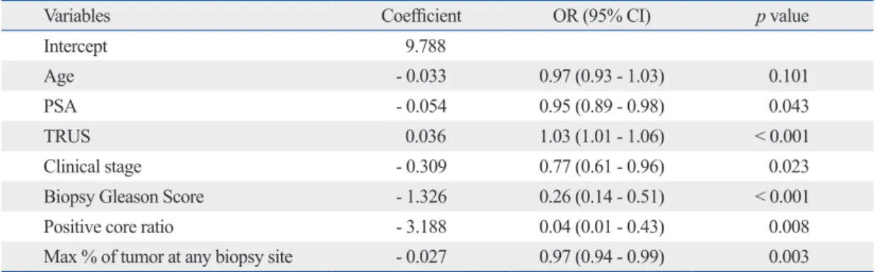
ables between significant and insignificant tumors, the ma- jority variables had highly statistically significant differences as shown in Table 1 (p < 0.001). Table 2 shows that the mul- tivariate regression analysis fitted in the development co- hort. PSA, prostate volume (TRUS), clinical stage, biopsy Gleason score, positive core ratio and maximum % of biop- sy tumor were observed to be independent predictors of the presence of IPC (all p values < 0.05). The performance (discrimination measurement) of the multivariate logistic regression model (MLRM) was demonstrated via the cal- culated area under the ROC curve (AUC), which was 0.847. Fig. 1A. shows the regression coefficient-based no- mogram, which was devised from these predictor variables.
Originally, we tried to fit PSAD into the nomogram; how- ever, because the calibration of the generated nomogram did not show good properties, we decided to include PSA and prostate volume (TRUS) instead of PSAD in our no- mogram. The nomogram is used by first locating the patient position on each predictor variable scale. Each scale posi- probability of IPC at radical prostatectomy. Calibration was
carried out for the constructed nomogram and internal vali- dation was performed from 200 additional bootstrap sam- ples to decrease the overfit bias. External validation was also performed in another cohort. Finally, various nomo- gram probability cutoffs were tested to assess the ability to identify patients with or without IPC. All analyses were per- formed using SAS version 9.1 statistical software (SAS In- stitute, Cary, NC, USA) and R version 2.8.1 (The R Founda- tion for Statistical Computing, Vienna, Austria).
RESULTS
Histopathologic evaluation of prostatectomy specimens re- vealed insignificant tumor in 67 (6.5%) of 1,031 patients.
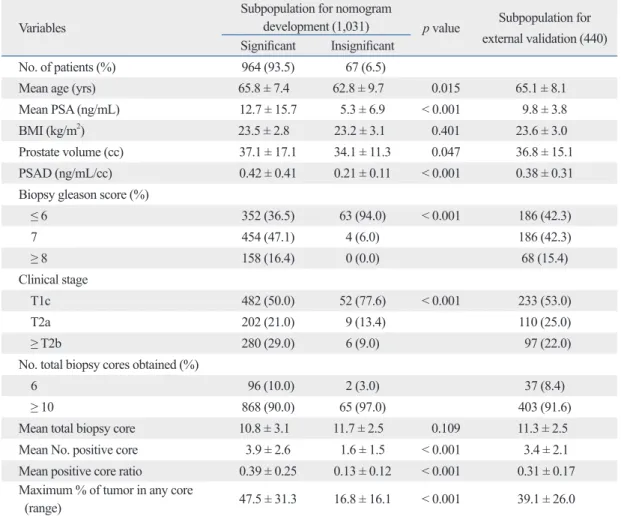
Of these 1031 patients, 98 (9%) underwent TRUS-guided preoperative sextant biopsy, while 933 (91%) underwent ex- tended biopsy (≥ 10 cores) schemes. When comparing vari-
Table 1. Comparison of Variables between Significant and Insignificant Prostate Cancer Variables
Subpopulation for nomogram
development (1,031) p value Subpopulation for external validation (440) Significant Insignificant
No. of patients (%) 964 (93.5) 67 (6.5)
Mean age (yrs) 65.8 ± 7.4 62.8 ± 9.7 0.015 65.1 ± 8.1
Mean PSA (ng/mL) 12.7 ± 15.7 5.3 ± 6.9 < 0.001 9.8 ± 3.8
BMI (kg/m2) 23.5 ± 2.8 23.2 ± 3.1 0.401 23.6 ± 3.0
Prostate volume (cc) 37.1 ± 17.1 34.1 ± 11.3 0.047 36.8 ± 15.1
PSAD (ng/mL/cc) 0.42 ± 0.41 0.21 ± 0.11 < 0.001 0.38 ± 0.31
Biopsy gleason score (%)
≤ 6 352 (36.5) 63 (94.0) < 0.001 186 (42.3)
7 454 (47.1) 4 (6.0) 186 (42.3)
≥ 8 158 (16.4) 0 (0.0) 68 (15.4)
Clinical stage
T1c 482 (50.0) 52 (77.6) < 0.001 233 (53.0)
T2a 202 (21.0) 9 (13.4) 110 (25.0)
≥ T2b 280 (29.0) 6 (9.0) 97 (22.0)
No. total biopsy cores obtained (%)
6 96 (10.0) 2 (3.0) 37 (8.4)
≥ 10 868 (90.0) 65 (97.0) 403 (91.6)
Mean total biopsy core 10.8 ± 3.1 11.7 ± 2.5 0.109 11.3 ± 2.5
Mean No. positive core 3.9 ± 2.6 1.6 ± 1.5 < 0.001 3.4 ± 2.1
Mean positive core ratio 0.39 ± 0.25 0.13 ± 0.12 < 0.001 0.31 ± 0.17 Maximum % of tumor in any core
(range) 47.5 ± 31.3 16.8 ± 16.1 < 0.001 39.1 ± 26.0
PSA, prostate specific antigen; BMI, body mass index; PSAD, PSA density.
Data are presented as number (%) or mean ± standard deviation.
(diagonal 45-degree dashed line). The solid line indicates bootstrap corrected nomogram performance. The internal validity of the multivariate model was evaluated by the bootstrap resampling method. The bootstrap-corrected AUC of the nomogram was 0.827. The external validation cohort data are represented by dotted line in Fig. 1C, where the curve represents the logistic calibration of the model.
tion has corresponding prognostic points (top axis). The points for each variable are added and the probability of IPC is estimated from the bottom line. The graphical repre- sentation of the accuracy of the nomogram is shown in Fig.
1B, where the nomogram predicted probability is represent- ed on the x-axis and the observed rate of IPC is plotted on the y-axis. Perfect prediction corresponded to a slope of 1
Table 2. Multivariate Logistic Regression Analysis of Preoperative Predictors for Insignificant Prostate Cancer
Variables Coefficient OR (95% CI) p value
Intercept 9.788
Age - 0.033 0.97 (0.93 - 1.03) 0.101
PSA - 0.054 0.95 (0.89 - 0.98) 0.043
TRUS 0.036 1.03 (1.01 - 1.06) < 0.001
Clinical stage - 0.309 0.77 (0.61 - 0.96) 0.023
Biopsy Gleason Score - 1.326 0.26 (0.14 - 0.51) < 0.001
Positive core ratio - 3.188 0.04 (0.01 - 0.43) 0.008
Max % of tumor at any biopsy site - 0.027 0.97 (0.94 - 0.99) 0.003
PSA, prostate specific antigen; TRUS, transrectal ultrasonograpy; OR, odds ratio; CI: confidence interval.
Points
0 10 20 30 40 50 60 70 80 90 100
PSA 50 45 40 35 30 25 20 15 10 5 0
Positive core ratio
1 0.7 0.3 0
Maxvol
100 70 40 10
Gleason score 1
≥ 7 6 5 4
Total points
0 50 100 150 200 250 300
T. stage 1
≥ T2b T2a T1c
Probabillty
0.001 0.01 0.1 0.5 0.9
TRUS 0 20 40 60 80 100 120 140 160 180 200
Fig. 1. (A) Nomogram for predicting IPC in localized prostate cancer. Find the position of each variable on the corresponding axis, draw a line to the ‘points’ axis for the number of points, add the points from all the variables together, and draw a line from the ‘total points’ axis to determine the IPC probabilities at the bottom. (B) Calibration curves of preoperative nomogram in internal validation cohort. The x-axis is the predicted probability and the y-axis is the actual probability of IPC from the nomogram. The dashed line represents the ideal calibra- tion curve for the nomogram (i.e., predicted probability completely corresponds with actual probability). The apparent accuracy without correction for over fit and bootstrap-corrected performance of our nomogram were represented by the dotted and solid line, respective- ly. (C) Calibration plot of nomogram in external validation cohort (n = 440). Solid line indicates logistic calibration curve and dotted line represent data for validation cohort. IPC, insignificant prostate cancer.
0.6 0.5 0.4 0.3 0.2 0.1 0.0
Actual probability
0.0 0.1 0.2 0.3
Predicted probability Internal validation
Apparent Bias-corrected Ideal
0.4 0.5 0.6
1.0
0.8
0.6
0.4
0.2
0.0
Actual probability
0.0 0.2
Predicted probability External validation
Ideal
Logistic corrected Nonparametric
0.4 0.6 0.8 1.0
A
B C
Generally, as not all prostate cancers pose a serious threat to life and consequently does not necessarily require therapy, it has been proposed that conservative management may be appropriate for those patients with IPC and a life expectan- cy of less than 10 years, or for younger men with a favor- able cancer, who are willing to accept the risk of metastases from an untreated tumor.3,12,13 However, men with IPC are often subjected to overtreatment with radical prostatectomy, because of difficulties in predicting the presence of IPC.
Draisma, et al.has revealed that the lead times were 12.3 and 6.0 years and overdetection rate were 27 and 56% in prostate-specific antigen screening tests at ages 55 and 75 years.14 Therefore, it is necessary to diagnosis patients with clinically low-risk prostate cancers to avoid the problem of overtreatment.
Over the past few years, a considerable number of nomo- grams have been constructed to predict IPC preoperatively.
For instance, Kattan, et al.3 showed several nomograms that predict the probability of IPC (range of c-index, 64-79%).
Those nomograms were externally validated in a screening cohort (range of c-index, 61-76%).15 Nakanishi, et al.4 at- tempted to further improve the accuracy of the existing tools, especially in patients with a single positive core at bi- opsy. In a cohort of 258 men, their model predicted with 73% accuracy. Chun, et al.5 also suggested another statisti- cal model with a predictive accuracy of 90.4% in a cohort of 1,132 men. These nomograms give clinicians many in- formations about the decision-making process between de- finitive therapy versus active surveillance.
In reality, however, most of the available reports on the The model curve and valiation cohort data show important
departures from ideal prediction. The overall discrimination measurement of the validation cohort was 0.771.
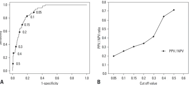
Fig. 2A illustrates the ROC curve, based on the fitted mul- tivariate logistic regression model. Fig. 2B illustrates the graph of positive predictive value (PPV)/negative predictive value (NPV) by various nomogram cutoffs. These figures are meant to assist clinicians in daily practice with identification of individuals with a particularly high chance for IPC, in whom active surveillance should be considered.
DISCUSSION
Today, the natural history of screen-detected prostate cancer remains poorly understood. Autopsy studies have revealed that 50% of men in the age group of 40-49 years harbor prostate cancer.9 It is also said that about one-third of men older than 50 years of age will harbor prostate cancer at au- topsy, whereas a man’s lifetime probability of developing invasive cancer is estimated to be 16.7% and the probabili- ty of dying from it is approximately 2.5%.10,11 Epstein, et al.7 reported that 80% of these cancers are of low volume (<
0.5 cc), and low grade, and can be classified as insignificant cancer. Insignificant cancer is usually defined as having a total tumor volume of less than 0.5 cc, being confined to the prostate with no focal or established extracapsular ex- tension and having no Gleason grade 4 or 5.
Traditionally, physician’s judgment has formed the basis for risk estimation, patient counseling and decision making.
Fig. 2. (A) ROC curve based on the fitted multivariate logistic regression model. Sensitivity and 1-specificity are represented based on a cut- off point (each dot) for IPC predicted probability. (B) The graph of PPV/NPV by nomogram cutoff. The x-axis indicates various cut off values and y-axis indicates the values of PPV/NPV. ROC, receiver operating characteristic; IPC, insignificant prostate cancer; PPV, positive predic- tive value; NPV, negative predictive value.
1.0
0.8
0.6
Sensitivity0.4
0.2
0.0 0.0
0.5
0.2 0.4
1-specificity
0.6 0.8 1.0
0.4 0.3
0.2 0.15
0.10.05
0.8 0.7 0.6 0.5
0.3 0.4
PPV / NPV ratio
0.2
0.0 0.1
0.05 0.1 0.15 0.2 Cut off value
0.3 0.4 0.5 0.6 PPV / NPV
A B
mor volume measurements were made primarily by visual estimation. Nevertheless, other studies have shown a strong correlation between the tumor volume estimates by visual inspection and grid morphometric analysis.24 Third, because our subjects were all Korean natives, the results of our study might have been different if our sample were more geo- graphically diverse. This factor may undermine the accura- cy of predictive nomogram in a specific population.
Choosing a therapy involves curing as well as morbidity and quality of life issues. As a consequence, individual in- formation on treatment and related morbidity are essential in treatment decision making for both doctor and patient. In that sense, this model can support patients and clinicians when various treatment options for screen-detected prostate cancer are considered.
In conclusions, our nomograms for the prediction of in- significant cancer provide more information than any cur- rently available diagnostic test alone, while the currently used tumor prognostic factors, such as serum PSA, Gleason score, and clinical stage, provide general prognostic infor- mation. The new nomogram, which is based on data from a large series of Korean patients, can predict insignificant cancer in Korean men more accurately than nomograms based on data from Western population.
ACKNOWLEDGEMENTS
The authors express their thanks to Korean Uropathology Society for providing pathological support. This study was supported by the grant of the Korean Urological Oncology Society sponsored by Korea Ipsen and the grant of the Korea Healthcare technology R&D Project, Ministry for Health, Welfare & Family Affairs, Republic of Korea (A084120).
REFERENCES
1. Klein EA. What is ‘insignificant’ prostate carcinoma? Cancer 2004;101:1923-5.
2. Klotz L. Active surveillance for prostate cancer: for whom? J Clin Oncol 2005;23:8165-9.
3. Kattan MW, Eastham JA, Wheeler TM, Maru N, Scardino PT, Er- bersdobler A, et al. Counseling men with prostate cancer: a nomo- gram for predicting the presence of small, moderately differentiat- ed, confined tumors. J Urol 2003;170:1792-7.
4. Nakanishi H, Wang X, Ochiai A, Trpkov K, Yilmaz A, Donnelly JB, et al. A nomogram for predicting low-volume/low-grade pros- tate cancer: a tool in selecting patients for active surveillance.
Cancer 2007;110:2441-7.
selection of patients for active surveillance, especially those predicting the presence of IPC, are based on data from Western samples. Little attention has been given to Asian populations. It is well known that geographical and ethnic differences exist in prostate cancer risk.16,17 A lower inci- dence of prostate cancer has been reported in Asian men, specifically in Korean men, compared to European men.18 In addition, prostate cancer screening in Korea is currently not as widespread as in Western countries, and prostate can- cer in Korea is generally not detected as early as in Western countries. Taken together, these differences may limit the usefulness of preexisting nomograms, which has prompted us to develop a new model for more accurate preoperative prediction of IPC in Korean men.
Our results revealed the presence of IPC in 67 (6.5%) pa- tients. Insignificant prostate cancer has been reported to be 6.4-26% of contemporary prostatectomy specimens, there- fore, so our results are similar to those of other reports.19-22 Our nomogram included five variables: serum PSA, clini- cal stage, biopsy Gleason score, biopsy positive core ratio and maximum percent of tumor in any core. Our nomo- gram is simple and easily applicable to patients because these variables are not difficult to be gathered clinically be- fore the surgery. These are also useful to predict prostate cancer stage.23 The strength of our nomogram for predict- ing IPC is constructed chiefly from analysis of a systematic extended biopsy scheme. We used a greater than 10-core extended biopsy scheme that included additional lateral cores from the base and the mid-zone, in addition to con- ventional six cores biopsy. Only 98 (9%) patients underwent sextant biopsy. Consequently, our results may be more pre- cise than those based on sextant biopsy-based data. More- over, our data came from 14 different institutions, which we think accurately reflects the Korean population. In addi- tion, pathologic slides in our study were re-evaluated by 9 uropathologists who had fully discussed the pathological assessment criteria, including tumor volume and biopsy Gleason grading, to improve interobserver reproducibility.
We believe that our prediction nomograms will assist phy- sicians in counseling Korean and Asian men with prostate cancer regarding the probability of the presence of IPC.
Limitations to the present study exist. First, as head to head comparison were not made with western tools, we did not demonstrate the performance of the nomogram in a west- ern population data set. Second, some prostatectomy speci- mens sectioned by different methods as previously described may affect the accuracy of tumor volume. In addition, tu-
West DW, et al. Serum androgens and sex hormone-binding glob- ulins in relation to lifestyle factors in older African-American, white, and Asian men in the United States and Canada. Cancer Epidemiol Biomarkers Prev 1995;4:735-41.
17. Lee SE, Kwak C, Park MS, Lee CH, Kang W, Oh SJ. Ethnic dif- ferences in the age-related distribution of serum prostate-specific antigen values: a study in a healthy Korean male population. Urol- ogy 2000;56:1007-10.
18. Robbins AS, Koppie TM, Gomez SL, Parikh-Patel A, Mills PK.
Differences in prognostic factors and survival among white and Asian men with prostate cancer, California, 1995-2004. Cancer 2007;110:1255-63.
19. Stamey TA, Freiha FS, McNeal JE, Redwine EA, Whittemore AS, Schmid HP. Localized prostate cancer. Relationship of tumor vol- ume to clinical significant for treatment of prostate cancer. Cancer 1993;71:933-8.
20. Stamey TA, Donaldson AN, Yemoto CE, McNeal JE, Sözen S, Gill H. Histological and clinical findings in 896 consecutive pros- tates treated only with radical retropubic prostatectomy: epidemio- logic significance of annual changes. J Urol 1998;160:2412-7.
21. Soh S, Kattan MW, Berkman S, Wheeler TM, Scardino PT. Has there been a recent shift in the pathological features and prognosis of patients treated with radical prostatectomy? J Urol 1997;157:
2212-8.
22. Noguchi M, Stamey TA, McNeal JE, Yemoto CM. Relationship between systematic biopsies and histological features of 222 radi- cal prostatectomy specimens: lack of prediction of tumor signifi- cance for men with nonpalpable prostate cancer. J Urol 2001;166:
104-9.
23. Lee HW, Seo SI, Jeon SS, Lee HM, Choi HY. Can we predict real T3 stage prostate cancer in patients with clinical T3 (cT3) disease before radical prostatectomy? Yonsei Med J 2010;51:700-7.
24. Humphrey PA, Vollmer RT. Percentage carcinoma as a measure of prostatic tumor size in radical prostatectomy tissues. Mod Pathol 1997;10:326-33.
5. Chun FK, Haese A, Ahyai SA, Walz J, Suardi N, Capitanio U, et al. Critical assessment of tools to predict clinically insignificant prostate cancer at radical prostatectomy in contemporary men.
Cancer 2008;113:701-9.
6. Shukla-Dave A, Hricak H, Scardino PT. Imaging low-risk prostate cancer. Curr Opin Urol 2008;18:78-86.
7. Epstein JI, Walsh PC, Carmichael M, Brendler CB. Pathologic and clinical findings to predict tumor extent of nonpalpable (stage T1c) prostate cancer. JAMA 1994;271:368-74.
8. Epstein JI, Chan DW, Sokoll LJ, Walsh PC, Cox JL, Rittenhouse H, et al. Nonpalpable stage T1c prostate cancer: prediction of in- significant disease using free/total prostate specific antigen levels and needle biopsy findings. J Urol 1998;160:2407-11.
9. Sakr WA, Grignon DJ, Crissman JD, Heilbrun LK, Cassin BJ, Pontes JJ, et al. High grade prostatic intraepithelial neoplasia (HG- PIN) and prostatic adenocarcinoma between the ages of 20-69: an autopsy study of 249 cases. In Vivo 1994;8:439-43.
10. Franks LM. Latent carcinoma of the prostate. J Pathol Bacteriol 1954;68:603-16.
11. Jemal A, Thomas A, Murray T, Thun M. Cancer Statistics, 2002.
CA Cancer J Clin 2002;52:23-47.
12. Steinberg GD, Bales GT, Brendler CB. An analysis of watchful waiting for clinically localized prostate cancer. J Urol 1998;159:
1431-6.
13. Walsh PC. Surgery and the reduction of mortality from prostate cancer. N Engl J Med 2002;347:839-40.
14. Draisma G, Boer R, Otto SJ, van der Cruijsen IW, Damhuis RA, Schröder FH, et al. Lead times and overdetection due to prostate- specific antigen screening: estimates from the European Random- ized Study of Screening for Prostate Cancer. J Natl Cancer Inst 2003;95:868-78.
15. Steyerberg EW, Roobol MJ, Kattan MW, van der Kwast TH, de Koning HJ, Schröder FH. Prediction of indolent prostate cancer:
validation and updating of a prognostic nomogram. J Urol 2007;
177:107-12.
16. Wu AH, Whittemore AS, Kolonel LN, John EM, Gallagher RP,