191
Multi-Detector Computed Tomography for Assessing the Left Ventricular Function, Perfusion and Viability
Soo Jin Lim MD1 and Ki Seok Choo MD2
1Department of Cardiology, Kim Hae Bokum Hospital, Kimhae,
2Department of Radiology, Pusan National University Hospital, Busan, Korea
ABSTRACT
MDCT has recently been used as a diagnostic tool to evaluate coronary artery stenosis and to detect coronary artery anomalies. The accuracy of MDCT has improved the assessment of stenosis as the number of detectors has increa- sed. In addition to its excellent role in evaluating coronary artery stenosis, MDCT can provide information re- garding the left ventricular function without having to perform additional scanning, and the myocardial viability of the left ventricle can be assessed on a delayed scan. MDCT has several disadvantages such as the amount of radiation exposure and the use of an iodine contrast medium, which might cause an adverse reaction, when combined with the reconstruction of the systolic and diastolic phases and the delayed scan. Yet MDCT may provide the opportu- nity to evaluate the coronary anatomy, the left ventricular function and the tissue characterization in one single imaging session that lasts less than 15 minutes. (Korean Circulation J 2007;37:191-195)
KEY WORDS:X-ray tomography, computed;Myocardium;Infarction.
Introduction
Ischemic heart disease is the leading cause of morbi- dity and mortality in most industrialized countries.1) The ventricular volume and myocardial mass are independent predictors of morbidity and mortality for patients suf- fering with coronary artery disease(CAD).2)3) Assessing the left ventricular function is important for arriving at a correct clinical diagnosis as well as for determining the optimal management and follow-up of patients with co- ronary artery disease.4) In addition, a diagnostic tool that can provide comprehensive information on the infarcted myocardium will not only help in the prognostic assess- ment of patients, it will also be valuable to identify those patients who will benefit from revascularization as well as to monitor the effects of new therapeutic strategies.5) To date, magnetic resonance imaging and nuclear medicine procedures seem to be optimal for assessing ischemic heart disease as these modalities can combine the assess- ment of myocardial perfusion and function and they are able to assess the myocardial viability.6)7) The recent, rapid technical development of CT scanner hardware has led to a rapid improvement of the spatial and temporal re-
solution and also to significantly faster cardiac scans.5) Therefore, MDCT has become an alternative tool to assess ischemic heart disease as it can evaluate coronary artery obstruction as well as assessing the left ventricu- lar function, perfusion and viability. In this article, we will review the MDCT studies that have evaluated the assessment of cardiac function, perfusion, and viability.
Assessment of Left Ventricular Function
With using sub-second gantry rotation times and de- dicated cardiac reconstruction algorithms on MDCT scanners, thin-section coronary angiograms have been able to depict significant proximal coronary artery ste- nosis in those patients who are known or suspected to have CAD.8)9) Data acquisition can cover the entire car- diac cycle with using a spiral computed tomography tech- nique. The diastolic and systolic image reconstructions can be generated from the same thin-section MDCT data sets with using a retrospective ECG-gating technique. A freely selectable distance from the preceding or follo- wing R-peak defines the data segment from the cardiac cycle that’s used for image reconstruction.10) For assessing only the global LV function, both the diastolic and sys- tolic phases are required to identify the proper image reconstructions windows, with a single axial image being reconstructed every 5% of the RR interval at a represen- tative mid-ventricular level. The appropriate reconstruc-
Correspondence:Ki Seok Choo, MD,Department of Radiology, Pusan Na- tional University Hospital, 1-10 Ami-dong, Seo-gu, Busan 602-739, Korea
Tel: 82-51-240-7354, Fax: 82-51-244-7534 E-mail: kschoo0618@naver.com
tion windows for the systolic and diastolic phases are visually identified as the images that show the minimum ventricular diameter(found at 25% of the RR interval) and the maximum ventricular diameter(found at 95% of
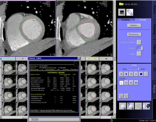
the RR interval), while the thin-section secondary refor- mations in a true short-axis orientation in the diastolic and systolic windows allow calculation of the LV volumes and consecutively, the functional parameters(Fig. 1).11)
Fig. 2. In this acute MI case, the findings on the early- and late-phase CT images (perfusion defect on the early phase (black arrow) and delayed hy- perenhancement (white arrow) and persistent perfusion defect (red arrow) in the papillary muscle) were well correlated with those findings on the perfusion and delayed MR images and on color-coded CT images, and so they helped to assess the infarcted area. CT: computed tomography, MI:
myocardiac infaretic, MR: magnets resonance imaging.
CT
Early CT Late CT Color coded
MR
Perfusion MR 5 min delay 15 min delay
Fig. 1. This screen-shot from a Wizard workstation (Siemens, Forchheim, Germany) shows the endocardial and epicardial borders of both the end-dia- stolic and the end-systolic phases; these were traced semi-automatically in the short axis orientation, by using dedicated analysis software, for eva- luating the left ventricular function.
However, it is not an easy task to determine the dedica- ted image sets that represent the appropriate end-diasto- lic and end-systolic phases on the axial image series, which are reconstructed in 5% increments of the R-R interval throughout the cardiac cycle. This is because the position of the heart changes continuously during con- traction; thus, a two-phase reconstruction method can be a useful alternative tool in this situation.12) Various invasive and noninvasive imaging modalities for the quan- titative and, in part, qualitative assessment of the left ventricular performance are available and they include x-ray angiography, 2-dimensional and 3-dimensional echocardiography, MRI, EBCT and gated SPECT.13) In addition, cardiac MRI provides excellent temporal and spatial resolution, it allows image acquisition in any de- sired plane and it has a high degree of accuracy and reproducibility for the quantitative measurements. The- refore, MRI is currently considered as a reference stan- dard for assessing cardiac function.14)15) Although MRI is already recognized as the preferred method for ana- lyzing cardiac volume and function among the existing modalities, the ability to obtain functional information with using MDCT may have a significant clinical im- pact.10) This is because MDCT can simultaneously obtain information regarding both coronary artery stenosis and the ventricular function without any additional scans.
The left ventricular volume measurements from the re- trospectively ECG-gated MDCT images enable volume- tric and global functional analysis that is well-correlated with cardiac MRI, which has been accepted as the refe- rence method for precisely analyzing the quantitative LV function as well as for detecting dyspnea and heart fai- lure. MDCT also has the advantage of being fast with regard to the breath-hold data acquisition and it is suita- ble for use in patients with pacemakers and implanted defibrillators. As respiratory motion during MDCT exa- minations can affect image quality and the subsequent volume measurements, another of its advantages is that data is usually acquired within one prolonged breath hold(8-15 s), as compared with the repetitive short brea- tholds that are employed for cine MRI. However, the images from both modalities are susceptible to degra- ded image quality that’s caused by any imperfect sinus rhythm. The MDCT images should theoretically be less susceptible to cardiac arrhythmias than the acquired MRI images because of the retrospective referencing of the ECG signal with performing MDCT versus the prospec- tive referencing with performing MRI.10) However, MDCT has several limitations: the need to administer contrast medium(which causes a volume load and might influ- ence the LV ejection fraction on cardiac MDCT), the radiation exposure to the patient and the limited tem- poral resolution.16)17) The radiation dose should be sub- stantially reduced by the introduction of ECG-triggered tube current modulation. Several studies have demon-
strated a 28% to 48% dose reduction with using ECG dose modulation, depending on the baseline heart rate.18)19) As the temporal resolution of cardiac 4-detec- tor row MDCT is not yet sufficient to image the coro- nary arteries at higher heart rates(70 bpm) without producing motion artifacts, cardiac MDCT is regularly performed after administering beta-blockers to the pati- ent in order to reduce the heart rate.20) Recent reports have shown that the use of beta blockers is effective to lower heart rates and reduce motion artifacts.21) A more rapid rotation time(up to 0.33 s per rotation) has been attained with MDCT,22) which makes it possible to shor- ten and stabilize the temporal resolution by using a seg- mental approach. Furthermore, with the introduction of dual source CT, the temporal resolution will decrease to 83 ms in the single segment reconstructions.23)
Assessment of Perfusion and Viability
Cardiac MRI can evaluate the cardiac morphology and function, and the myocardial perfusion, viability and me- tabolism, as well as the coronary status.24) An enhan- cing defect on the first-pass image involving the ventri- cular wall thickness, as noted on both the first-pass image and the delayed image of contrast-enhanced MRI, may allow the prediction of myocardial viability.25) Park et al.26) conducted an in vivo analysis of myocardial necrosis, as was determined by performing MRI in patients with acute myocardial infarction; they found that an infarct transmurality greater than 50% and the average necro- sis index of the dysfunctional segments(ANI) might repre- sent significant factors in the genesis of a pathologic Q wave and the endocardial sparing pattern of myocardial injury, and the latter was demonstrated by delayed enhan- cement. MR imaging was very useful for predicting the presence of an infarct-related artery in patients with myocardial necrosis, and this myocardial necrosis was determined by elevated levels of cardiac enzymes.27) Early experiments performed in animal models and subse- quently in human subjects have suggested that in vivo imaging for the detection, sizing and dating of myocar- dial infarctions is possible with using single-slice non- spiral CT systems.28-31) The principal limitations of these early techniques were the low temporal and spatial resolution and the inadequate imaging of the inferior wall because of the inability to obtain short-axis views of the left ventricle.32) The improved spatial and tempo- ral resolution of MDCT has made possible using this technique to assess the myocardium. Because an MDCT coronary angiographic examination is performed during maximal enhancement of the coronary artery, assessment of the myocardium in the same scan may reflect the myo- cardial perfusion.33) Gerber et al.34) recently reported that MDCT can identify two distinct contrast-enhan- cement patterns of MI, that is, the early hypoenhance-
ment as observed on the images of tissue perfusion that are obtained shortly after contrast injection, and the delayed hyperenhancement as seen on the images acqui- red 10 minutes after contrast injection. The location and extent of these two contrast enhancement patterns as noted on MDCT images are in good agreement with those seen on MR images, and these patterns have good interobserver, intraobserver and intrasubject reprodu- cibility(Fig. 2). Even though the composition of the con- trast materials used for MRI and CT imaging differs, a similar phenomenon occurs at the infarcted myocardium because of the similar pharmacokinetic properties of the CT and MRI contrast agents.35)36) In one study, because the CT examination was obtained during the rest phase, any perfusion defect distal to the coronary stenosis may not be obvious due to the compensatory vasodilatation of the distal segment.37) Kurata et al.38) demonstrated that adenosine triphosphate stress MDCT can describe both adenosine-triphosphate-induced myocardial ische- mia and coronary artery stenoses in patients suffering with coronary artery disease. However, assessing myo- cardial perfusion by MDCT is not currently widely accepted and this is still a controversial challenge in the field of cardiac CT.39) Similar to MR, analysis of the early and late contrast-enhancement patterns on CT also provides valuable information on tissue viability and therefore on the likelihood of functional recovery after revascularization.40)41) In patients suffering with acute MI, both the extent of the early hypoenhanced region, which reflects the extent of the microvascular obstruction, and the extent of the delayed hyperenhanced region, which reflects the infarct size, have indeed been associated with an increased risk of developing compli- cations during follow-up, including the development of adverse LV remodeling.42)43) Although performing a dual- phase study requires a double dose contrast media com- pared to a single-phase study, the X-ray exposure seems acceptable for those patients with heart disease that may be life-threatening, when considering the useful infor- mation that’s obtained from the myocardial studies.39)
Conclusion
MDCT can provide valuable information about both the left ventricular function and the left ventricular myo- cardial viability, and this modality has been proven to be in excellent agreement with MRI, which is the gold stan- dard for the assessment of both the function and via- bility of the left ventricular myocardium. CT coronary angiography with using MDCT has become a very re- liable method for evaluating coronary artery stenosis.
MDCT can give additional information regarding the left ventricular function in patients undergoing MDCT coronary angiography, as well as information on left ventricular myocardial viability if a delayed scan is per-
formed. Therefore, MDCT can be the optimal modality for assessing ischemic heart disease.
■ Acknoewledgments
We thank Bonnie Hami, Department of Radiology, University Hos- pitals of Cleveland, OH, for her editorial assistance.
REFERENCES
1) Murray CJ, Lopez AD. Alternative projections of mortality and disability by cause 1990-2020. Lancet 1997;349:1498-504.
2) White HD, Norris RM, Brown MA, Brandt PW, Whitlock RM, Wild CJ. Left ventricular end-systolic volume as the major deter- minant of survival after recovery from myocardial infarction.
Circulation 1987;76:44-51.
3) Hammermeister KE, DeRouen TA, Dodge HT. Variables predic- tive of survival in patients with coronary disease: selection by univariate and multivariate analyses from the clinical, electrocar- diographic, exercise, arteriographic, and quantitative angiogra- phic evaluations. Circulation 1979;59:421-30.
4) The Multicenter Postinfarction Research Group. Risk stratifica- tion and survival after myocardial infarction. N Engl J Med 1983;
309:331-6.
5) Nikolaou K, Knez A, Sagmeister S, et al. Assessment of myocar- dial infarctions using multidetector-row computed tomography. J Comput Assist Tomogr 2004;28:286-92.
6) Kim RJ, Fieno DS, Parrish TB, et al. Relationship of MRI dela- yed contrast enhancement to irreversible injury, infarct age, and contractile function. Circulation 1999;100:1992-2002.
7) Kitagawa K, Sakuma H, Hirano T, Okamoto S, Makino K, Takeda K. Acute myocardial infarction: myocardial viability assessment in patients early thereafter comparison of contrast-enhanced MR imaging with resting (201)Tl SPECT: single photon emission computed tomography. Radiology 2003;226:138-44.
8) Raff GL, Gallagher MJ, O`Neill WW, Goldstein JA. Diagnostic accuracy of noninvasive coronary angiography using 64-slice spiral computed tomography. J Am Coll Cardiol 2005;46:552-7.
9) Leber AW, Knez A, von Ziegler F, et al. Quantification of obstruc- tive and nonobstructive coronary lesions by 64-slice computed tomography: a comparative study with quantitative coronary an- giography and intravascular ultrasound. J Am Coll Cardiol 2005;
46:147-54.
10) Orakzai SH, Orakzai RH, Nasir K, Budoff MJ. Assessment of cardiac function using multidetector row computed tomography.
J Comput Assist Tomogr 2006;30:555-63.
11) Juergens KU, Grude M, Maintz D, et al. Multi-detector row CT of left ventricular function with dedicated analysis software versus MR imaging: initial experience. Radiology 2004;230:403-10.
12) Kim TH, Hur J, Kim SJ, et al. Two-phase reconstruction for the assessment of left ventricular volume and function using retro- spective ECG-gated MDCT: comparison with echocardiography.
AJR Am J Roentgenol 2005;185:319-25.
13) Greenberg SB, Sandhu SK. Ventricular function. Radiol Clin North Am 1999;37:341-59.
14) Pattynama PM, Lamb HJ, van der Velde EA, van der Wall EE, de Roos A. Left ventricular measurements with cine and spin-echo MR imaging: a study of reproducibility with variance component analysis. Radiology 1993;187:261-8.
15) Bellenger NG, Burgess MI, Ray SG, et al. Comparison of left ventricular ejection fraction and volumes in heart failure by echo- cardiography, radionuclide ventriculography and cardiovascular magnetic resonance: are they interchangeable? Eur Heart J 2000;
21:1387-96.
16) Grude M, Juergens KU, Wichter T, et al. Evaluation of global left
ventricular myocardial function with electrocardiogram-gated mul- tidetector computed tomography: comparison with magnetic reso- nance imaging. Invest Radiol 2003;38:653-61.
17) Ritchie CJ, Godwin JD, Crawford CR, Stanford W, Anno H, Kim Y. Minimum scan speeds for suppression of motion artifacts in CT. Radiology 1992;185:37-42.
18) Jakobs TF, Becker CR, Ohnesorge B, et al. Multislice helical CT of the heart with retrospective ECG gating: reduction of radiation exposure by ECG-controlled tube current modulation. Eur Radiol 2002;12:1081-6.
19) Gerber TC, Stratmann BP, Kuzo RS, Kantor B, Morin RL. Effect of acquisition technique on radiation dose and image quality in multidetector row computed tomography coronary angiography with submillimeter collimation. Invest Radiol 2005;40:556-63.
20) Schroeder S, Kopp AF, Kuettner A, et al. Influence of heart rate on vessel visibility in noninvasive coronary angiography using new multislice computed tomography: experience in 94 patients. Clin Imaging 2002;26:106-11.
21) Ropers D, Baum U, Pohle K, et al. Detection of coronary artery stenoses with thin-slice multi-detector row spiral computed tomo- graphy and multiplanar reconstruction. Circulation 2003;107:
664-6.
22) Nieman K, Cademartiri F, Lemos PA, Raaijmakers R, Pattynama PM, de Feyter PJ. Reliable noninvasive coronary angiography with fast submillimeter multislice spiral computed tomography. Cir- culation 2002;106:2051-4.
23) Achenbach S, Ropers D, Kuettner A, et al. Contrast-enhanced coronary artery visualization by dual-source computed tomogra- phy: initial experience. Eur J Radiol 2006;57:331-5.
24) Chang HJ, Choi SI. Era of multimodality imaging: where do we stand? Korean Circ J 2006;36:717-22.
25) Jung SE, Youn HJ, Lee KH, Hahn ST, Hong SJ, Kim CY. Use- fulness of contrast-enhanced magnetic resonance imaging in the prediction of myocardial viability after acute myocardial infarction.
Korean Circ J 2000;30:1257-63.
26) Park YH, Kim JH, Jung JH, et al. The meaning of pathologic Q wave in myocardial infarction assessed by magnetic resonance imaging. Korean Circ J 2004;34:945-52.
27) Song SG, Kim JH, Kim CW, et al. The diagnostic usefulness of cardiovscular magnetic resonance imaging for non-ischemic myo- cardial injury: the value of the endocardial sparing pattern on delayed enhancement. Korean Circ J 2004;34:1174-81.
28) Huber DJ, Lapray JF, Hessel SJ. In vivo evaluation of experimen- tal myocardial infarcts by ungated computed tomography. AJR Am J Roentgenol 1981;136:469-73.
29) Adams DF, Hessel SJ, Judy PF, Stein JA, Abrams HL. Computed tomography of the normal and infarcted myocardium. AJR Am J
Roentgenol 1976;126:786-91.
30) Kramer PH, Goldstein JA, Herkens RJ, Lipton MJ, Brundage BH.
Imaging of acute myocardial infarction in man with contrast-en- hanced computed transmission tomography. Am Heart J 1984;
108:1514-23.
31) Masuda Y, Yoshida H, Morooka N, Watanabe S, Inagaki Y. The usefulness of x-ray computed tomography for the diagnosis of myocardial infarction. Circulation 1984;70:217-25.
32) Georgiou D, Bleiweis M, Brundage BH. Conventional and ultra- fast computed tomography in the detection of viable versus infarc- ted myocardium. Am J Cardiol Imaging 1992;6:228-36.
33) Ko SM, Seo JB, Hong MK, et al. Myocardial enhancement pat- tern in patients with acute myocardial infarction on two-phase contrast-enhanced ECG-gated multidetector-row computed tomo- graphy. Clin Radiol 2006;61:417-22.
34) Gerber BL, Belge B, Legros GJ, et al. Characterization of acute and chronic myocardial infarcts by multidetector computed tomo- graphy: comparison with contrast-enhanced magnetic resonance.
Circulation 2006;113:823-33.
35) Mutzel W, Speck U, Weinmann HJ. Pharmacokinetics of iopro- mide in rat and dog. Fortschr Geb Rontgenstrahlen Nuklearmed Erganzungsbd 1983;118:85-90.
36) Allard M, Doucet D, Kien P, Bonnemain B, Caille JM. Experi- mental study of DOTA-gadolinium: pharmacokinetics and phar- macologic properties. Invest Radiol 1988;23(Suppl):S271-4.
37) Gould KL, Lipscomb K. Effects of coronary stenoses on coronary flow reserve and resistance. Am J Cardiol 1974;34:48-55.
38) Kurata A, Mochizuki T, Koyama Y, et al. Perfusion imaging using adenosine triphosphate stress multi-slice spiral computed tomo- graphy: alternative to stress myocardial perfusion scintigraphy.
Circ J 2005;69:550-7.
39) Koyama Y, Mochizuki T, Higaki J. Computed tomography assess- ment of myocardial perfusion, viability, and function. J Magn Reson Imaging 2004;19:800-15.
40) Gerber BL, Garot J, Bluemke DA, Wu KC, Lima JA. Accuracy of contrast-enhanced magnetic resonance imaging in predicting improvement of regional myocardial function in patients after acute myocardial infarction. Circulation 2002;106:1083-9.
41) Kim RJ, Wu E, Rafael A, et al. The use of contrast-enhanced magnetic resonance imaging to identify reversible myocardial dys- function. N Engl J Med 2000;343:1445-53.
42) Gerber BL, Rochitte CE, Melin JA, et al. Microvascular obstruc- tion and left ventricular remodeling early after acute myocardial infarction. Circulation 2000;101:2734-41.
43) Wu K, Zerhouni EA, Judd R, et al. Prognostic significance of microvascular obstruction by magnetic resonance imaging in pati- ents with acute myocardial infarction. Circulation 1998;97:765-72.