INTRODUCTION
Recent studies suggest the role of hypoxia in the tubuloin- terstitium as a common final pathway to end-stage renal dis- ease (1-3). Hypoxia has been shown to induce cellular prolif- eration and extracellular matrix (ECM) synthesis by cultured mesangial cells (4, 5) and fibroblasts (6). Hypoxia has also been shown to increase ECM synthesis in renal tubular epithe- lial cells as well as inducing epithelial-to-mesenchymal trans- differentiation (EMT) and apoptosis (1, 7). Chronic renal hy- poxia may occur by changes of oxygen supply (hypoxic injury) or impairment of blood flow to tubulointerstitium. Histo- logic studies of animal model and human kidney suggest that insufficient oxygenation resulting from peritubular capillary loss has a pivotal role in the pathogenesis of renal disease (8, 9). But, the underlying signaling mechanisms whereby hypox- ia alters cellular behaviors remain poorly defined. Hypoxia can regulate expressions of a variety of growth factors and cytokines in a cell and tissue-specific manner. These include transform- ing growth factor-β(TGF-β), vascular endothelial growth factor (VEGF), and endothelin-1 (ET-1) (10).
Connective tissue growth factor (CTGF) is a 38 kD cysteine- rich heparin-binding protein belonging to the CCN family and involves in stimulation of proliferation, angiogenesis, mi-
gration, ECM production, cell attachment, cell survival and apoptosis (11). TGF-βenhances CTGF mRNA and protein expression in mesangial cells (12). CTGF has been proposed to play an important role in tubulointerstitial fibrosis as one of the major mediators of TGF-β. It has been shown to be hypoxia-inducible in human breast cancer cells (13). Higgins et al. were the first to show hypoxic induction of CTGF in renal tubular epithelial cell cultures (14). But, the precise sig- naling mechanisms of the hypoxia-induced expression of CTGF remain unclear.
In the present study, we investigated: 1) the effect of hypoxia on TGF-βconcentration in conditioned medium and CTGF gene expression in cultured renal tubular cells; 2) what kind of mitogen-activated protein (MAP) kinase is involved in hypoxia-stimulated CTGF mRNA expression; and 3) whether hypoxia-induced CTGF mRNA expression is mediated by the release of TGF-β1.
MATERIALS AND METHODS Cell culture and hypoxic condition
Mouse tubular cells (MTC) are a proximal epithelial cell
S176
Young Ki Lee, Eun-Ji Kim*, Jung Eun Lee�, Jung Woo Noh, and Yoon-Goo Kim�
Department of Internal Medicine, Hallym University, Seoul; Cardiology Center*, Seoul National University Bundang Hospital, Seongnam; Department of Medicine�, Division of Nephrology, Samsung Medical Center, Sungkyunkwan University School of Medicine, Seoul, Korea
Address for correspondence Yoon-Goo Kim, M.D.
Department of Medicine, Division of Nephrology, Samsung Medical Center, Sungkyunkwan University School of Medicine, 50 Irwon-dong, Gangnam-gu, Seoul 135-710, Korea
Tel : +82.2-3410-3442, Fax : +82.2-3410-3849 E-mail : [email protected]
DOI: 10.3346/jkms.2009.24.S1.S176
Hypoxia Induces Connective Tissue Growth Factor mRNA Expression
Connective tissue growth factor (CTGF) is known to be a profibrotic growth factor, which mediate the fibrotic effect of transforming growth factor-β(TGF-β) and to stim- ulate cell proliferation and matrix production. CTGF has been shown to be hypoxia- inducible in several cell types. Here we investigated the effect of hypoxia on CTGF gene expression in cultured mouse renal tubular cells (MTC). Quiescent cultures of MTC were exposed to hypoxia (1% O2) or normoxia in serum-free medium. The effects on hypoxia-induced CTGF expression were evaluated by Northern blot and real-time PCR. The roles of mitogen-activated protein kinase (MAPK) and TGF-β were also determined using specific biochemical inhibitors. Exposure of quiescent tubular cells to hypoxia for 24 hr in a conditioned medium resulted in a significant increase TGF-β. Hypoxia caused a significant increase in CTGF mRNA expression in MTC. Either JNK or ERK inhibitor did not block the hypoxia-induced stimulation of CTGF, whereas an inhibitor of p38 MAPK reduced the hypoxia-induced changes of CTGF. Although hypoxia stimulated TGF-βproduction, neutralizing anti-TGF-β1 antibody did not abolish the hypoxia-induced CTGF mRNA expression. The data suggest that hypoxia up-regulates CTGF gene expression, and that p38 MAPK plays a role in hypoxic-stimulation of CTGF. We also demonstrated that hypoxia induces CTGF mRNA expression via a TGF-β1-independent mechanism.
Key Words : Cell Hypoxia; Connective Tissue Growth Factor; Transforming Growth Factor Beta 1; Mitogen- activated Protein Kinase
Received : 11 August 2008 Accepted : 19 November 2008
line isolated from 8-10-week-old naive SJL/J (H-2s) mice as previously described (15) and cultured in renal epithelial cell basal medium (REBM) (Clonetics, San Diego, CA, U.S.A.) containing 5% fetal bovine serum (FBS), 5,000 U/mL peni- cillin, 5,000 μg/mL streptomycin, and L-glutamine. Cultures were maintained in 75 cm3flasks at 37℃under a humidified atmosphere of 5% CO2/95% air. Cells were passed by tripsi- nization after they reached 80% confluency and utilized be- tween passages three and seven for all of the studies. Conflu- ent cells were made quiescent by 24 hr incubation in serum- free medium.
Fresh quiescence medium was added and cells were exposed to 1% O2, 4% CO2, 95% N2in an Anaerobic system 1,029 (Forma Scientific, Marietta, OH, U.S.A.) in open dishes. Cells were overlaid with medium to a depth of 3 mm sufficient to prevent dehydration. Control dishes were incubated for equiv- alent periods under normoxic conditions (21% O2, 5% CO2, 37℃). The tetrazolium dye-reduction assay (MTT; 3-[4,5- dimethylthiazol-2-yl]-2,5-diphenyl tetrazolium bromide;
Sigma) was used to test cell viability before and at end of treat- ment and did not reveal any signs of increased cell death in hypoxia (data not shown).
RNA extraction and Northern blot analysis
To determine the effect of hypoxia on CTGF gene expres- sion, MTC were grown in hypoxic condition for up to 10 hr.
At the end of each incubation time, cultures were harvested and total cellular RNA was extracted using TRIZOLRReagent (Gibco-BRL, MD, U.S.A.) according to the manufacturer’s instructions. Thirty micrograms of total RNA were electro- phoresed on a 1.2% formaldehyde-agarose gel and ethidium- bromide-stained gels were photographed under UV illumi- nation. RNA was transferred to nitrocellulose membranes for 3 hr. Membranes were prehybridized for 1 hr at 68℃in Quick Hyb Buffer (Stratagene, La Jolla, CA, U.S.A.) and hybridized for 3 hr in the same solution containing the 32P- labeled cDNA probes for human CTGF at 68℃. The blots were then washed two times, 30 min each in 4×SSC, 0.1%
SDS at 68℃followed by three 1 hr washes with 0.2×SSC, 0.1% SDS at 55℃. The blots were exposed to Kodak XAR- 5 film (Eastman Kodak, Rochester, NY, U.S.A.). Photographs of ethidium bromide-stained gels and autoradiograms were scanned and quantified by densitometry. To control for rela- tive equivalence of RNA loading, the blots were hybridized with β-actin cDNA (Clothech) probe or an oligonucleotide probe corresponding to the 18S rRNA. Data are expressed as relative mRNA levels calculated as a relative ratio of con- trol value from arbitrary densitometry units.
Real-time PCR
CTGF mRNA transcripts were detected by a real-time quantitative real-time PCR procedure using ABI Prism 7000
Sequence Detection System (PE Applied Biosystems, Foster City, CA, U.S.A.). This system is based on the ability of the 5′nuclease activity of Taq polymerase to cleave a dual-labeled fluorogenic hybridization probe during DNA chain exten- sion. The probe is labeled with a reporter fluorescent dye FAM at the 5′end and a quencher fluorescent dye TAMRA (6-carboxy-tetramethyl-rhodamine) at the 3′end. During the extension phase of PCR, the nucleolytic activity of the DNA polymerase cleaves the hybridization probe and releases the reporter dye from the probe with concomitant increase in reporter fluorescence. The following sequence-specific primers and probes were designed using Primer Express software (PE Applied Biosystems, Foster City, CA, U.S.A.): Rat CTGF forward primer: 5′-CAA GCT GCC CGG GAA AT-3′, reverse primer: 5′-CGG TCC TTG GGC TCA TCA-3′, and probe: 5′FAM-CTG TGA GGA GTG GGT-MGBNFQ- 3′; 18S rRNA forward primer 5′-CGG CTA CCA CAT CCA AGG AA-3′, reverse primer: 5′-GCT GGA ATT ACC GCG GCT-3′: probe 5′VIC-TGC TGG CAC CAG ACT TGC CCT C-TAMRA 3′. Primers were used at a concentra- tion of 900 nM and probes at 100 nM at each reaction. Reac- tions were assembled in a 96-well optical reaction plate. Each reaction contained 1×master mix from the kit (TaqMan One- Step RT-PCR Master Mix; Applied Biosystems, Inc.), forward primer, reverse primer, fluorescent probe, and RNA sample to a final volume of 25 μL per reaction. The plate was analyzed on a sequence-detection system, which simultaneously per- forms RT-PCR and detects fluorescence signal.
TGF-ββ1 ELISA
The cell supernatant was collected at each time point, clar- ified by centrifugation at 1,500×g for 5 min at 4℃and stored at -80℃. Release of TGF-β1 into culture supernatant of cells was measured using corresponding ELISA kits according to the manufacturer’s instructions (R&D Systems, Minneapolis, MN, U.S.A.). As the ELISA only measured levels of the active protein, activation of the supernatant was performed to mea- sure the total levels of both proteins. Briefly, cell supernatants were activated with 1.0 N HCl and subsequently neutralized with 1.2 N NaOH/0.5 M HEPES. Acid-activated samples were added to the precoated microplates and incubated at room temperature for 2 hr. A second antibody, anti-TGF-β1 poly- clonal antibody, was then added followed by TGF-β1 horseradish peroxidase conjugate. TGF-β1 was detected by adding the chromogenic substrate and this was followed by stop solution.
Absorbance was determined at optical density 450 nm.
Effect of MAP kinase inhibitors on CTGF gene expression
We also examined the role of MAP kinase in stimulation of CTGF mRNA induced by hypoxia. Cells were pretreated with MAP kinase inhibitors for 30 min, and incubated under
. .
hypoxic or normoxic conditions for 4 hr. Inhibitors were: PD- 098059, SB-203580 and JNK inhibitor (all from Calbiochem, La Jolla, CA, U.S.A.). At the end of incubation, the hypox- ia-induced CTGF expression was evaluated by Northern blot and RT-PCR.
Neutralization TGF-ββ1 activity
To determine whether the effects of hypoxia on CTGF gene expression are mediated by TGF-β1, anti-TGF-β1 antibody (2 μg/mL; R&D Systems, Minneapolis, MN, U.S.A.) was added to cells for 30 min immediately before hypoxia. Parallel exper- iments were undertaken without anti-TGF-β1 antibody. Lev- els of CTGF mRNA expression were measured by RT-PCR as described above.
Statistical analysis
Results are expressed as mean±SD of the mean. Statisti- cal significance (p<0.05) was evaluated using Student’s t-test or one-way analysis of variance. All experiments were repeat- ed at least three times with reproducible results.
RESULTS
Effect of hypoxia on CTGF mRNA expression
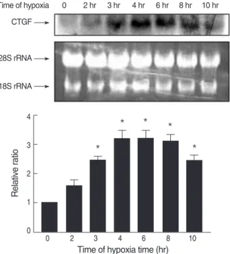
We then examined the effect of hypoxia on CTGF gene expression in MTC. Quiescent cultures were exposed in serum- free medium to hypoxia for 2, 3, 4, 6, 8, or 10 hr. At the end of each incubation time, CTGF gene expression was assessed by Northern blot analysis. As shown in Fig. 1, hypoxia caused a significant increase in CTGF mRNA expression in MTC.
The steady-state level of CTGF mRNA was maximally up regulated by 3-fold within 4 hr as compared with the cells cultured under the normoxic condition. Subsequently, there was a decline, but a 2-fold elevated level was maintained for 10 hr.
Role of MAP kinase in hypoxia-induced stimulation of CTGF mRNA
Since TGF-βhas been reported to activate the MAP kinase pathway (16-18), we tested the possible participation of MAP kinases in hypoxia-induced CTGF expression. We examined the activation of MAP kinase in response to hypoxia over 4-hr period. To inhibit MAP kinase, ERK inhibitors (PD-098059, 50 μM/L), p38 inhibitor (SB-203580, 20 μM/L) or JNK inhibitor (20 μM/L) was incubated 30 min before hypoxic stimulation. The effects of MAP kinase on hypoxia-induced CTGF gene expression were studied by RT-PCR (Fig. 2). Inter- estingly, ERK, p38 and JNK showed a different response to hypoxia on CTGF mRNA expression. Either JNK or ERK inhibitor did not block the up-regulation by hypoxia of CTGF
Fig. 1. Time course of CTGF mRNA expression in response to hypox- ia. MTC were isolated, cultured until 80% confluent, and exposed to 1% oxygen for 0-10 hr. CTGF transcripts were detected by North- ern blot analysis; a representative blot is shown along with ethidi- um bromide stained 28S and 18S rRNA for loading. Densitomet- ric analysis for CTGF mRNA levels is shown normalized to 18S rRNA level. Data are representative of three separate experiments.
*p<0.05 compared with 0 hr sample.
Time of hypoxia 0 2 hr 3 hr 4 hr 6 hr 8 hr 10 hr CTGF
28S rRNA
18S rRNA
Relative ratio
4
3
2
1
0
0 2 3 4 6 8 10
Time of hypoxia time (hr)
*
* * *
*
Fig. 2. Effects of MAPK inhibitors on hypoxia-induced CTGF expres- sion. Quiescent cells were pretreated for 30 min either with PD- 098059 (PD, 50 μM/L), SB-203580 (SB, 20 μM/L), or JNK inhibitor (20 μM/L) followed by exposure to hypoxia or normoxia for 4 hr.
At the end of incubations, total RNA was prepared from the cells and subjected to and RT-PCR. *p<0.05 compared with hypoxia.
Relative ratio
12
10
8
6
4
2
0
1 2 3 4 5 6 7 8
1. Control 2. Hypoxia
3. PD 4. PD+hypoxia
5. SB 6. SB+hypoxia
7. JNK inhibitor 8. JNK inhibitor+hypoxia PD: ERK inhibitor SB: p38 inhibitor
*
expression, whereas an inhibitor of p38 MAP kinase reduced the hypoxia-induced stimulation of CTGF.
TGF-ββstimulation of CTGF expression
TGF-βhas been shown to increase the expression of CTGF by a variety of cells (12, 19, 20). As a foundation for defining the TGF-β1 regulating CTGF expression in our system, we first determined the response of cultured renal tubular cells to TGF-β1. Quiescent cultures of MTC were exposed to normox- ic condition in serum-free medium, and then treated with 10 ng/mL TGF-β1 for 8 hr. A time-course study of the response demonstrated that there was a little delay before the level of CTGF mRNA began to increase after the addition of TGF-β1 (Fig. 3). TGF-β1 induced the expression of CTGF mRNA that began within 30 min, peaked at 2 hr and then declined by 8 hr.
Time-dependent effect of hypoxia on TGF-ββsynthesis
TGF-β1 is the major fibrogenic cytokine within the kid- ney and induced by hypoxia in a variety of cell types. Con- centrations of TGF-β1 by MTC was examined using ELISA to measure TGF-β1 in the cell supernatant (Fig. 4). Hypox- ia induced a 91% increase in the amount of TGF-β1 after 4 hr of hypoxia: 30.14±13.30 ng/mL vs. 15.74±2.92 ng/mL
in control. Assay of the cell supernatant in the 24-hr hypoxia showed a 161% increase in the amount of TGF-β1 (41.16± 6.31 ng/mL).
Effect of anti-TGF-ββ1 antibody on hypoxia-induced CTGF gene expression
Since CTGF is an response gene to TGF-βstimulation, we tested whether TGF-βsynthesis was required for the enhanced expression of CTGF by hypoxia, MTC were exposed to hypox- ia for 4 or 8 hr in the presence of anti-TGF-β1 antibody (2 μg/
mL) to block TGF-βproduced by the cells after hypoxia. The results showed that hypoxia produced substantial increases in
Fig. 3. TGF-β1 stimulation of CTGF in normoxic condition. TGF-β1 induces CTGF mRNA in cultured MTC. Tubular cells were serum starved for 24 hr before being treated with 10 ng/mL TGF-β1. Cells were harvested at various time points and RNA was extracted. No- rthern hybridization for CTGF mRNA levels were performed and normalized against endogenous 18S ribosomal RNA. *p<0.05 com- pared with 0 hr sample.
0 0.5 hr 1 hr 2 hr 4 hr 8 hr
CTGF TGF-β
Actin
18S RNA
Relative ratio
25
20
15
10
5
0
0 0.5 hr 1 hr 2 hr 4 hr 8 hr
Time
*
*
* Fig. 4. Effect of hypoxia on levels of TGF-β1 in MTC. MTC were iso- lated, cultured until 80% confluent, and exposed to 1% oxygen for 0-24 hr. Conditioned medium was collected at the end of hypoxia.
TGF-βlevel in the cell medium was measured by a TGF-βELISA kit. *p<0.05 compared with 0 hr sample.
TGF-beta (ng/mL)
50
40
30
20
10
0 0 4 hr 8 hr 12 hr 24 hr
Time of hypoxia
* *
*
Fig. 5. Effects of anti-TGF-β1 antibody on CTGF activity exposed to hypoxia. MTC were exposed to hypoxia in the presence of 2 μg/
mL of anti-TGF-β1 antibody for 30 min, followed by exposure to hy- poxia for 0-8 hr. Relative CTGF levels were calculated as a percent- age of the relevant control values from arbitrary densitometry units.
NS, not significant.
CTGF mRNA (% of normoxia)
600
500
400
300
200
100
0
Normoxia Hypoxia Hypoxia+ Normoxia Hypoxia Hypoxia+
anti-TGF- β1 anti-TGF- β1
4 hr 8 hr
NS
NS
the level of CTGF mRNA in both the absence and the pres- ence of anti-TGF-β1 antibody (Fig. 5). These findings suggest- ed that TGF-β1 synthesis was not required for hypoxia-induced stimulation of CTGF and TGF-β1 is not the primary medi- ator of the hypoxia-induced CTGF gene expression.
DISCUSSION
In this study, we found that hypoxia stimulated TGF-β1 and CTGF expression in cultured renal tubular cells. We also demonstrated that either JNK or ERK inhibitor did not block hypoxia-mediated CTGF expression, whereas an inhibitor of p38 MAP kinase reduced the expression of CTGF. Although hypoxia stimulated TGF-βproduction, neutralizing anti- TGF-β1 antibody did not abolish the hypoxia-induced CTGF mRNA expression. These data suggest that hypoxia regulates CTGF expression through TGF-β1-independent mechanism.
A number of studies have shown that hypoxia leads to pro- fibrotic responses in tubular epithelial cells and renal fibrob- lasts (2, 5, 6). Since tubular hypoxia is present in the early stage of disease in a progressive glomerulonephritis model (8), hypoxia from peritubular capillary loss may contribute to the process of renal fibrosis. Although numerous signal- ing pathways have been implicated in hypoxic signal trans- duction, the specific mechanisms underlying the fibrogenic response to hypoxia remain unknown. These could be medi- ated by direct effects on gene expression via hypoxia response elements (HRE) present in the gene promoters or secondary factors induced by hypoxia (14, 21). At the center of this cel- lular response to hypoxia is hypoxia-inducible factor (HIF) (22, 23). HIF is composed of two subunits, an oxygen-sensitive HIF-a subunit and a constitutively expressed HIF-b subunit (1). Hypoxia also modulates the activation of Nuclear factor- kappaB (NF-κB) in cells through decreased oxygen-depen- dent suppression of the key regulators of this pathway (24).
Hypoxia has been reported to stimulate the production of a variety of growth factors including the profibrotic factor, TGF-β, in multiple cell types (10, 25, 26). In our study, hypoxic treatment produced a 2- to 3-fold increase in the level of TGF-β1 in the cell supernatant. The period of hypoxia in this study (24 hr, 1% O2) was based on data showing that in vitro exposure of various cell lines to 1% O2for a minimum of 16 hr alters gene expression (25). Although previous studies showed that 24 hr of hypoxia induced a increase in the level of TGF-β1 (27), we have shown the shorter period of hypox- ia also increased TGF-β1 production.
CTGF has recently received much attention as a key deter- minant of progressive fibrosis and also wound repair, neoan- giogenesis, bone formation and embryonic development (28).
Comparison of the mouse and rat CTGF proteins showed a 95% identity and the rat CTGF protein also showed 91%
identity to the human CTGF protein (29). TGF-βinduces CTGF through different signaling pathways and a specific
TGF-βresponsive element in the CTGF promotor (12). CTGF is thought to act both as a profibrotic marker and as a down- stream mediator of TGF-β. In the present cell culture system, TGF-β1 produced a 15-fold increase in the level of CTGF mRNA. The induction of CTGF mRNA by TGF-β1 was also found in renal fibroblasts (unpublished observations). These results are consistent with observations in other cell systems where TGF-βinduces CTGF expression (12, 19, 20). It has previously been found that CTGF was regulated by hypoxia in a human breast cancer cell line, MDA231 (13). Our pre- sent study demonstrated that hypoxic treatment lead to sig- nificant elevation of CTGF mRNA in renal tubular cells. But, we have not measured CTGF protein levels. The steady-state level of CTGF mRNA in MTC was maximally up-regulated by 3-fold within 4 hr as compared with that from the normox- ic condition. Subsequently, there was a decline, but a 2-fold elevated level was maintained for 10 hr. Similarly, hypoxia (0.5% O2) has been shown to stimulate CTGF mRNA by 2-fold in renal tubular epithelial cells (14).
Since transcription is the major level of modulation of CTGF expression by TGF-β1 and since TGF-βhas been reported to activate the MAP kinase pathway (16-18), we tested what kind of kinase was involved in hypoxia-induced CTGF expres- sion. We found distinct difference in the role of MAP kinas- es in mediating CTGF expression in response to hypoxia. An inhibitor of p38 MAP kinase (SB-203580) reduced hypoxia- stimulated CTGF expression, whereas either JNK or ERK inhibitor (PD-098059) did not block the expression of CTGF.
The roles of MAP kinases under hypoxia are complicated, which depend on the specific cell types. ERK activation has been shown to be critical in hypoxia-induced VEGF expres- sion in HepG2 cells (30). In contrast, the study using NRK52 cells, ERK activation was not observed in response to hypox- ia, whereas activation of p38 could be demonstrated (26). In terms of p38, several studies have reported its critical role under hypoxia (26, 31), which is compatible with our data. To our knowledge, the roles of MAP kinases in CTGF regulation of renal tubular cells in response to hypoxia have not been pre- viously examined. Therefore the one new finding is that hypox- ia induces the expression of CTGF in a p38 MAPK-dependent manner. Demonstration of different roles of MAP kinases under hypoxic condition would be important in the investigation of the mechanisms regulating CTGF expression.
The present work has focused on TGF-βas a mediator to increase the expression of CTGF gene by hypoxia in cultured renal tubular cells. Since CTGF acts as a mediator of TGF-β1 signaling, we hypothesized that hypoxia-induced TGF-βcon- tributes to CTGF expression. Although TGF-β1 activity in- creased in response to hypoxia, the addition of the neutraliz- ing anti-TGF-β1 antibody did not block the hypoxia-induced CTGF expression. These data suggest that TGF-β1 is not the primary mediator of the hypoxia-induced CTGF expression in renal tubular cells. Also, hypoxia-induced CTGF expres- sion seems not to be from the increased bioactivity of TGF-β1.
Our data examining signaling pathways involved in CTGF gene expression are in good agreement with some recent stud- ies. Recent study showed that TGF-β1 signaling was not re- quired for hypoxic induction of CTGF promotor activity in renal tubular cells (14). The mechanisms by which hypoxia stimulates CTGF gene expression remain to be worked out but appear to be independent of TGF-β1. Limitations of our study were that the level of CTGF protein and in particular secreted protein were not measured, while mRNA expression was important.
In summary, this study demonstrates that hypoxia up-reg- ulates CTGF gene expression in cultured tubular cells and hypoxia-induced CTGF activation is dependent on p38 MAP kinase. Based on our study on CTGF gene regulation, we have identified that hypoxia regulates CTGF expression through TGF-β1-independent pathway. The data suggest an important role of CTGF in fibrogenic effect by hypoxia. Understand- ing the molecular mechanisms by which hypoxia induced fibrosis may open new avenues to the treatment of progres- sive renal diseases.
ACKNOWLEDGMENTS
The authors thank Dr Dong-Chul Han (Department of Internal Medicine, Soonchunhyang University, Seoul, Korea) for cell culture preparation. We thank Dr Dae Joong Kim, Dr Wooseong Huh and Dr Ha Young Oh (Samsung Medi- cal Center, Sungkyunkwan University School of Medicine, Seoul, Korea) for their helpful advice on this study. We are also grateful to Young Ok Kim and Hyun Joong Kim for their technical assistance and their useful comments on the paper.
REFERENCES
1. Nangaku M. Chronic hypoxia and tubulointerstitial injury: a final common pathway to end-stage renal failure. J Am Soc Nephrol 2006;
17: 17-25.
2. Fine LG, Bandyopadhay D, Norman JT. Is there a common mecha- nism for the progression of different types of renal diseases other than proteinuria? Towards the unifying theme of chronic hypoxia. Kidney Int Suppl 2000; 75: S22-6.
3. Norman JT, Orphanides C, Garcia P, Fine LG. Hypoxia-induced ch- anges in extracellular matrix metabolism in renal cells. Exp Nephrol 1999; 7: 463-9.
4. Kim SB, Kang SA, Park JS, Lee JS, Hong CD. Effect of hypoxia on the extracellular matrix production of cultured rat mesangial cells.
Nephron 1996; 72: 275-80.
5. Sahai A, Mei C, Pattison TA, Tannen RL. Chronic hypoxia induces proliferation of cultured mesangial cells: role of calcium and protein kinase C. Am J Physiol 1997; 273: F954-60.
6. Norman JT, Clark IM, Garcia PL. Hypoxia promotes fibrogenesis in
human renal fibroblasts. Kidney Int 2000; 58: 2351-66.
7. Manotham K, Tanaka T, Matsumoto M, Ohse T, Inagi R, Miyata T, Kurokawa K, Fujita T, Ingelfinger JR, Nangaku M. Transdifferenti- ation of cultured tubular cells induced by hypoxia. Kidney Int 2004;
65: 871-80.
8. Matsumoto M, Tanaka T, Yamamoto T, Noiri E, Miyata T, Inagi R, Fujita T, Nangaku M. Hypoperfusion of peritubular capillaries induces chronic hypoxia before progression of tubulointerstitial injury in a progressive model of rat glomerulonephritis. J Am Soc Nephrol 2004;
15: 1574-81.
9. Choi YJ, Chakraborty S, Nguyen V, Nguyen C, Kim BK, Shim SI, Suki WN, Truong LD. Peritubular capillary loss is associated with chronic tubulointerstitial injury in human kidney: altered expression of vascular endothelial growth factor. Hum Pathol 2000; 31: 1491-7.
10. Sahai A, Mei C, Schrier RW, Tannen RL. Mechanisms of chronic hypoxia-induced renal cell growth. Kidney Int 1999; 56: 1277-81.
11. Blom IE, Goldschmeding R, Leask A. Gene regulation of connec- tive growth factor: new targets for antifibrotic therapy? Matrix Biol 2002; 21: 473-82.
12. Chen Y, Blom IE, Sa S, Goldschmeding R, Abraham DJ, Leask A.
CTGF expression in mesangial cells: involvement of SMADs, MAP kinase, and PKC. Kidney Int 2002; 62: 1149-59.
13. Kondo S, Kubota S, Shimo T, Nishida T, Yosimichi G, Eguchi T, Sugahara T, Takigawa M. Connective tissue growth factor increased by hypoxia may initiate angiogenesis in collaboration with matrix metalloproteinases. Carcinogenesis 2002; 23: 769-76.
14. Higgins DF, Biju MP, Akai Y, Wutz A, Johnson RS, Haase VH. Hypox- ic induction of CTGF is directly mediated by Hif-1. Am J Physiol Renal Physiol 2004; 287: F1223-32.
15. Haverty TP, Kelly CJ, Hines WH, Amenta PS, Watanabe M, Harp- er RA, Kefalides NA, Neilson EG. Characterization of a renal tubu- lar epithelial cell line which secretes theautologous target antigen of autoimmune experimental interstitial nephritis. J Cell Biol 1988; 107:
1359-68.
16. Hayashida T, Poncelet AC, Hubchak SC, Schnaper HW. TGF-beta1 activates MAP kinase in human mesangial cells: a possible role in collagen expression. Kidney Int 1999; 56: 1710-20.
17. Hartsough MT, Mulder KM. Transforming growth factor beta acti- vation of p44mapk in proliferating cultures of epithelial cells. J Biol Chem 1995; 270: 7117-24.
18. Ravanti L, Hakkinen L, Larjava H, Saarialho-Kere U, Foschi M, Han J, Kahari VM. Transforming growth factor-beta induces collagenase- 3 expression by human gingival fibroblasts via p38 mitogen-activat- ed protein kinase. Biol Chem 1999; 274: 37292-300.
19. Kucich U, Rosenbloom JC, Herrick DJ, Abrams WR, Hamilton AD, Sebti SM, Rosenbloom J. Signaling events required for transforming growth factor-βstimulation of connective tissue growth factor expres- sion by cultured human lung fibroblasts. Arch Biochem Biophys 2001;
395: 103-12.
20. Paradis V, Dargere D, Bonvoust F, Vidaud M, Segarini P, Bedossa P. Effects and regulation of connective tissue growth factor on hep- atic stellate cells. Lab Invest 2002: 82: 767-74.
21. Zhang H, Akman HO, Smith EL, Zhao J, Murphy-Ullrich JE, Batu- man OA. Cellular response to hypoxia involves signaling via Smad
proteins. Blood 2003; 101: 2253-60.
22. Maxwell P. HIF-1: an oxygen response system with special relevance to the kidney. J Am Soc Nephrol 2003; 14: 2712-22.
23. Marx J. Cell biology. How cells endure low oxygen. Science 2004; 303:
1454-6.
24. Cummins EP, Comerford KM, Scholz C, Bruning U, Taylor CT. Hy- poxic regulation of NF-kappaB signaling. Methods Enzymol 2007;
435: 479-92.
25. Minchenko A, Bauer T, Salceda S, Caro J. Hypoxic stimulation of vascular endothelial growth factor expression in vitro and in vivo. Lab Invest 1994; 71: 374-9.
26. Nakagawa T, Lan HY, Zhu HJ, Kang DH, Schreiner GF, Johnson RJ.
Differential regulation of VEGF by TGF-βand hypoxia in rat prox- imal tubular cells. Am J Physiol Renal Physiol 2004; 287: F658-64.
27. Orphanides C, Fine LG, Norman JT. Hypoxia stimulates proximal
tubular cell matrix production via a TGF-beta1-independent mecha- nism. Kidney Int 1997; 52: 637-47.
28. Gupta S, Clarkson MR, Duggan J, Brady HR. Connective tissue growth factor: potential role in glomerulosclerosis and tubulointerstitial fibro- sis. Kidney Int 2000; 58: 1389-99.
29. Xu J, Smock SL, Safadi FF, Rosenzweig AB, Odgren PR, Marks SC Jr, Owen TA, Popoff SN. Cloning the full-length cDNA for rat con- nective tissue growth factor: implications for skeletal development.
J Cell Biochem 2000; 77: 103-15.
30. Mottet D, Michel G, Renard P, Ninane N, Raes M, Michiels C. Role of ERK and calcium in the hypoxia-induced activation of HIF-1. J Cell Physiol 2002; 194: 30-44.
31. Sodhi CP, Batlle D, Sahai A. Osteopontin mediates hypoxia-induced proliferation of cultured mesangial cells: role of PKC and p38 MAPK.
Kidney Int 2000; 58: 691-700.