INTRODUCTION
Osteoporosis is characterized by low bone mass and the microarchitectural deterioration of bone tissue with a conse- quent increase in bone fragility and susceptibility to fracture (1). The diagnosis and the clinical management of osteoporosis have been based on the measurement of bone mineral density (BMD) using dual X-ray absorbtiometry (DXA) as a stan- dard method. However, BMD measurements alone do not provide information about the microarchitecture or the me- chanical properties of bone. Quantitative ultrasound (QUS) methods have been introduced in recent years for the assess- ment of the skeletal status in osteoporosis. It has been sug- gested that QUS may provide information about not only bone density but also the microarchitecture and elastic prop- erties of bone (2). The QUS measurements of bone status have been shown to be related to fracture risk, independently of BMD (3-5). However, calcaneal QUS thresholds for the diag- nosis or treatment of osteoporosis have not as yet been defined.
In addition, there are several unresolved issues concerning the accuracy, long-term stability and precision of the technique, cross-calibration between the various QUS devices (6).
In 1994, the World Health Organization (WHO) proposed guidelines for the diagnosis of osteoporosis based on BMD measurements. According to the WHO criteria, osteoporosis is defined as a BMD of 2.5 or more standard deviations (SD) below the peak mass of a young, healthy, sex - and race-match- ed reference population (7). However, the reference range may vary in different populations (8-10). Therefore, the construc- tion of reference ranges that accurately represent the popula- tion at large is essential for the correct identification of osteo- porosis.
The number of QUS devices in clinical use is scheduled to increase in Korea. However, there are currently no QUS ref- erence data available for Korean population. This study aimed to establish reference data for the QUS measurements of the calcaneus and for the BMDs of the forearm and calcaneus, and to examine the correlations between the QUS parameters and
Min-Ho Shin, Sun-Seog Kweon, Kyeong-Soo Park, Heon Heo, Seung-joon Kim, Hae-Sung Nam*, Seul-Ki Jeong�, Eun-Kyung Chung�, Jin-Su Choi�
Department of Preventive Medicine, Seonam University College of Medicine, Namwon; Department of Preventive Medicine*, Chungnam National University College of Medicine, Daejeon; Department of Neurology�, Chonbuk National University Hospital, Jeonju, Department of Preventive Medicine�, Chonnam National University College of Medicine, Chonnam National University Research Center of Medical Sciences, Gwangju, Korea
Address for correspondence Eun-Kyung Chung, M.D.
Department of Preventive Medicine, Chonnam National University College of Medicine, Chonnam National University, Research Center of Medical Sciences, 5 Hak 1-dong, Dong-gu, Gwangju 501-746, Korea
Tel : +82.62-220-4175, Fax : +82.62-233-0305 E-mail : [email protected]
1011
Quantitative Ultrasound of the Calcaneus in a Korean Population:
Reference Data and Relationship to Bone Mineral Density Determined by Peripheral Dual X-ray Absorptiometry
The aim of this study was to establish reference data for the quantitative ultrasound (QUS) of the calcaneus and for the bone mineral densities (BMD) of the calcaneus and distal forearm, and to evaluate the correlation between QUS parameters and BMD in a Korean population. We performed a cross-sectional study involving 3,053 subjects (1,225 men and 1,828 women). QUS was conducted on the calcaneus and was quantified as speed of sound (SOS, m/sec), broadband ultrasound attenuation (BUA, dB/MHz), and stiffness index. The BMD of the calcaneus and distal forearm were measured using dual X-ray absorptiometry. The peak mean values for the QUS parameters occurred in the 20 to 29-yr-old subjects of both sexes, with the exception of the BUA, which reached the highest values in women of 30-39 yr. For both sexes, the mean BMD of the calcaneus was highest in those 20-29 yr old and that of the distal forearm was highest in those 40-49 yr old. The correlations between the QUS and BMD results were found to be 0.41 to 0.73 in men and 0.51 to 0.76 in women. Theses data can serve as a reference values for both sexes in Korea.
Key Words : Absorptionmetry; Photon Ultrasonography; Quantitative Ultrasound; Bone Density; Bone Mineral Density; Reference Data; Calcaneus; Korea
Received : 14 October 2004 Accepted : 14 June 2005
the BMD in a Korean population.
MATERIALS AND METHODS Subjects
The study population was composed of participants in the Namwon Study and the Thyroid Disease Prevalence Study.
Out of 4,697 eligible subjects aged 45 to 74 yr in the Nam- won Study which is an ongoing prospective study that was designed to investigate the determinants of the occurrence and progression of cardiovascular disease, osteoporosis, and dementia in elderly Koreans, 1,964 subjects (41.8% of eligi- ble subjects; 694 men and 1,253 women) underwent clini- cal examinations following interviews. Out of the Thyroid Disease Prevalence Study participants, 3,000 subjects aged 20 to 74 yr were randomly selected by 5-yr age strata and sex from the Yeonggwang and Muan counties in Jeollanam- do province of Korea. Of these, 1,175 subjects (39.2% of eligible subjects; 575 men and 600 women) underwent clini- cal examinations. In all, a total of 3,139 subjects (1,269 men and 1,853 women) aged 20 to 74 yr were recruited. Of these, 3,053 subjects who underwent both QUS and BMD mea- surements (1,225 men and 1,828 women; 20 to 74 yr old) formed the final study sample for the principal analysis. All participants provided informed consent, and the study was approved by the institutional research ethics committee.
Anthropometric measurements
Anthropometric measurements of subjects wearing light clothing and no shoes were conducted by experienced research staff. Height was measured to the nearest 0.1 cm, and weight was measured in the upright position to the nearest 0.1 kg.
The body mass index (BMI) was calculated as weight divid- ed by height squared (kg/m2).
Measurement of risk factors
The questionnaire administered to all participants was de- signed to determine the history of type 2 diabetes mellitus, hypertension, medication and lifestyle factors such as alcohol consumption and smoking habits. For women, information on menstrual status and use of hormone replace therapy was also collected. The subjects were categorized according to their smoking habits as current smokers, ex-smokers, and non-smo- kers. Type 2 diabetes mellitus was defined as a self-reported history (having been informed by a physician that diabetes was present) or as a fasting blood glucose level ≥126 mg/dL.
Subjects with persistent elevated blood pressure (≥140/90 mmHg) or who were on antihypertensive medication were classified as hypertensive according to the criteria of the report of the Joint National Committee on Prevention, Detection,
Evaluation, and Treatment of High Blood Pressure (JNC 7) (11).
Measurements of quantitative ultrasound and bone mineral densitometry
The QUS parameters of the right calcaneus were determined in all participants using the Achilles InSight ultrasonometer (GE Lunar Corporation, Madison, WI, U.S.A.), according to the instruction manual provided by the manufacturer. Two QUS parameters were measured: broadband ultrasound atten- uation (BUA) and speed of sound (SOS). A third parameter, the stiffness index (SI), was calculated using the formula, SI=
(0.67×BUA)+(0.28×SOS)-420. The within-day coefficients of variation (CV) for duplicate measurements in 30 adults were 1.93% for SI, 1.74% for BUA, and 0.20% for SOS. The bone mineral densities (BMD; g/cm2) of the right calcaneus and the nondominant forearm were measured by dual energy X-ray absorptiometry (DXA) using a PIXI densitometer (GE Lunar Corporation). Calibration was performed daily with a physi- cal phantom. The within-day CV for duplicate measurements in 60 adults were 0.19% and 0.17% for the forearm and cal- caneus BMD, respectively.
Statistical analysis
Data are expressed as means±standard deviations (SD).
The correlations were examined using Pearson’s correlation and partial correlation, with age held constant. The T-scores for each subject were calculated using the means and SDs for the QUS parameters and BMD using the 20-39 age group in this study as the normal young adult comparison popula- tion. All analyses were performed using SPSS software version 11.0 (SPSS, Chicago, IL, U.S.A.).
RESULTS
The demographic and clinical features of the study popu- lation are presented in Table 1. The mean ages of the partic- ipants were 57.6±12.0 yr for the men and 57.6±11.3 yr for the women. The percentages of smoking, alcohol intake, and diabetes mellitus were significantly higher in men than in women; the percentage of hypertension was not signifi- cantly different between the sexes. In women, the percent- ages of menopause and use of hormone replacement therapy were 64.9% and 10.8%, respectively.
Table 2 and 3 present the reference data for the QUS and BMD values according to age and sex for the population. The peak mean values for the QUS parameters occurred in the 20 to 29-yr-old subjects of both sexes, with the exception of the BUA, which reached the highest values in women of 30-39 yr. For both sexes, the mean BMD of the calcaneus was the highest in 20 to 29-yr-old subjects and that of the distal fore-
arm was the highest in 40 to 49-yr-old subjects. From the peak values to the values in the 70 to 74-yr-old group, there were overall declines of 10.5% for BUA, 1.9% for SOS, 17.1% for SI, 23.4% for forearm BMD, and 24.2% for calcaneus BMD in men, and 20.8% for BUA, 3.1% for SOS, 31.6% for SI, 37.6% for forearm BMD, and 41.6% for calcaneus BMD in women.
The mean ages of the young normal controls were 31.1± 6.1 yr for the men (n=127) and 31.7±5.6 yr for the women (n=164). The means and SDs for the BUA, SOS and SI were 121.3±15.6 dB/MHz, 1,555.2±43.2 m/sec and 96.2± 20.7, respectively for the men, and 113.7±13.8 dB/MHz,
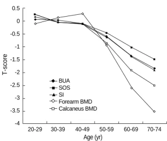
1,551.0±29.1 m/sec and 90.0±15.4, respectively for the women. The means and SDs for forearm and calcaneus BMD were 0.589±0.071 g/cm2and 0.623±0.113 g/cm2, respecti- vely for the men and 0.496±0.052 g/cm2and 0.493±0.081 g/cm2, respectively for the women. The age-related declines in T-scores for both the BMD and QUS measurements are shown in Fig. 1 and 2. The mean T-scores for the BMD mea- surements were more negative than those for the QUS mea- surements. In the 70-74 yr group, the mean T-scores for BUA, SOS, SI, forearm BMD, and calcaneus BMD were -0.8, -0.6, -0.8, -1.7, and -1.3, respectively, in men, and -1.7, -1.5, -1.8, -3.4, and -2.4, respectively, in women.
Table 4 shows the correlations between the QUS parame- ters and the BMD results. The correlations between BUA and SOS were found to be 0.69 in men and 0.61 in women. The correlations between the QUS parameters and BMD were 0.41 to 0.73 in men and 0.51 to 0.76 in women. The highest corre- lations were found between SI and calcaneus BMD in women (r=0.76) and between BUA and calcaneus BMD in men (r=
0.73). The correlations between the BMDs of the distal fore- arm and the calcaneus were found to be 0.63 in men and 0.74 in women.
DISCUSSION
This study provides population-based reference data for the QUS parameters and the forearm and calcaneus BMDs in a Korean population. This is the first large study to present ref- erence data for QUS parameters in both sexes in Korea. Kim
Age range (yr)
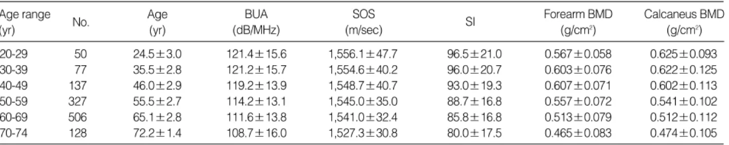
20-29 50 24.5±3.0 121.4±15.6 1,556.1±47.7 96.5±21.0 0.567±0.058 0.625±0.093
30-39 77 35.5±2.8 121.2±15.7 1,554.6±40.2 96.0±20.7 0.603±0.076 0.622±0.125
40-49 137 46.0±2.9 119.2±13.9 1,548.7±40.7 93.0±19.3 0.607±0.071 0.602±0.113 50-59 327 55.5±2.7 114.2±13.1 1,545.0±35.0 88.7±16.8 0.557±0.072 0.541±0.102 60-69 506 65.1±2.8 111.6±13.8 1,541.0±32.4 85.8±16.8 0.513±0.079 0.512±0.112 70-74 128 72.2±1.4 108.7±16.0 1,527.3±30.8 80.0±17.5 0.465±0.083 0.474±0.105 Table 2.Quantitative ultrasound indices and bone mineral density reference data in Korean men (n=1,225)
Data are given as mean±standard deviation.
BUA, broadband ultrasound attenuation; SOS, speed of sound; SI, stiffness index; BMD, bone mineral density.
SOS (m/sec) BUA
(dB/MHz)
Calcaneus BMD (g/cm2) Forearm BMD
(g/cm2) Age SI
No. (yr)
Age range (yr)
20-29 55 25.0±3.0 113.2±13.6 1,556.1±28.2 91.1±14.7 0.486±0.048 0.507±0.084
30-39 109 35.1±2.9 113.9±13.9 1,548.5±29.4 89.4±15.8 0.501±0.053 0.486±0.078 40-49 228 46.6±2.5 111.1±13.8 1,547.3±32.1 87.2±15.7 0.510±0.060 0.483±0.087 50-59 519 55.4±2.7 105.2±12.9 1,537.3±28.0 80.5±14.2 0.446±0.067 0.422±0.087
60-69 756 65.0±2.9 95.4±11.7 1,520.8±23.0 69.4±12.2 0.364±0.066 0.344±0.080
70-74 161 72.0±1.4 90.2±11.7 1,507.6±20.5 62.3±11.5 0.318±0.057 0.296±0.074
Table 3.Quantitative ultrasound indices and bone mineral density reference data in Korean women (n=1,828)
Data are given as mean±standard deviation.
BUA, broadband ultrasound attenuation; SOS, speed of sound; SI, stiffness index; BMD, bone mineral density.
SOS (m/sec) BUA
(dB/MHz)
Calcaneus BMD (g/cm2) Forearm BMD
(g/cm2) Age SI
No. (yr)
Data are given as mean±standard deviation.
*p value by t-test or chi-square test as appropriate.
Characteristics
Age, (yr) 57.6±12.0 57.6±11.3 0.92
Height, (cm) 165.5±6.4 152.7±6.0 0.00 Body weight, (kg) 65.8±10.1 57.6±8.6 0.00
Body mass 24.0±3.0 24.7±3.2 0.00
index, (kg/m2)
Current smoker, (%) 38.7 3.5 0.00
Alcohol intake, (%) 63.5 28.3 0.00
Hypertension, (%) 31.8 33.4 0.36
Diabetes mellitus, (%) 11.5 6.9 0.00
Hormone replacement - 10.8 -
treatment, (%)
Menopausal state (%) 64.9 -
Table 1.Characteristics of the subjects by sex
p*
(n=1,828) (n=1,225)
Female Male
et al. (12) presented reference data for the QUS of the calca- neus from 552 Korean women and 238 young women. Their data showed lower mean values for both normal young women and for women over 50 yr old, compared with the data pre- sented here. When compared with the reference values report- ed in a Japanese study (13), our BUA data values were simi-
lar for both sexes, and our SOS data were similar for the 20- 29 age group of men and the 20-39 age group of women;
however, our SOS values for men over 30 yr and for women over 40 yr were higher than the Japanese values. The data values in the present study were lower than those reported in Brazilian women (14). The SOS data values in the present stu- dy were similar to and the BUA data were lower than the ref- erence data reported for Nigerian women (15).
Shin et al. (16) presented the mean BMD values for a total of 317 healthy Korean women and 183 healthy Korean men aged 20 to 29 yr. The authors reported mean and SD values for calcaneus BMD in young males and females of 0.613± 0.100 g/cm2and 0.494±0.076 g/cm2, respectively; the values for forearm BMD in young males and females were 0.550± 0.064 g/cm2and 0.465±0.061 g/cm2, respectively. These values are lower than those in the present study; however, our data were similar to the reference values for Asia supplied by the manufacturer of the densitometer (0.580±0.06 g/cm2 for the forearm and 0.620±0.09 g/cm2for the calcaneus in men; 0.490±0.06 g/cm2 for the forearm and 0.500±0.08 g/cm2for the calcaneus in women).
In our study, the mean BMD of the distal forearm was the highest in 40 to 49-yr-old subjects. This finding is in accor- dance with previous studies (17, 18), but three large popula- tion studies (19-21) showed that the mean BMD of the dis- tal forearm was highest in 30 to 34-yr-old subjects. This may be attributable to the ethnic difference or to use of the differ- ent devices to determine the BMD at the forearm. This also may be attributable to the relatively small sample of normal young adults in our study.
The age-related declines in the T-scores based on the QUS parameters were approximately half those based on the BMD measured by DXA. This is consistent with other studies (22, 23). When the WHO definition of osteoporosis is applied to the T-scores based on QUS measurements, the prevalence of
T-score
0.5 0 -0.5 -1 -1.5 -2 -2.5 -3 -3.5 -4
20-29 30-39 40-49 50-59 60-69 70-74
T-score
0.5 0 -0.5 -1 -1.5 -2 -2.5 -3 -3.5 -4
20-29 30-39 40-49 50-59 60-69 70-74
Fig. 1.Age-related decline of T-score for QUS parameters and BMD in men.
BUA SOS SI
Forearm BMD Calcaneus BMD
Fig. 2.Age-related decline of T-score for QUS parameters and BMD in women.
Data are given as Pearson’s correlation coefficient (partial correlation coefficient).
BUA, broadband ultrasound attenuation; SOS, speed of sound; SI, stiffness index; BMD, bone mineral density.
All value are statistically significant (p<0.001) by Pearson’s correlation and partial correlation analysis.
Men BUA 1.00
(1.00)
SOS 0.69 1.00
(0.68) (1.00)
SI 0.92 0.92 1.00
(0.91) (0.92) (1.00) Forearm 0.49 0.41 0.49 1.00
BMD (0.43) (0.37) (0.44) (1.00) Calcaneus 0.73 0.59 0.71 0.63 1.00
BMD (0.70) (0.57) (0.69) (0.56) (1.00)
Women BUA 1.00
(1.00)
SOS 0.61 1.00
(0.50) (1.00)
SI 0.92 0.88 1.00
(0.89) (0.84) (1.00) Forearm 0.59 0.51 0.62 1.00
BMD (0.41) (0.33) (0.43) (1.00) Calcaneus 0.72 0.65 0.76 0.74 1.00
BMD (0.60) (0.53) (0.66) (0.57) (1.00) Table 4.Pearson’s and partial correlation coefficients between quantitative ultrasound and bone mineral density
BUA
Cal- caneus
BMD Forearm SI BMD SOS
Age (yr) Age (yr)
BUA SOS SI
Forearm BMD Calcaneus BMD
osteoporosis appears to be lower than that determined based on BMD measurements. It is clear that the T-score cannot be used interchangeably with measurements derived from differ- ent techniques and different sites (24). Frost et al. suggested that the WHO T-score threshold of -2.5 for the diagnosis of osteoporosis requires modification when using QUS to assess the skeletal state and that a T-score threshold of -1.8, based on the BUA and SOS of the calcaneus, may be appropriate for identifying postmenopausal women at risk for osteoporosis (22).
In this study, the correlations between QUS parameters and BMD measurements were found to be 0.41 to 0.73 in men and 0.51 to 0.76 in women. The correlation coefficients bet- ween the QUS parameters and BMD values reported in other studies have ranged from 0.14 to 0.88, with most studies fin- ding correlations in the range of 0.4 to 0.7 (25), suggesting that a considerable part of the variabilities of QUS and BMD are unrelated. The very high correlations we found between BUA and calcaneal BMD, and between SI and calcaneal BMD, with coefficients ranging from 0.71 to 0.76, were similar to those in other reports (26-28) because of same measurement site (25). The SI parameter, which had a high correlation with BMD in this and other studies, has been previously proposed by the manufacturer as a useful means of improving the cor- relation between QUS and BMD measurements.
The present study has some limitations. First, the response rate in our study was relatively low. However, because the sample size is sufficiently large, we believe the data are repre- sentative of the local population. Second, the relatively small sample of normal young adults prevented us from determin- ing the young adult mean and SD representative of the Kore- an population. Thus, further larger studies are required to vali- date our findings.
In summary, we present data for the QUS and BMD mea- surements in a normal Korean population that can serve as reference data for studies examining the QUS of the calcaneus and the BMD of the forearm and calcaneus. Our result sug- gest that WHO T-score threshold of -2.5 for the diagnosis of osteoporosis requires modification when using QUS, and further larger studies are required to establish diagnostic thre- shold for QUS and peripheral DXA.
REFERENCES
1. Consensus development conference. Diagnosis, prophylaxis, and trea- tment of osteoporosis. Am J Med 1993; 94: 646-50.
2. Brandenburger GH. Clinical determination of bone quality: is ultra- sound an answer? Calcif Tissue Int 1993; 53: S151-6.
3. Hans D, Dargent-Molina P, Schott AM, Sebert JL, Cormier C, Kotz- ki PO, Delmas PD, Pouilles JM, Breart G, Meunier PJ. Ultrasono- graphic heel measurements to predict hip fracture in elderly women:
the EPIDOS prospective study. Lancet 1996; 348: 511-4.
4. Bauer DC, Gluer CC, Cauley JA, Vogt TM, Ensrud KE, Genant HK,
Black DM. Broadband ultrasound attenuation predicts fractures strongly and independently of densitometry in older women. A pros- pective study. Study of Osteoporotic Fractures Research Group. Arch Intern Med 1997; 157: 629-34.
5. Khaw KT, Reeve J, Luben R, Bingham S, Welch A, Wareham N, Oakes S, Day N. Prediction of total and hip fracture risk in men and women by quantitative ultrasound of the calcaneus: EPIC-Norfolk prospective population study. Lancet 2004; 363: 197-202.
6. Magkos F, Manios Y, Babaroutsi E, Sidossis LS. Contralateral dif- ferences in quantitative ultrasound of the heel: the importance of side in clinical practice. Osteoporos Int 2005; 16: 879-86.
7. Assessment of fracture risk and its application to screening for post- menopausal osteoporosis. Report of a WHO Study Group. World Health Organ Tech Rep Ser 1994; 843: 1-129.
8. Petley GW, Cotton AM, Murrills AJ, Taylor PA, Cooper C, Cawley MI, Wilkin TJ. Reference ranges of bone mineral density for women in southern England: the impact of local data on the diagnosis of osteoporosis. Br J Radiol 1996; 69: 655-60.
9. Diaz Curiel M, Carrasco de la Pena JL, Honorato Perez J, Perez Cano R, Rapado A, Ruiz Martinez I. Study of bone mineral density in lum- bar spine and femoral neck in a Spanish population. Multicentre Re- search Project on Osteoporosis. Osteoporos Int 1997; 7: 59-64.
10. Looker AC, Wahner HW, Dunn WL, Calvo MS, Harris TB, Heyse SP, Johnston CC Jr, Lindsay R. Updated data on proximal femur bone mineral levels of US adults. Osteoporos Int 1998; 8: 468-89.
11. Chobanian AV, Bakris GL, Black HR, Cushman WC, Green LA, Izzo JL Jr, Jones DW, Materson BJ, Oparil S, Wright JT Jr, Roccel- la EJ. The seventh report of the joint national committee on preven- tion, detection, evaluation, and treatment of high blood pressure:
the JNC 7 report. JAMA 2003; 289: 2560-72.
12. Kim CH, Kim YI, Choi CS, Park JY, Lee MS, Lee SI, Kim GS. Pre- valence and risk factors of low quantitative ultrasound values of cal- caneus in Korean elderly women. Ultrasound Med Biol 2000; 26: 35- 40.
13. Takeda N, Miyake M, Kita S, Tomomitsu T, Fukunaga M. Sex and age patterns of quantitative ultrasound densitometry of the calcaneus in normal Japanese subjects. Calcif Tissue Int 1996; 59: 84-8.
14. Heldan de Moura Castro C, Medeiros Pinheiro M, Lucia Szejnfeld V. Quantitative ultrasound of the calcaneus in Brazilian Caucasian women: normative data are similar to the manufacturer’s normal
range. Osteoporos Int 2000; 11: 923-8.
15. VanderJagt DJ, Damiani LA, Goodman TM, Ujah IO, Obadofin MO, Imade GE, Shatima DR, Glew RH. Assessment of the skeletal health of healthy Nigerian men and women using quantitative ultrasound.
Bone 2004; 35: 387-94.
16. Shin A, Choi JY, Chung HW, Park SK, Shin CS, Choi YH, Cho SI, Kim DS, Kim DI, Lee KM, Lee KH, Yoo KY, Kang D. Prevalence and risk factors of distal radius and calcaneus bone mineral density in Korean population. Osteoporos Int 2004; 15: 639-44.
17. Gallagher JC, Goldgar D, Moy A. Total bone calcium in normal wo- men: effect of age and menopause status. J Bone Miner Res 1987; 2:
491-6.
18. Nuti R, Righi G, Martini G, Turchetti V, Lepore C, Caniggia A. Meth- ods and clinical applications of total body absorptiometry. J Nucl
Med Allied Sci 1987; 31: 213-21.
19. Kaneko M, Miyake T, Yokoyama E, Harano S, Toki T, Komine Y, Nozaki N, Nozaki S, Takeda N, Miyake M, Fukunaga M. Standard radial bone mineral density and physical factors in ordinary Japanese women. J Bone Miner Metab 2000; 18: 31-5.
20. Berntsen GK, Fonnebo V, Tollan A, Sogaard AJ, Magnus JH. Fore- arm bone mineral density by age in 7,620 men and women: the Trom- so study, a population-based study. Am J Epidemiol 2001; 153: 465- 73.
21. Forsmo S, Langhammer A, Forsen L, Schei B. Forearm bone min- eral density in an unselected population of 2,779 men and women:
The HUNT Study, Norway. Osteoporos Int 2005; 16: 562-7.
22. Frost ML, Blake GM, Fogelman I. Quantitative ultrasound and bone mineral density are equally strongly associated with risk factors for osteoporosis. J Bone Miner Res 2001; 16: 406-16.
23. Knapp KM, Blake GM, Spector TD, Fogelman I. Can the WHO def- inition of osteoporosis be applied to multi-site axial transmission quantitative ultrasound? Osteoporos Int 2004; 15: 367-74.
24. Kanis JA, Gluer CC. An update on the diagnosis and assessment of osteoporosis with densitometry. Committee of scientific advisors, international osteoporosis foundation. Osteoporos Int 2000; 11:
192-202.
25. Gregg EW, Kriska AM, Salamone LM, Roberts MM, Anderson SJ, Ferrell RE, Kuller LH, Cauley JA. The epidemiology of quantitative ultrasound: a review of the relationships with bone mass, osteoporo- sis and fracture risk. Osteoporos Int 1997; 7: 89-99.
26. Waud CE, Lew R, Baran DT. The relationship between ultrasound and densitometric measurements of bone mass at the calcaneus in women. Calcif Tissue Int 1992; 51: 415-8.
27. Salamone LM, Krall EA, Harris S, Dawson-Hughes B. Comparison of broadband ultrasound attenuation to single X-ray absorptiometry measurements at the calcaneus in postmenopausal women. Calcif Tissue Int 1994; 54: 87-90.
28. Ross P, Huang C, Davis J, Imose K, Yates J, Vogel J, Wasnich R.
Predicting vertebral deformity using bone densitometry at various skeletal sites and calcaneus ultrasound. Bone 1995; 16: 325-32.