564
©The Korean Society of Food Science and Technology
우슬 추출물의
Clostridium difficile
에 대한 항균 효과
정선미·최수임·박상민·허태련*
인하대학교 생물공학과
Antimicrobial Effect of
Achyranthes japonica
Nakai Extracts
against
Clostridium difficile
Sun-Mi Jung, Soo-Im Choi, Sang-Min Park, and Tae-Ryeon Heo*
Department of Biological Engineering, Inha UniversityAbstract
In this study, the ethanolic extracts of 40 species of traditional herbal medicines were examined for their
antimicrobial activities against Clostridium difficile. Among the 43 screened traditional herbal medicines, Achyranthes
japonica Nakai (AJN), Siegesbeckia glabrescens Makino, and Phelloedendron amurense Ruprecht showed antimicrobialactivities greater than 90% at a concentration of 500 ppm. According to the minimum inhibitory concentration (MIC) test,
the ethyl acetate soluble fraction of the AJN ethanolic extracts (AJNEA) showed the highest growth inhibitory activity
against C. difficile, with a MIC of 625
µg/mL. In addition, the effect of AJNEA on the growth of lactic acid bacteria was
investigated. AJNEA did not inhibit the growth of the tested Bifidobacterium spp. or Lactobacillus spp., with the exception
of
B. longum, Streptococcus thermophilus, and L. helveticus. These results indicate that AJNEA can be utilized as apotential antimicrobial agent against C. difficile related disease.
Key words:
Achyranthes japonica Nakai, antimicrobial effect, lactic acid bacteria, Clostridium difficile, minimum inhibitory
concentration
서
론
Clostridium difficile은혐기성 아포 형성그람양성균의 중요한 병원균이다. 우리나라에서는 1997년위막성대장염
(pseudomembra-nous colitis) 환자에게서 C. difficile의 toxin이발견되면서 항생제 성대장염의 원인이밝혀졌고, 특히, 생후 1개월이내의 신생아 에게서많이발견된다(1,2). C. difficile의주요감염원인은
ampi-cillin, amoxiampi-cillin, cephalosporins, clindamycin 등을포함한광범위
한항생제의 사용에의한것으로 이들은 C. difficile의 과증식을
유도한다. 증식된 C. difficile은대장내에존재하는유익균을억 제하며 대장점막에 염증을 유도하고, 항생제유발설사
(antibiotic-associated diarrhea)와 위막성대장염, C. difficile에 의한 설사(C.
difficile-associated diarrhea: CDAD) 질환을 유발시킨다(3-5). C.
difficile는병독성인자로서장독소(enterotoxin)인 toxin A와세포독 소(cytotoxin)인 toxin B를생산하여설사와장염을일으키는데
(6-8), 이중 toxin A는 인체대장내상피세포를 자극하는데 관여
한다(9,10). 특히, CDAD는 일반적으로 vancomycin이나
metron-idazole 항생제를 사용하여 일차적으로 치료하는데 이들 항생제
의투여에 의한재발률이 크게급증하고 있어항생제를 대체할 수있는새로운치료제제로서의천연소재개발이시급히요구되
는실정이다.
우슬(Achyranthes japonica Nakai, AJN)은 쇠무릎(Achyranthes bidentate또는 Achyranthes fauriei)의뿌리로한국, 중국, 일본등 의 아시아 지역에 광범위하게 분포하는 다년초 식물로 비름과
(Amaranthaceae)에속한다. 한방에서이뇨, 진통, 소중, 구어혈등 의치료에사용되고있으며, 항염증, 항암작용, 간보호, 항산화등
의생물학적 활성도보고되고있다(11-13). 우슬의 활성성분으로
는 oleanolic acid, saponin, metamorphosis hormone,
inokoster-one, ecdysinokoster-one, polysaccharide, 20-hydroxyecdysinokoster-one, steroid 계열 인 β-sitosterol, stigmasterol, rubrosterone 등이 보고되어 있다
(14,15). 본연구에서는 문헌조사를 통해항염증 및항균효과가알려 진 40여가지의생약재를선정하여대장염의원인균인 C. difficile 에 대한 항균활성을 조사하였고, 가장 높은 항균활성을 나타낸 우슬에대하여 C. difficile에대한생장억제활성을측정하였다. 또한장내유익균인유산균의생육에미치는영향을조사함으로 서새로운 C. difficile치료제또는기능성소재로서의개발 가능 성을확인하였기에 보고하고자하였다.
재료 및 방법
실험재료 본실험에사용한 40여가지의생약재는 서울경동시장약재 상가에서구입하였으며, 건조및마쇄하여시료로사용하였다. 시 료추출에사용한용매는 일급또는특급을사용하였다. 항생제 로는 CJ 제약(Seoul, Korea)의 vancomycin-HCl을 구입하여대조 군으로사용하였다.*Corresponding author: Tae-Ryeon Heo, Department of Biological Engineering, Inha University, Yonghyun-dong 253, Nam-gu, Incheon, 402-751, Korea
Tel: 82-32-860-7511 Fax: 82-32-872-4046 E-mail: [email protected]
사용균주 및 배지
본실험에사용한 C. difficile ATCC 9689는한국미생물보존센 터(KCCM, Seoul, Korea)에서분양받아사용하였다. 유산균은본 실험실에서 보유하고 있던 Bifidobacterium adolescentis ATCC
15703, B. bifidum ATCC 29521, B. breve ATCC 15700, B. infantis ATCC 15697, B. longum ATCC 15707, Lactobacillus brevis ATCC 14869, L. bulgaricus CH-2, L. helveticus ATCC 15009와 Denmark의 C.H.R. Hansen’s 실험실에서분양 받은
Sta-phylococcus thermophilus KCTC 21185, KCCM에서분양받은 L. acidophilus ATCC 4356, L. rhamnosus GG IFO 3863을 사용하 였다. C. difficile의전배양및본배양을위한생육배지는 reinforced
clostridial medium(RCM) 액체배지(Difco, Detroit, MI, USA)를사 용하였으며, 평판배지로 brucella agar with hemin/vitamin K1(BA,
Sigma, St. Louis, MO, USA)을사용하였다. Bifidobacterium spp. 는 trypticase-proteose peptone-yeast extract(TPY) 액체배지를사용 하였고, Lactobacillus spp.는 Lactobacilli MRS broth(Difco)를 사 용하였다.
시료 추출 및 분획
각각의시료 50 g에 10배 용량의 95% 에탄올을 가하여 침지 한뒤, 24시간동안 정지추출하고, 이를총 3회반복하여추출하 였다. 추출액은 여과지(Whatman No. 2, Whatman Ltd., Kent,
UK)를사용하여 여과하였고, 감압농축기(EYELA, Tokyo, Japan) 를사용하여감압농축한후건조하여냉동보관하면서활성검 증을위한에탄올추출물시료로사용하였다. 이에탄올조추출 물(44.67 g)을 증류수에 현탁시킨 후 n-hexane, chloroform, ethyl
acetate, n-butanol 및 water 순으로분획을실시하였고, 얻어진각 각의분획은 감압농축한후건조하여 시료로사용하였다. 에탄 올추출물로부터 얻어진각각의용매별분획물의함량은헥산층 0.4 g, 클로로포름층 0.9 g, 에틸아세테이트층 1.2 g, 부탄올층 1.5 g, 물층 19.5 g이였다. 생약재 추출물의 C. difficile에 대한 생육억제 활성 C. difficile에대한생약재추출물의항균활성은다음 2가지방 법으로실시하였다. 액체배지희석법:활성화된균액을 optical density(O.D.) 값이 0.3 이되도록희석하여 RCM 액체배지에 1% 접종하였다. Membrane
syringe filter(0.45µm, Millipore Co., Bedford, MA, USA)로제균 한추출물을 10 mL RCM broth 배지에첨가하였다. C. difficile을
12시간배양한후, O.D.(600 nm) 값을측정하였고생장억제효과 는다음과같은식에 의하여산출하였다. 이때 blank는각시료
만첨가하고, 균액을접종하지않은것으로하였다(16).
C. difficile에대한생육억제활성(%) =
[(Control−Control blank)−(Treatment−Treatment blank)] ×100
(Control − Control blank)
C. difficile에대한우슬추출물의최소저해농도(minimum
inhib-itory concentration, MIC)는액체배지희석법에의하여측정하였다. 각각의 시료를첨가하여 배양된 액체배지로부터 500µL씩취해 서 agar plate에접종한후균의생육을확인하였고, 균이생성되 지않은최소농도를 MIC로하였다.
Disc법: BA 평판배지에 활성화된 균액을 O.D.가 0.3이되도 록 희석하여 100µL 도말 후 membrane syringe filter로 제균한
시료를 원통에 100 µL 흡수시켰다. 37oC incubator에서 12-24시 간배양후원통(직경 7 mm) 주위에형성된억제환의크기에 따 라항균활성을비교하였다(17). 우슬 추출물의 유산균에 대한 생육 증식 효과 유산균에대한증식활성은 각각의유산균을 10 mL TPY 또는 MRS 액체배지에 1% 접종하고 우슬의 에틸아세테이트분획물을
membrane syringe filter로제균한후 500 ppm의농도로처리하여
37oC incubator에서 18시간배양한후 O.D.(600 nm)를측정하였다.
결과 및 고찰
생약재 에탄올 추출물의 항균활성
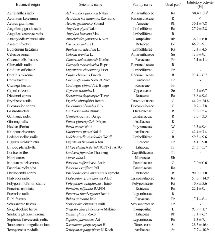
40여종의생약재 에탄올추출물의 C. difficile에대한항균 활 성을 검색한결과, 우슬(Achyranthes japonica Nakai, AJN), 희첨
(Siegesbeckia glabrescens Makino), 황백(Phelloedendron amurense
Ruprecht)은 1,000 ppm 농도에서 90% 이상의 높은 활성을보였 고, 지각(Citrus aurantium L.), 길경(Platycodon grandiflorum
ADC) 등은 50% 이상의 활성을 나타내었으며, 위령선(Clematis
mandshurica Rupr), 산사(Crataegus pinnatifida Bunge), 인삼
(Panax ginseng C.A. Meyer), 금은화(Lonicera japonica Thun-berg) 등은 C. difficile에대한항균활성을보이지않았다(Table 1). 각각의 추출물은 C. difficile에대하여 12시간 배양하였을 때가 장 높은 항균효과를 나타내었다(data not shown). Cai 등(18)은
Staphylococcus aureus와 Escherichia coli O157:H7에 대한 우슬, 목단피, 지실, 황백등의항균활성을보고하였고, Park 등(19)은 황백추출물의 L. plantarum및Leuconostoc mesenteroides에대한 항균 효과를보고한 바 있다. 그러나 우슬, 황백 및 희첨의 C. difficile에대한 생육억제 활성에 대한연구는 보고된 바 없어, 이들의 CDAD 관련치료제로서의 개발가능성을확인하였고, 가 장높은항균활성을나타낸우슬추출물을최종시료로서사용하였다. 생약재 에탄올 추출물의 용매분획별 항균활성 항균활성이 우수한 우슬, 희첨, 황백의 에탄올 추출물에대하 여 극성에 따른 용매분획을 실시하였다. 각각의 분리된 분획물 (500 ppm)에대하여액체배지희석법에의해 C. difficile에대한 생 육억제활성을측정한결과(Fig. 1), 우슬에틸아세테이트분획물에 서 95%로가장높은 항균력을나타냈으며, 희첨의클로로포름분 획물에서는 80% 이상을나타내었다. 황백은헥산분획물을 제외 한나머지용매분획물에서 60% 이상의활성을나타내었으나물 분획물에서는 활성을 보이지않았다. 따라서 우슬에틸아세테이 트분획물을 C. difficile에대한최종항균활성시료로선정하였다. 우슬 에틸아세테이트분획물의 농도별 항균활성 우슬에틸아세테이트분획물의첨가농도에따른항균활성을 측 정한결과 2가지방법에서모두농도의존적으로높은항균활성 을나타내었으며(Table 2), 500 ppm의농도에서는 현재 CDAD 치 료에 사용되고있는 vancomycin(12.5 ppm, 98.4%)과유사한 항균 활성(96.8%)을나타내었고, 이때 액체배지희석법에 의한 MIC는 625µg/mL였다. Vancomycin은그람양성균과관련된경험적치료 에 사용되는데 Staphylococcus에 의한 소장결장염이나 항생제의 사용과관련이있는C. difficile에의한위막성대장염에사용되고 있다. 본 연구에 사용된 vancomycin의 유효최저혈청농도는 5-12µg/mL이고, 유효최고혈청농도는 25-40µg/mL로이농도를 기 준으로 12.5µg/mL을처리농도를 하였다. Kim 등(14)은우슬이
다양한 진균성 식물병해들에 대하여 항진균력을 보인다고 보고 한바있으나 아직까지항생제 내성균주에 대해서는 보고된바 는없다. 우슬 에틸아세테이트 분획물의 C. difficile에 대한 시간에 따 른 생장 저해효과 현재 CDAD의치료를위한항생제사용의문제점을보완하기
Table 1. Screening of antimicrobial activity of herbal plant extracts against Clostridium difficile. Each ethanolic extracts of herbal
medicines was treated at 1,000 ppm in RCM broth as final concentration
Botanical origin Scientific name Family name Used parta Inhibitory activity
(%)
Achyranthes radix Achyranthes japonica Nakai Amaranthaceae Ra 98.4 ± 0.7b
Aconitum koreanum Aconitum koreanum R. Raymond Ranunculaceae R -c
Acorus gramineus Acorus gramineus Soland Araceae Rh 30.1 ± 7.80
Angelica gigantis radix Angelica gigas Nakai Umbelliferae Ra 27.9 ± 2.80
Angelica koreanae radix Angelica koreana Max. Umbelliferae R
-Atractylodis rhizoma alba Atractylodes japonica Koidz Compositae Rh 36.2 ± 6.00
Aurantii fructus Citrus aurantium L. Rutaceae Fc 66.9 ± 9.10
Bupleurum falcatum Bupleurum falcatum L. Umbelliferae Ra 12.4 ± 4.50
Celosiae semen Celosia arentea L. Amaranthaceae Se 13.9 ± 9.10
Chaenomelis fructus Chaenomeles sinensis Koehn Rosaceae Fr 13.1 ± 11.4
Clematidis radix Clematis mandshurica Rupr Ranunculaceae R
-Cnidium officinale Ligusticum chuanxiong Hort Umbelliferae Fr
-Coptidis rhizoma Coptis chinensis Franch Ranunculaceae R 27.4 ± 6.70
Corni fructus Corus officinalis Sieb. et Zucc Cornaceae Fr
-Crataegi fructus Crataegus pinnatifida Bunge Rosaceae Fr
-Cyperi rhizoma Cyperus rotundus L Cyperaceae Se 15.4 ± 6.70
Dictamni cortex Dictamnus dasycarpus Turcz Rutaceae Ra 15.8 ± 9.50
Erycibeae caulis Erycibe obtusifolia Benth Convolvulaceae C 44.9 ± 24.8
Eucommiae cortex Eucommia ulmoides Oliv Eucommiaceae C 10.7 ± 3.80
Gastrodia elata Gastrodia elata Blume Orchidaceae R 22.6 ± 5.40
Gentianae radix Gentiana scabra Bunge Gentianaceae R 12.0 ± 3.30
Ginseng radix Panax ginseng C.A. Meyer Araliaceae R
-Hoelen (Poria) Poria cocos Wolf Polyporaceae W 13.3 ± 9.40
Kalopanacis cortex Kalopanax pictus Nakai Araliaceae C 42.4 ± 7.40
Ledebouriellae radix Ledebouriella seseloides Wolff Umbelliferae R 59.5 ± 9.60
Ligustri lucidirfructus Ligustrum lucidum Aiton Oleaceae Fr 18.1 ± 9.00
Liriope platyphylla Liriope plathyphylla WANGET et TANG Liliaceae Fr 27.3 ± 5.70
Lonicerae flos Lonicera japonica Thunberg Caprifoliaceae Fl
-Mori cortex Morus alba L Moraceae Sb
-Moutan radicis cortex Paeonia suffruticosa Andr Paeoniaceae C 17.0 ± 0.60
Paeoniae radix alba Paeonia lactiflora Pall Paeoniaceae Ra
-Phellodendri cortex Phelloedendron amurense Ruprecht Rutaceae R 90.0 ± 3.00
Platycodi radix Platycodon grandiflorum ADC Campanulaceae Ra 57.6 ± 14.9
Polygoni multiflori caulis Polygonum multiflorum Thunb Polygonaceae Ra 10.8 ± 3.60
Poncirus trifoliata Poncirus trifoliata RAFIN Rutaceae Ra 22.1 ± 9.10
Puerariae radix Pueraria thunbergiana Benth Leguminosae R
-Rubi fructus Rubus coreanus Miq Rosaceae Fr 17.1 ± 6.40
Schisandrae fructus Schisandra chinensis Baill Schisandraceae Fr
-Siegesbeckiae herba Siegesbeckia glabrescens Makino Compositae L 93.9 ± 1.70
Smilacis glabrae rhizoma Smilax glabra Roxb Liliaceae Rh 12.4 ± 6.70
Sophorae flavescentis radix Sophora flavescens Ait Leguminosae Ra 6.3 ± 7.1
Taraxacum mongolicum hand Taraxacum platycarpum H Taraxacum St 28.5 ± 16.4
Tetrapanacis medulla Tetrapanax papyriferus K.Koch Araliaceae St 17.7 ± 10.9
aC: cortex, Fl: flower, Fc: fruit cortex, Fr: fruit, L: leaf, R: root, Ra: radix, Rh: rhizome, Sb: stem bark, Se: seed, St: stem, T: tuber, W: wolf. bResult was presented as mean±S.D. (n = 4).
위한대체방법으로서 probiotics 또는 yogurt가치료제로서 이용 되고있으며(20,21), 특히 Saccharomyces boularidii, L. rhamnosus
GG, B. breve, B. longum과L. acidophilus등이 항생제성유발설 사에예방 및치료에 효과를나타내는 것으로보고되어있다 (22-24). 또한 Lee 등(25)은유아분변으로부터 C. difficile에 대한생 육을저해하는균주를분리하여 B. infantis와 L. salivarius로동정 한바 있다. 이미활성이 알려진 L. rhamnosus GG 균주배양액 또는 우슬 에틸아세테이트분획물 시료를 첨가하여 배양한 C. difficile을 20시간배양동안 4시간간격으로생장정도를확인한 결과(Fig. 2), 12시간까지그저해활성이가장높았고, 20시간까 지도유지되면서우수한효과를나타내었다. AJNEA(500 ppm)는 L. rhamnosus GG(100 ppm) 보다는높은 생장억제활성을나타내 었으나 vanco-mycins(12.5 ppm)과유사한높은효과를보였다. 우슬 에틸아세테이트분획물의 유산균에 대한 생육 증식 효과 우슬의새로운 C. difficile 치료제 또는기능성소재로서의 개 발가능성을확인하기 위해서장내유익균총인 Bifidobacterium spp.와 Lactobacillus spp.의생육에미치는영향을조사하였다(Fig. 3). 우슬 에틸아세테이트분획물은 실험에 사용한 균주 중 B. longum(25%)을제외한나머지 Bifidobacterium spp.에대해서는생 장에 거의 영향을 미치지 않았고, Lactobacillus spp. 중에는 L.
helveticus(12.1%)와 Streptococcus thermophilus(25%)에 대해서 약 간의생육저해활성을보였다. 따라서우슬은이들 probiotics 균
주들의 생육에는영향을 미치지않으면서 C. difficile에대한 항
균-상승효과(synergistic antimicrobial effect)를나타낼것으로 사료
Fig. 1. Antimicrobial activity of each solvent fractionates from
Achyranthes japonica Nakai (AJN), Siegesbeckia glabrescensMakino and Phelloedendron amurense Ruprecht ethanolic
extracts against Clostridium difficile.
C. difficile (0.3 at O.D. 600 nm) was inoculated into 10 mL of RCM broth containing 500 ppm of each of the filtered fractions separated from AJN, Siegesbeckiae herba and Phellodendri cortex was incubated at 37oCfor 12 h.
Table 2. Antimicrobial effect of ethyl acetate fractionates from
Achyranthes japonica Nakai ethanolic extracts (AJNEA) withdifferent concentrations against Clostridium difficile
Concentration
(ppm) Growth inhibition (%) Clear zone (mm)Inhibition activity
AJNEA 050 - -a AJNEA 100 05.69 ± 1.5 +b AJNEA 200 11.13 ± 1.3 + AJNEA 300 22.89 ± 1.3 ++c AJNEA 400 62.50 ± 1.6 ++ AJNEA 500 96.80 ± 1.2 +++d Vancomycin 12.5 98.23 ± 1.2 +++
a: No antimicrobial activity, C.Z.(clear zone) of sample <10 mm. b: C.Z. of sample 10-13 mm.
c: C.Z. of sample 13-16 mm. d: C.Z. of sample >16 mm.
Fig. 2. Growth inhibitory effect of ethyl acetate fractionates from
Achyranthes japonica Nakai ethanolic extracts (AJNEA) against Clostridium difficile. The control was inoculated C. difficile (0.3 at O.D. 600 nm) without any treat and incubated at 37oC for 20 h.Tested concentration of filtered supernatant from Lactobacillus spp. was 100 ppm at the final concentration.
Fig. 3. Growth effect of ethyl acetate fractionates from
Achyranthes japonica Nakai ethanolic extracts (AJNEA) on Bifidobacterium spp. (A) and Lactobacillus spp. (B). These species were inoculated into 10 mL of TPY broth or MRS broth (2 %, v/v) with or without AJNEA (500 ppm) and incubated at 37oC for 12 h.되어이에 대한추가실험을 진행중이며, 이들결과로부터우슬 추출물의 CDAD 관련질환에 대한치료제제 또는기능성 소재 로서활용가능성을확인하였다.
요
약
본연구에서는대장염유발균인C. difficile에대한항균활성이 우수한천연항균물질을검색하기위하여 40여가지생약재에탄 올추출물에대하여 C. difficile에대한생육저해활성을측정하였 다. 결과, 우슬, 희첨, 황백은 1,000 ppm 농도에서 90% 이상의높 은항균활성을나타내었고, 우슬에틸아세테이트분획물(AJNEA) 은 가장 높은 활성을 나타내었으며 이의 최소저해농도(MIC)는 625µg/mL였다. 또한기능성 소재로의활용가능성을확인하기위 하여 AJNEA의 유산균 생육에 미치는 영향을 조사한 결과, B.longum, L. helveticus와 S. thermophilus를제외한 나머지 균주들 에대해서생장억제효과를나타내지않았다. 이들결과로부터 우슬추출물은 C. difficile관련질환에 대하여우수한 항균제제 로이용될 수있을것으로확인되었다.
감사의 글
본연구는인하대 교내연구비에의해수행되었으며, 이에저자 는감사드립니다.문
헌
1. Kelly CP, Pothoulakis C, Lamont JT. Clostridium difficile colitis. New Engl. J. Med. 330: 257-262 (1994)
2. McFarland LV, Mulligan ME, Kwok RY, Stamm WE. Nocoso-mial acquisition of Clostridium difficile infection. New Engl. J. Med. 320: 204-210 (1989)
3. Bartlett JG. Clostridium difficile: history of its role as an enteric pathogen and the current state of knowledge about the organism. Clin. Infect. Dis. 18: 5265-5275 (1994)
4. Cartmill TD, Panigrahi H, Worsley MA, McCann DC, Nice CN, Keith E. Management of diarrhoea due to Clostridium difficile. J. Hosp. Infect. 27: 1-15 (1994)
5. Freeman J, Wilcox MH. Antibiotics and Clostridium difficile. Microbes Infect. 1: 377-384 (1999)
6. Ackermann G, Loffler B, Tang-Feldman YJ, Cohen SH, Silva J, Rodloff AC. Cloning and expression of Clostridium difficile toxin A gene(tcdA) by PCR amplication and use of an expression vec-tor. Mol. Cell. Probe 18: 271-274 (2004)
7. Pothoulakis C. Effect of Clostridium difficile toxins on epithelial cell barrier. Ann. NY Acad. Sci. 915: 347-356 (2000)
8. Anderson TL, McGregor A. Evaluation of the clearview Clostrid-ium difficile toxin A test and various selective culture media in comparison with the cytotoxin assay for the diagnosis of
Clostridium difficile-associated diarrhoea. Pathology 35: 244-247 (2003)
9. Lyerly DM, Saum KE, Macdonald DK, Wilkins TD. Effects of
Clostridium difficile toxins given intragastrically to animals. Infect. Immun. 47: 349-352 (1985)
10. Lee JY, Yoon YM, Roh HC, Kim JM. Nuclear factor-kappa B activation and chemokine genes expression in HT-29 intestinal epithelial cells in response to Clostridium difficile toxin A stimu-lation. J. Bacteriol. Virol. 35: 217-226 (2005)
11. Marcone MF, Jahaniaval F, Aliee H, Kakuda Y. Chemical charac-terization of Achyranthes bidentata seed. Food Chem. 81: 7-12 (2003)
12. Kiso Y, Suzuki Y, Konno C, Hikino H, Hashimoto I, Yagi Y. Liver-protective drugs. 3. The viability of the oriental medicines, 38. Application of carton tetrachloride-induced liver lesion in mice for screening of liver protective crude drugs. Shoyakugaku Zasshi 36: 238-244 (1982)
13. Shimomura H, Sashida Y, Nakata H. Plant growth regulating activities of crude drugs and medicinal plants. Shoyakugaku Zasshi 35: 173-179 (1981)
14. Kim JC, Choi GJ, Lee SW, Kim JS, Chung KY, Cho KY. Screening extracts of Achyranthes japonica and Rumex crispus
for activity against various plant pathogenic fungi and control of powdery mildew. Pest Manag. Sci. 60: 803-808 (2004)
15. Son KH, Hwang JH, Lee SH, Park JH, Kang SJ, Chang SY, Lee KS. Isolation and quantitative determination of 20-hydroxyecdys-one from Achyranthes radix. Korean J. Pharmacogn. 30: 335-339 (1999)
16. Gavidson PH, Parish ME. Methods for testing the efficacy of food antimicrobials. Food Technol.-Chicago 43: 148-152 (1989) 17. Shin DH, Kim MS, Han JS. Antimicrobial effect of ethanol
extracts from some medicinal herbs and their fractionates against foodborn bacteria. Korean J. Food Sci. Technol. 29: 808-816 (1997)
18. Cai H, Choi SI, Lee YM, Heo TR. Antimicrobial effects of herbal medicine extracts on Staphylococcus aureus and Escheri-chia coli O157:H7. Korean J. Biotechnol. Bioeng. 17: 537-542 (2002)
19. Park MG, Jeong GS, In MJ. Effect of Scutellaria baicalensis and
Phellodendron amurense extracts on growth of lactic acid bacteria and kimchi fermentation. J. Korean Soc. Food Sci. Nutr. 33: 420-426 (2004)
20. D'Souza AL, Rajkumar C, Cooke J, Bulpitt J. Probiotics in pre-vention of antibiotic-associated diarrhoea: meta analysis. Brit. Med. J. 324: 1361-1364 (2002)
21. Surawicz CM. Probiotics, antibiotic-associated diarrhoea and
Clostridium difficile diarrhoea in human. Best Pract. Res. Cl. Ga. 17: 775-783 (2003)
22. Tasteyre A, Barc MC, Karjalainen T, Bourlioux P, Collignon A. Inhibition of in vitro cell adherence of Clostridium difficile by
Saccharomyces boulardii. Microb. Pathogenesis 32: 219-225 (2002)
23. Van-Niel CW, Feudtner C, Garrison MN, Christakis DA. Lacto-bacillus therapy for acute infectious diarrhoea in children: a meta analysis. Pediatrics 109: 678-684 (2002)
24. Araki T, Shinozaki T, Irie Y, Miyazawa Y. Trial of oral adminis-tration of Bifidobacterium breve for the prevention of rotavirus infection. Kansenshogaku Zasshi 73: 305-310 (1999)
25. Lee YJ, Yu WK, Heo TR. Identification and screening for antimi-crobial activity against Clostridium difficile of Bifidobacterium
and Lactobacillus species isolated from heathy infant faeces. Int. J. Antimicrob. Ag. 21: 340-246 (2003)