INTRODUCTION
Acquired aplastic anemia is characterized by the depletion of hematopoietic stem cells and progenitor cells, the hypocel- lularity of bone marrow and blood pancytopenia, and it com- monly occurs in children and young adults (1). The standard treatments are hematopoietic stem cell transplantation (HSCT) or immunosuppressive therapy in patients with life-threat- ening cytopenia. HSCT is the treatment of choice in young patients with an human leukocyte antigen (HLA)-matched sibling or unrelated stem cell donor and immunosuppressive therapy is indicated for patients without HLA-matched donor or older patients (1, 2). However, patients who relapsed despite these treatments or those who did not receive such active therapies depend on continuous blood transfusions, and they are ultimately fatal because of its natural clinical course, clon- al evolution to other malignant diseases (3), or secondary complications such as hemochromatosis (4).
A very poor prognosis may be predicted in patients with severe aplastic anemia (SAA) in whom major organ failures occurred by multiple blood transfusions. Can the only sup- portive therapies such as blood component transfusions including packed red blood cell (PRBC) and continuous
iron chelation be possible in normalizing the disease and recovering organ failures? The answer may be no. However, we experienced an interesting finding in a patient with SAA using a continuous iron chelation therapy. We expect that the therapy will be an alternative answer to this problem on the aspect of stem cell immunobiology. In addition, the same treatment protocol has been applied to other similar patients from our hospital, in whom a gradual improvement is being noted and a follow-up monitoring is needed.
CASE REPORT
In 1998, a 16-yr-old male patient visited our emergency center with a chief complaint of severe dyspnea. He was diag- nosed with SAA at 11 yr of age. According to his medical record, his bone marrow was markedly hypocellular (<10%
cellularity) without dysplastic features, and the Ham’s and sucrose tests were negative. He received twice of ALG im- munotherapies and supportive erythropoietin injections, but did not have any responses. He neither had an HLA-matched sibling stem cell donor nor found an unrelated stem cell donor despite continuous search. He was given oral oxymetholone
320
Soo-Jeong Park and Chi-Wha Han
Division of Hematology and Oncology, Our Lady of Mercy Hospital, The Catholic University of Korea, Incheon, Korea
Address for correspondence Soo-Jeong Park, M.D.
Division of Hematology and Oncology, Our Lady of Mercy Hospital, 665 Bupyeng 6-dong, Bupyeng-gu, Incheon 403-720, Korea
Tel : +82.32-510-5507, Fax : +82.32-510-5683 E-mail : hellenpark@hanmail.net
J Korean Med Sci 2008; 23: 320-3 ISSN 1011-8934
DOI: 10.3346/jkms.2008.23.2.320
Copyright � The Korean Academy of Medical Sciences
Complete Hematopoietic Recovery after Continuous Iron Chelation Therapy in a Patient with Severe Aplastic Anemia with Secondary Hemochromatosis
A 16-yr-old male patient with hemochromatosis due to multiple packed red blood cell transfusions was referred to our emergency center for the treatment of severe aplas- tic anemia and dyspnea. He was diagnosed with aplastic anemia at 11-yr of age.
He had received continuous transfusions because an HLA-matched marrow donor was unavailable. Following a continuous, approximately 5-yr transfusion, he was noted to develop hemochromatosis. He had a dilated cardiomyopathy and required diuretics and digitalis, multiple endocrine and liver dysfunction, generalized bleed- ing, and skin pigmentation. A total volume of red blood cell transfusion before defer- oxamine therapy was about 96,000 mL. He received a regular iron chelation thera- py (continuous intravenous infusion of deferoxamine, 50 mg/kg/day for 5 days q 3-4 weeks) for approximately seven years after the onset of multiple organ failures. His cytopenia and organ dysfunctions began to be gradually recovered since about 2002, following a 4-yr deferoxamine treatment. He showed completely normal ranges of peripheral blood cell counts, heart size, and liver function two years ago.
He has not received any transfusions for the last four years. This finding suggests that a continuous deferoxamine infusion may play a role in the immune regulation in addition to iron chelation effect.
Key Words : Aplastic Anemia; Hemochromatosis; Deferoxamine
Received : 25 January 2007 Accepted : 26 June 2007
Hematopoietic Recovery after Iron Chelation Therapy in a Patient with SAA 321
and continuously had two pints of PRBC transfusion at a 1- to 2-week interval for five years while awaiting an HLA- matched unrelated stem cell donor. The total volume of PRBC (320-400 mL/pack) transfusion was approximately 96,000 mL during that period. Initial electrocardiography showed atrial fibrillation with a heart rate of 150/min. The chest radiography showed huge cardiomegaly with 80% of car- dio-thoracic ratio and both pleural effusions (Fig. 1), and the erect and supine view of abdomen radiography showed a severe hepatomegaly. An echocardiographic examination presented 4% of left ventricular ejection fraction with severe- ly dilated left ventricle and nearly akinetic cardiac wall move- ment. Laboratory tests showed 4.5 g/dL of hemoglobin, 20×
109/L of absolute reticulocyte count, 2.7×10⁹/L of white blood cell count (absolute neutrophil count, 0.3×10⁹/L), 3
×109/L of platelet count, 407 mg/dL of fasting blood glu- cose-level, and 68,800 ng/mL of serum ferritin.
We immediately admitted him to our intensive care unit.
He received appropriate doses of oxygen, digitalis, diuretics, and a continuous intravenous infusion of insulin in addition to levothyroxine, calcium, and vitamin D of his previous pre- scriptions. Other laboratory tests showed 0.37 ng/dL of serum free thyroxine (normal range, 0.8 to 1.9 ng/dL), 142.2 mIU/L of thyrotropin (normal range, 0.4 to 4 mIU/L), 6.0 mg/dL of serum calcium level (normal range, 8.2 to 10.2 mg/dL), 13 pg/mL of intact PTH (normal range, 15 to 70 pg/mL), and 8.4% of hemoglobin A1C(normal range, 4 to 6%). These find- ings were suggestive of multiple endocrine dysfunctions includ- ing hypothyroidism, hypoparathyroidism with hypocalcemia, and diabetes. Liver function tests were 2.5 mg/dL of serum total bilirubin, 2.8 g/dL of serum albumin, prolonged pro-
thrombin time of 16.4 sec (normal range, 11.1 to 13.1 sec) and 1.437 INR (normal range, 0.9 to 1.12), 35.1 sec of acti- vated partial thromboplastin time (normal range, 25 to 38 sec), 649 IU/L of serum aspartate aminotransferase (normal range, 8 to 40 IU/L), and 539 IU/L of serum alanine amino- transferase (normal range, 5 to 40 IU/L). Ophthalmologic examination confirmed a cataract in both eyes. Generalized skin pigmentation was seen in his skin. Abdominal ultrasonog- raphy showed severe hepatomegaly with increased parenchy- mal echogenecity. Based on these findings, he was diagnosed with secondary hemochromatosis accompanied by multiple organ failures which resulted from iron deposition in body organs due to multiple PRBC transfusions. Based on some published reports that iron chelation plays a prominent role in hemochromatosis (5, 6), he received a 12-hr continuous intra- venous infusion of deferoxamine (50 mg/kg/day) for five days at a 4-week interval. He received PRBC during deferoxamine treatment, if necessary. He also received innumerable units of platelet concentrates. Thereafter, he was frequently admitted to the medical intensive care unit through our emergency cen- ter because of the aggravated symptoms and signs of heart fail- ure and diabetes-related complications, multiple cerebral and subdural hematomas, and seizure. Meanwhile, he was being given a 7-yr deferoxamine treatment. The frequency of his hospitalization was markedly being lowered, and his hema- tologic profiles (Fig. 2) and heart size were beginning to nearly normalize after an approximately 4-yr deferoxamine treatment.
His insulin requirement also decreased, and from then, a low- dose erythropoietin injection was initiated. Since then, he has only received outpatient care. He was noted to have completely normal ranges of peripheral blood cell counts, heart size, and
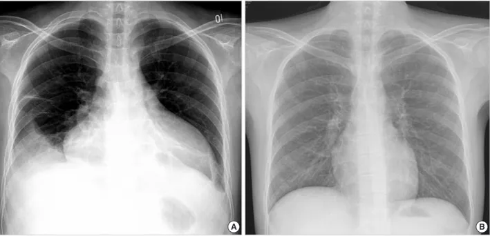
Fig. 1. Posteroanterior chest radiographs reveal a marked cardiomegaly with right pleural effusion before the treatment (A) and a normal- ized heart after the deferoxamine therapy (B).
A B
322 S.-J. Park, C.-W. Han
liver functions two years ago. A defective expression of CD55/
59 was not observed on either granulocytes or erythrocytes of peripheral blood. Since the event of complete response, his bone marrow aspirate and biopsy for the evaluation of bone marrow status has not been performed because of his strong refusal. He has not required any blood transfusions over the past four years (Fig. 3). He currently shows normal reference ranges of all hematologic and other laboratory tests.
DISCUSSION
The prognosis of SAA is very fatal if its therapy is the only supportive one such as transfusions. A few patients with aplastic anemia have been reported to recover spontaneously without specific therapy, but these remissions usually occur within the first 7 weeks and the remainder followed a rapidly fatal course (7). Only 1/3-1/2 of them may be alive six months after the diagnosis, and about 80% die within 18 months to two years as described in many reports. Even patients who survive infections or bleeding also lead to a dangerous situa- tion because of secondary hemochromatosis associated with multiple major organ failures. It is very surprising that a continuous, several-year iron chelation combined with low- dose erythropoietin injection improves major organ dys- functions and produces a complete recovery of hematologic abnormalities. Moreover, these promising results are being noted gradually in other two patients undergoing the simi- lar disease situations.
It has been reported that iron chelator not only removes iron deposited in the tissues but also plays a role in immunologic mechanisms in spite of some controversy. Carotenuto et al.
reported that deferoxamine acts as a blocker for IL-2 recep- tor of human T-lymphocytes, and Hoffbrand et al. indicated the immunosuppressive effect of deferoxamine in inhibiting the activity of ribonucleotide reductase in human lymphocytes (8, 9). These reports indicate the immunologic inhibitory effects of deferoxamine.
We have postulated that the deferoxamine treatment grad- ually had an immunologic influence on the suppressive envi- ronment of aplastic marrow and changed bone marrow stro- mal environment into positive hematopoiesis. In addition, erythropoietin therapy might have enhanced hematopoietic proliferation (10, 11). There are some evidences supporting our postulates: according to Aucella et al., the combination therapy using deferoxamine and recombinant erythropoietin promotes synergistically erythropoiesis in patients with chron- ic renal failure. Kling et al. also noted that iron deprivation increases erythropoietin production in normal subjects and patients with malignancy (12, 13). It is very surprising that a continuous iron chelation induces the recovery of organ dys- functions. This was previously supported by Tsironi et al. who reported that combined iron chelation reversed heart failure in patients with thalassemia major (14). This makes us spec- ulate that a continuous, long-term infusion of iron chelator can cause a partial or complete recovery of multiple organ fail- ures due to iron deposition.
We suggest that an early introduction of continuous iron chelation could prevent secondary hemochromatosis due to multiple transfusions and also reverse their hematopoiesis by its immune modulation effect in patients with refractory or relapsed aplastic anemia who did not respond to any treatments, those who did not find an available HLA-matched stem cell donor, or those who relapsed into the original disease despite the treatments.
REFERENCES
1. Keohane EM. Acquired aplastic anemia. Clin Lab Sci 2004; 17: 165- 71.
2. Geissler K. Pathophysiology and treatment of aplastic anemia. Wien Klin Wochenschr 2003; 115: 444-50.
3. Maciejewski JP, Risitano AM. Aplastic anemia: management of adult patients. Hematolgoy Am Soc Hematol Educ 2005; 110-7.
4. Mwanda OW, Otieno CF, Abdalla FK. Transfusion haemosiderosis inspite of regular use of desferrioxamine: case report. East Afr Med J 2004; 81: 326-8.
5. Girot R, Thevenin M, Bouveret JP, Jeannel F, Rymer JC. Treatment Fig. 2. Hemoglobin changes before and after deferoxamine therapy.
Hemoglobin (g/dL)
16 14 12 10 8 6 4 2
96/6/18 97/6/18 98/6/18 99/6/18 00/6/18 01/6/18 02/6/18 03/6/18 04/6/18 05/6/180 Deferoxamine therapy
Packed RBC transfusion
Date (y/m/d)
Fig. 3. The full clinical course of the patient: Pre- and post-deferox- amine therapy. ALG, anti-lymphocyte globulin; EPO, erythropoi- etin; PRC, packed red blood cell; PC, platelet concentrate; CR, complete response.
PRC/PC transfusion
Deferoxamine therapy Response (+)
CR ALG×2.
EPO.
Oxymethelone
1993 1994 1995 1996 1997 1998 1999 2000 2001 2002 2003 2004 2005 2006 (yr)
11 12 13 14 15 16 17 18 19 20 21 22 23 24 (age)
′
Hematopoietic Recovery after Iron Chelation Therapy in a Patient with SAA 323
of iron overload due to repeated transfusions with subcutaneous infu- sions of desferrioxamine. Arch Fr Pediatr 1980; 37: 241-7.
6. Cohen AR, Mizanin J, Schwartz E. Rapid removal of excessive iron with daily, high-dose intravenous chelation therapy. J Pediatr 1989;
115: 151-5.
7. Lee JH, Lee JH, Shin YR, Lee JS, Kim WK, Chi HS, Park CJ, Lee KH. Spontaneous remission of aplastic anemia: a retrospective anal- ysis. Haematologica 2001; 86: 928-33.
8. Carotenuto P, Pontesilli O, Cambier JC, Hayward AR. Desferoxamine blocks IL 2 receptor expression on human T lymphocytes. J Immunol 1986; 136: 2342-7.
9. Hoffbrand AV, Ganeshaguru K, Hooton JW, Tattersall MH. Effect of iron deficiency and desferrioxamine on DNA synthesis in human cells.
Br J Haematol 1976; 33: 517-26.
10. Shao Z, Chu Y, Zhang Y, Chen G, Zheng Y. Treatment of severe aplas- tic anemia with an immunosuppressive agent plus recombinant human granulocyte-macrophage colony-stimulating factor and erythropoi- etin. Am J Hematol 1998; 59: 185-91.
11. Bessho M, Hirashima K, Asano S, Ikeda Y, Ogawa N, Tomonaga M, Toyama K, Nakahata T, Nomura T, Mizoguchi H, Yoshida Y, Niit- su Y, Kohgo Y. Treatment of the anemia of aplastic anemia patients with recombinant human erythropoietin in combination with granu- locyte colony-stimulating factor: a multicenter randomized controlled study. Multicenter Study Group. Eur J Haematol 1997; 58: 265-72.
12. Aucella F, Vigilante M, Scalzulli P, Musto P, Prencipe M, Valente GL, Carotenuto M, Stallone C. Synergistic effect of desferrioxamine and recombinant erythropoietin on erythroid precursor proliferation in chronic renal failure. Nephrol Dial Transplant 1999; 14: 1171-5.
13. Kling PJ, Dragsten PR, Roberts RA, Dos Santos B, Brooks DJ, Hed- lund BE, Taetle R. Iron deprivation increases erythropoietin produc- tion in vitro, in normal subjects and patients with malignancy. Br J Haematol 1996; 95: 241-8.
14. Tsironi M, Deftereos S, Andriopoulos P, Farmakis D, Meletis J, Aes- sopos A. Reversal of heart failure in thalassemia major by combined chelation therapy: a case report. Eur J Haematol 2005; 74: 84-5.