A Higher Burden of Small Low-density Lipoprotein Particles is Associated with Profound Changes in the Free Androgen Index in Male Adolescents
From a young age, males are at higher cardiovascular risk than females. Dyslipidemia, including a higher burden related to small low-density lipoproteins (LDL), plays an important role in precipitating atherosclerosis in both males and females. We investigated sex differences in atherogenic lipoprotein burden and the independent predictors of LDL particle size in children and adolescents. We measured the concentrations of total testosterone, sex hormone-binding globulin, estradiol, total cholesterol, triglyceride, LDL cholesterol, HDL cholesterol, and LDL particle size in 135 children and adolescents (67 boys, 68 girls). The free androgen index was significantly and negatively correlated with LDL particle size (r = –0.273, P = 0.026) in boys, but estrogen and LDL particle size were not related. In a stepwise multiple regression analysis adjusted for body mass index, age, and homeostasis model assessment for insulin resistance, free androgen index was still an independent predictor of LDL particle size in boys (R2 = 0.075, P = 0.026).
The prominent decrease in LDL particle size along with increased testosterone concentrations in males might explain why they are more likely to display atherogenic dyslipidemia from adolescence.
Key Words: Adolescent; Small dense LDL; Testosterone Yong Jun Choi1,*, Sung Hee Choi2,*,
Hae Jin Kim1, Seung Jin Han1, Jin Soon Hwang3, Yoon-Sok Chung1, Kwan Woo Lee1, Hong Keun Cho4, and Dae Jung Kim1
1Department of Endocrinology and Metabolism, Ajou University School of Medicine, Suwon;
2Department of Internal Medicine, Seoul National University Bundang Hospital, Seoul National University College of Medicine, Seongnam;
3Department of Pediatrics, Ajou University School of Medicine, Suwon; 4Dr. Cho’s Internal Medicine Clinic, Seoul, Korea
*Yong Jun Choiand Sung Hee Choi contributed equally to this work.
Received: 12 July 2010 Accepted: 25 January 2011 Address for Correspondence:
Dae Jung Kim, MD
Department of Endocrinology and Metabolism, Ajou University School of Medicine, 164 Worldcup-ro, Yeongtong-gu, Suwon 443-721, Korea
Tel: +82.31-219-5128, Fax: +82.31-219-4497 E-mail: [email protected]
This study was supported by a grant from the Korean Health 21 R&D Project, Ministry of Health and Welfare, Korea (A050463), and a 2004 grant from Ajou University School of Medicine.
There is no conflict of interests with these financial supports relating to publishing this article.
DOI: 10.3346/jkms.2011.26.4.534 • J Korean Med Sci 2011; 26: 534-539
INTRODUCTION
Plasma low-density lipoprotein cholesterol (LDL-C) is associat- ed with an increased risk of coronary heart disease (CHD) (1, 2);
however, more than 30% of patients with CHD events have nor- mal LDL-C level (3, 4). LDL particles are heterogeneous and can be divided into two subclasses based on variations in density, size, and chemical composition: large buoyant LDL (lb-LDL, pattern A) and small dense LDL (sd-LDL, pattern B) (5). Stud- ies have shown that sd-LDL has greater atherogenic potential than lb-LDL in various ethnic groups (6-9).
Several studies have reported that LDL particle size tends to be smaller in both young and adult males compared to females (10-12). Hormonal differences have been hypothesized to ac- count for these sex differences, but few studies have examined the association between sex steroid hormones and LDL particle
size in young people. A recent clinical trial showed that hormone replacement therapy (HT) does not reduce the risk of coronary disease in postmenopausal women (13). Furthermore, HT users reportedly do not have a better LDL subclass distribution, which may explain the failure of HT to reduce the incidence of heart disease events (14). These results suggest that estrogens might not be the key determinant of this sex difference.
Children and adolescents experience profound changes in sex steroid hormones during puberty. Moreover, adverse pat- terns of atherosclerosis begin during childhood (15). Therefore, children and adolescents are appropriate candidates to study the relationship between sex steroid hormones and LDL size.
The purpose of this study was to compare LDL particle size in boys and girls, and to investigate whether changes in male sex hormone concentrations are related to LDL particle size in chil- dren and adolescents.
MATERIALS AND METHODS
A detailed description of the study design and methods is out- lined in a previous report (16). Briefly, 135 children and adoles- cents (67 boys, 68 girls; mean age, 11.6 ± 2.0 yr) participated after guardians’ permission in this study from January to February in 2004; their ages ranged from 7 to 16 yr. The subjects were recruit- ed through newspaper advertisements in Suwon, Korea. Puber- tal stage was assessed by physical examination, which was per- formed by a pediatrician at our institute, according to the Tan- ner criteria. We assigned 33 boys and 19 girls to the child group (Tanner stage = 1) and 34 boys and 49 girls to the adolescent group (Tanner stage ≥ 2).
Anthropometric and body fat measurements
We measured the weight and height of all subjects in the morn- ing after minimum 8-hr fasting while they were wearing light clothing. Body mass index (BMI) was calculated as weight (kg)/
height (m2). Blood pressure was measured twice in the sitting po- sition at 3-min intervals using a standard sphygmomanometer.
To assess the distribution of body fat in the abdomen, we per- formed computed tomography (High Speed Advantage; General Electric Co., Fairfield, CT, USA) at the umbilical level and mea- sured the total abdominal fat area (cm2) equivalent to a Houn- sfield unit range of -50 to -250. We identified the visceral fat area and subcutaneous fat area by applying the peritoneum as a bound- ary. The clinical status of the participants was made unknown to the investigator who collected and analyzed these data.
Laboratory measurements
After fasting for 8 hr or more, all subjects visited laboratory be- tween 08:00 and 10:00, and a blood sample was drawn from the antecubital vein. Plasma was separated from the collected blood and stored at -70°C until analyzed. Glucose, insulin, total cho- lesterol, triglyceride (TG), and high-density lipoprotein (HDL)- cholesterol concentrations were measured by standard meth- ods. LDL-C concentration was calculated using the Friedewald equation. As a marker of insulin resistance, a homeostasis mod- el assessment (HOMA) index was calculated from the fasting insulin and glucose concentrations as (insulin [μIU/mL] × glu- cose [mM/L])/22.5.
Estradiol (E2) and total testosterone concentrations were mea- sured with commercial solid-phase radioimmunoassays (Diag- nostic Products Corp., Los Angeles, CA, USA). Sex hormone- binding globulin (SHBG) concentrations were measured using a commercial immunoradiometric assay (Diagnostic Products Corp.). The free androgen index (FAI) was used to estimate the amount of testosterone unbound by SHBG and thus, immedi- ately biologically active. FAI was calculated as (100 Total testos- terone/SHBG). To convert testosterone to nM, the ng per mL value was multiplied by 3.467.
LDL particles were isolated by sequential flotation ultracen- trifugation, and LDL particle size distribution (d 1.019-1.063 g/
mL) was examined using a pore gradient lipoprotein system (CBS Scientific, Del Mar, CA, USA) with commercially available non-denaturing polyacrylamide slab gels containing a linear gradient of 2% to 16% acrylamide (Alamo Gels Inc., San Anto- nio, TX, USA). Standards, including latex beads (34 nm), thyro- globulin (17 nm), apoferritin (12.2 nm), and catalase (10.4 nm), were used to estimate the relative migration (Rf) rates of each band. The gels were scanned using a GS-800 Calibrated Imaging Densitometer (Bio-Rad Laboratories, Graz, Austria). LDL parti- cle size was calculated with reference to the Rf value of the stan- dards. LDL subclasses were classified as sd-LDL (pattern B; diam- eter < 25.5 nm) and lb-LDL (pattern A; diameter < 25.5 nm) (5).
Statistical analysis
The subjects were divided into two groups based on the Tanner stage: children (Tanner stage = 1) and adolescents (Tanner stage
≥ 2). In each group, boys and girls were analyzed separately to better understand the association between LDL particle size and sex steroid hormone concentrations. All data are presented as means ± standard deviation and number. Since HOMA for in- sulin resistance (IR) and concentrations of insulin, TG, testos- terone, SHBG, FAI, and E2 were not normally distributed, these variables were log-transformed prior to analysis.
The clinical characteristics and laboratory findings were com- pared for boys and girls using Student’s t test. Because BMI and age differed significantly between boys and girls in both children and adolescents, each factor was compared using an ANCOVA after adjusting for BMI and age. We used correlation analyses to examine the relationship between LDL particle size and each factor in boys and girls, separately. Multiple stepwise regression analysis was performed to investigate whether the FAI was re- lated to LDL particle size in boys, after adjusting for age, BMI, and HOMA-IR.
Statistical analyses were performed using PASW for Windows (v. 18.0; SPSS Inc., Chicago, IL, USA), and P < 0.05 was consid- ered significant.
Ethics statement
This study was conducted according to the ethical guidelines of Ajou University Hospital and Ajou University School of Medi- cine and written informed consent was obtained from all guard- ians of the participants.
RESULTS
Sex differences in baseline characteristics and LDL particle size in children and adolescents
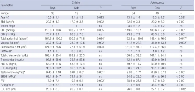
Table 1 shows the data for clinical and biochemical parameters and LDL particle size grouped by sex in children and adolescents.
Boys were older than girls in both the child (P = 0.013) and ado- lescent groups (P = 0.021). No significant sex difference in Tan- ner stage existed within adolescents. Systolic blood pressure (SBP) was significantly higher in boys for both children (P = 0.035) and adolescents (P < 0.001). BMI differed significantly between boys and girls, and was higher in both groups of boys (P = 0.002 for children, P < 0.001 for adolescents). Visceral fat area was sig- nificantly higher in boys of both groups (P = 0.002 for children, P = 0.022 for adolescents), although the subcutaneous fat area was significantly higher only in male children (P = 0.023). FAI and serum testosterone concentrations were significantly higher in boys for both children (P = 0.002) and adolescents (P < 0.001).
SHBG and E2 concentrations were significantly higher in girls than in boys, but only for adolescents (P < 0.001).
The concentrations of total cholesterol, HDL-C, LDL-C, and TG did not differ significantly between boys and girls in either age group. Of note, LDL particle size did not differ between the sexes in children, but was significantly smaller in male adoles- cents as compared to female adolescents (P = 0.012). After ad- justing for BMI and age, the difference between sexes was still significant for SBP in both age groups (P < 0.05). In adolescents, the serum concentrations of testosterone, SHBG, and E2, and LDL particle size remained significantly different after adjust- ment (P < 0.05). However, FAI and serum testosterone concen-
Table 1. Comparison of clinical and biochemical parameters and LDL particle size by sex in children and adolescents
Parameters Children Adolescents
Boys Girls P Boys Girls P
Number 33 19 - 34 49 -
Age (yr) 10.5 ± 1.4 9.4 ± 1.3 0.013 13.1 ± 1.4 12.3 ± 1.7 0.021
BMI (kg/m2) 20.7 ± 4.2 17.0 ± 3.3 0.002 22.9 ± 3.3 20.2 ± 3.2 < 0.001
Tanner stage 1 1 ns 3.0 ± 1.2 3.4 ± 1.2 ns
SBP (mmHg) 110.0 ± 10.6 103.2 ± 11.1 0.035 113.6 ± 10.1 100.6 ± 9.2 < 0.001
DBP (mmHg) 70.7 ± 8.1 66.3 ± 7.6 ns 73.2 ± 7.3 63.3 ± 6.6 < 0.001
Total abdominal fat (cm2) 164.6 ± 100.2 100.2 ± 71.8 0.014† 192.8 ± 110.6 148.4 ± 76.0 0.047† Visceral fat (cm2) 39.7 ± 23.3 23.2 ± 12.6 0.002† 41.2 ± 22.3 31.0 ± 13.8 0.022†
Subcutaneous fat (cm2) 124.9 ± 76.6 77.1 ± 59.9 0.023 151.6 ± 91.9 117.4 ± 66.6 ns
HOMA-IR* 1.1 ± 1.0 0.8 ± 0.8 ns 1.7 ± 1.3 1.8 ± 1.2 ns
Total cholesterol (mg/dL) 168.4 ± 25.4 168.9 ± 25.3 ns 160.6 ± 33.2 161.1 ± 24.1 ns
Triglycerides (mg/dL)* 92.8 ± 56.6 75.7 ± 55.6 ns 112.1 ± 67.1 89.9 ± 59.4 ns
HDL-C (mg/dL) 53.0 ± 11.5 58.3 ± 17.4 ns 49.7 ± 14.7 53.0 ± 10.0 ns
LDL-C (mg/dL) 96.8 ± 20.2 95.5 ± 20.6 ns 88.5 ± 24.6 90.1 ± 20.0 ns
Testosterone (ng/mL)* 0.43 ± 1.18 0.04 ± 0.01 0.001† 2.88 ± 1.71 0.20 ± 0.13 < 0.001
SHBG (nM/L)* 63.1 ± 24.7 79.1 ± 34.1 ns 34.6 ± 23.0 57.4 ± 26.5 < 0.001
FAI* 3.0 ± 7.4 0.3 ± 0.4 0.002† 38.6 ± 25.6 1.7 ± 1.9 < 0.001
E2 (pg/mL)* 12.9 ± 3.8 12.5 ± 5.1 ns 21.1 ± 8.9 46.4 ± 46.2 < 0.001
LDL size (nm) 26.8 ± 0.8 26.9 ± 0.7 ns 26.6 ± 0.8 27.1 ± 0.7 0.012
Data are presented as means ± SD; P values by Student’s t-test for continuous variables and χ2 tests for categorical variables are given. *log-transformed; †not significant after adjustment for BMI and age by ANCOVA. BMI, body mass index; HOMA-IR, homeostasis model assessment for insulin resistance; HDL-C, high density lipoprotein cholesterol;
LDL-C, low density lipoprotein cholesterol; SBP, systolic blood pressure; DBP, diastolic blood pressure; SHBG, sex hormone-binding globulin; FAI, free androgen index; E2, estradiol;
LDL, low density lipoprotein; ns, not significant.
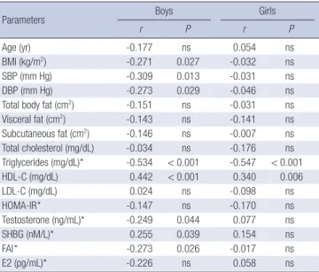
Fig. 1. Correlation between low density lipoprotein (LDL) particle size and sex hormones in children (A) and adolescents (B).
LDL particle size (nm) LDL particle size (nm)
Log free androgen index Log estradiol
r = -0.273, P = 0.026 r = 0.058, P = 0.645
-4.0 -2.0 0.0 2.0 4.0 2.0 3.0 4.0 5.0 6.0
28.0
27.0
26.0
25.0
29.0
28.0
27.0
26.0
25.0
A B
trations were not significantly different in children after adjust- ment.
Correlation between sex hormones and LDL particle size in boys and girls
In boys, univariate analysis showed significant associations be- tween LDL particle size and BMI, SBP, DBP, TG, HDL-C, testos- terone, SHBG, and FAI (P < 0.05). In girls, only TG and HDL-C were significantly associated with LDL particle size (P < 0.05);
none of sex hormones were associated with LDL particle size.
The relationships between LDL size and sex hormones in boys and girls are shown in Fig. 1.
Because LDL particle size was significantly associated with FAI only in boys, we performed the stepwise multivariable re- gression analysis in boys only. Since age, BMI, and HOMA-IR were significantly associated with FAI (data not shown), these parameters were defined as confounding variables in the anal- ysis. As a result, FAI remained an independent predictor of LDL particle size in boys (R2 = 0.075, P = 0.026).
LDL particle size before and after puberty in relation to sex hormone concentrations
LDL particle size was significantly different only in adolescents, not in children (Table 1), and FAI was independently correlated with LDL particle size only in boys. E2 concentration and LDL particle size were not significantly correlated in either age group (Table 2). We hypothesized that LDL particle size is affected by more profound changes in testosterone concentrations. To test this hypothesis, we categorized individuals as either before or
after puberty in both boys and girls according to their testoster- one and E2 concentrations, respectively. We arbitrarily deter- mined a cutoff level as the prepubertal upper normal values of testosterone (0.8 nM/L) and E2 (10 pg/mL) concentrations and lower normal value of SHBG (62.4 nM/L) concentrations (17).
We also calculated the FAI cutoff value (1.28) using the arbitrari- ly determined testosterone and SHBG concentrations. The anal- ysis showed that LDL particle size differed significantly before and after puberty (P = 0.025) in boys, but not in girls (P = 0.982) (Fig. 2).
DISCUSSION
In this study, we showed that LDL particle size was significantly smaller only in adolescents, and that the particle size decreases as FAI increases in boys. Even though LDL-C concentrations were within the normal range and did not differ between boys and girls in children and adolescents, the higher burden of small dense LDL during puberty in boys could imply a higher cardio- vascular risk in males starting at a young age.
The factors affecting LDL particle size have been studied wide- ly in adults, but few studies have included children (12, 18-20).
Among these factors, sex has been suggested to be important, although few studies have found sex differences in LDL size in children (12, 21). In contrast, some investigations of adults have reported significant sex differences in LDL size (10, 11). These studies found larger LDL particle sizes in women, and our re- sults extend this previously reported sex difference to include children and adolescents.
The mechanisms responsible for the sex differences in LDL particle size remain unclear. Studies have suggested that differ- ences in visceral fat accumulation and TG metabolism between men and women are responsible for this sex difference (10-12).
However, we found no sex differences in visceral fat or TG con- centrations in either age group. In addition, we found no associ- ation between visceral fat and LDL particle size in boys and girls.
The relationship between testosterone concentrations and Table 2. Anthropometric and biochemical parameters associated with LDL particle
size in boys and girls
Parameters Boys Girls
r P r P
Age (yr) -0.177 ns 0.054 ns
BMI (kg/m2) -0.271 0.027 -0.032 ns
SBP (mm Hg) -0.309 0.013 -0.031 ns
DBP (mm Hg) -0.273 0.029 -0.046 ns
Total body fat (cm2) -0.151 ns -0.031 ns
Visceral fat (cm2) -0.143 ns -0.141 ns
Subcutaneous fat (cm2) -0.146 ns -0.007 ns
Total cholesterol (mg/dL) -0.034 ns -0.176 ns Triglycerides (mg/dL)* -0.534 < 0.001 -0.547 < 0.001
HDL-C (mg/dL) 0.442 < 0.001 0.340 0.006
LDL-C (mg/dL) 0.024 ns -0.098 ns
HOMA-IR* -0.147 ns -0.170 ns
Testosterone (ng/mL)* -0.249 0.044 0.077 ns
SHBG (nM/L)* 0.255 0.039 0.154 ns
FAI* -0.273 0.026 -0.017 ns
E2 (pg/mL)* -0.226 ns 0.058 ns
*log-transformed. r, correlation coefficient; BMI, body mass index; SBP, systolic blood pressure; DBP, diastolic blood pressure; HDL-C, high density lipoprotein cholesterol;
LDL-C, low density lipoprotein cholesterol; HOMA-IR, homeostasis model assessment for insulin resistance; SHBG, sex hormone-binding globulin; FAI, free androgen index;
E2, estradiol; ns, not significant.
Fig. 2. Mean low density lipoprotein (LDL) particle size before and after mid-puberty in boys and girls according to testosterone and estrogen levels, respectively.
LDL particle size (nm)
27.1 27.0 26.9 26.8 26.7 26.6 26.5 26.4 26.3 26.2
P = 0.025
26.93 27.03
26.49
27.02 P = 0.982
Boys Girls
Pre-puberty Puberty
the lipid profile is controversial. Few studies have examined the association between LDL particle size and testosterone concen- tration. In one adult study, low levels of SHBG and testosterone were associated with small dense LDL in normoglycemic mid- dle-aged men (22). These data are inconsistent with our results.
However, testosterone administration in men decreased LDL size in other studies (23, 24). In vivo studies found that testoster- one administration increases the activity of hepatic lipase, which hydrolyzes TG in LDL and produces small dense LDL particles.
This mechanism may explain why testosterone is associated with smaller LDL particle size (23-25).
In this study, we found a difference in LDL particle size only in adolescents and not in children. Moreover, FAI was associat- ed with LDL particle size only in boys. This led us to hypothesize that some threshold in testosterone concentration must be reached for this association. To test this hypothesis, we compared LDL particle size before and after puberty according to sex hormone concentrations and found a significant difference between the two groups, but only in boys. These results were consistent with the hypothesis that testosterone had a more potent effect on LDL particle size than estrogen, and that profound changes in testosterone concentration were involved in the regulation of LDL particle size.
In general, estrogen has been regarded as a favorable factor on lipid profiles and anti-atherogenic effects, and is one of the factors responsible for the sex difference in cardiovascular risk (26). However, the Women’s Health Initiative clinical trial failed to find a reduction in coronary heart disease events among es- trogen HT users (13). They also found that HT users did not have a better LDL subclass distribution, which may explain the fail- ure of HT to reduce the incidence of heart disease events (14).
Furthermore, the SWAN study reported that FAI was related to cardiovascular risk factors, including unfavorable lipid profiles in multiethnic premenopausal and perimenopausal women (27). Based on our results and the reasons cited above, we sug- gest that testosterone might have a greater effect on LDL parti- cle size than estrogen. More studies are required to confirm this conclusion.
This study has several limitations. First, the study was per- formed on a small number of subjects and thus did not repre- sent all children and adolescents in the population. In addition, the study design was cross-sectional, and therefore, we cannot speculate on the association between LDL particle size and se- rial changes in FAI. Specifically, we cannot evaluate the longitu- dinal association between testosterone concentrations and LDL particle size before and after puberty in the same individuals.
However, we could estimate longitudinal associations from this study, including a broad spectrum of ages from prepuberty to late puberty. In this study, we could not measure free testoster- one, which is more informative as a male sex hormone. FAI is also known to have a poor correlation with actual free testoster-
one concentrations in male adults (28, 29). However, free testos- terone can be approximated by FAI if, and only if, the total tes- tosterone concentration is negligible in relation to the concentra- tions of SHBG-binding sites (28). For that reason, this is a reason- able approximation for samples from children in whom blood testosterone concentrations are rarely above 10% of the SHBG concentration (29). In addition, we could not draw blood at the same point in the menstrual cycle in girls. Given the variation in serum estradiol concentrations over the menstrual cycle, whether estradiol concentrations are associated with LDL par- ticle size is not clear. Further studies are required to elucidate the association between estrogen and LDL particle size in chil- dren and adolescents.
Our study, however, is the first to show that FAI is significant- ly related to LDL particle size in boys, and our findings suggest that testosterone might contribute to higher cardiovascular risk in males by lowering LDL particle size. Further prospective stud- ies should focus on the effects of sex hormones on the lipid pro- file and the associated development of atherosclerosis.
REFERENCES
1. Castelli WP, Garrison RJ, Wilson PW, Abbott RD, Kalousdian S, Kannel WB. Incidence of coronary heart disease and lipoprotein cholesterol lev- els. The Framingham Study. JAMA 1986; 256: 2835-8.
2. Kwon SW, Yoon SJ, Kang TS, Kwon HM, Kim JH, Rhee J, Lee SJ, Park JK, Lim JY, Yoon YW, Hong BK. Significance of small dense low-density li- poprotein as a risk factor for coronary artery disease and acute coronary syndrome. Yonsei Med J 2006; 47: 405-14.
3. Genest JJ, McNamara JR, Salem DN, Schaefer EJ. Prevalence of risk fac- tors in men with premature coronary artery disease. Am J Cardiol 1991;
67: 1185-9.
4. Hamsten A, Walldius G, Szamosi A, Dahlen G, de Faire U. Relationship of angiographically defined coronary artery disease to serum lipopro- teins and apolipoproteins in young survivors of myocardial infarction.
Circulation 1986; 73: 1097-110.
5. Austin MA, King MC, Vranizan KM, Krauss RM. Atherogenic lipopro- tein phenotype. A proposed genetic marker for coronary heart disease risk. Circulation 1990; 82: 495-506.
6. Gardner CD, Fortmann SP, Krauss RM. Association of small low-density lipoprotein particles with the incidence of coronary artery disease in men and women. JAMA 1996; 276: 875-81.
7. Krauss RM. Dense low density lipoproteins and coronary artery disease.
Am J Cardiol 1995; 75: 53B-7B.
8. Lamarche B, Tchernof A, Moorjani S, Cantin B, Dagenais GR, Lupien PJ, Després JP. Small, dense low-density lipoprotein particles as a predic- tor of the risk of ischemic heart disease in men. Prospective results from the Quebec Cardiovascular Study. Circulation 1997; 95: 69-75.
9. Slyper AH. Low-density lipoprotein density and atherosclerosis. Unrav- eling the connection. JAMA 1994; 272: 305-8.
10. Carr MC, Hokanson JE, Zambon A, Deeb SS, Barrett PH, Purnell JQ, Brunzell JD. The contribution of intraabdominal fat to gender differenc- es in hepatic lipase activity and low/high density lipoprotein heteroge-
neity. J Clin Endocrinol Metab 2001; 86: 2831-7.
11. McNamara JR, Campos H, Ordovas JM, Peterson J, Wilson PW, Schae- fer EJ. Effect of gender, age, and lipid status on low density lipoprotein subfraction distribution. Results from the Framingham Offspring Study.
Arteriosclerosis 1987; 7: 483-90.
12. Shimabukuro T, Sunagawa M, Ohta T. Low-density lipoprotein particle size and its regulatory factors in school children. J Clin Endocrinol Metab 2004; 89: 2923-7.
13. Anderson GL, Limacher M, Assaf AR, Bassford T, Beresford SA, Black H, Bonds D, Brunner R, Brzyski R, Caan B, Chlebowski R, Curb D, Gass M, Hays J, Heiss G, Hendrix S, Howard BV, Hsia J, Hubbell A, Jackson R, Johnson KC, Judd H, Kotchen JM, Kuller L, LaCroix AZ, Lane D, Langer RD, Lasser N, Lewis CE, Manson J, Margolis K, Ockene J, O’Sullivan MJ, Phillips L, Prentice RL, Ritenbaugh C, Robbins J, Rossouw JE, Sarto G, Stefanick ML, Van Horn L, Wactawski-Wende J, Wallace R, Wassertheil- Smoller S; Women’s Health Initiative Steering Committee. Effects of con- jugated equine estrogen in postmenopausal women with hysterectomy:
The Women’s Health Initiative Randomized Controlled Trial. JAMA 2004;
291: 1701-12.
14. Mackey RH, Kuller LH, Sutton-Tyrrell K, Evans RW, Holubkov R, Mat- thews KA. Hormone therapy, lipoprotein subclasses, and coronary calci- fication: the Healthy Women Study. Arch Intern Med 2005; 165: 510-5.
15. Reinehr T, Kiess W, de Sousa G, Stoffel-Wagner B, Wunsch R. Intima media thickness in childhood obesity: relations to inflammatory marker, glucose metabolism, and blood pressure. Metabolism 2006; 55: 113-8.
16. Choi YJ, Jo YE, Kim YK, Ahn SM, Jung SH, Kim HJ, Chung YS, Lee KW, Kim DJ. High plasma concentration of remnant lipoprotein cholesterol in obese children and adolescents. Diabetes Care 2006; 29: 2305-10.
17. Alan HB. Tietz clinical guide to laboratory tests, 4th ed. St. Louis, MO:
Saunders; 2006.
18. Miyashita M, Okada T, Kuromori Y, Harada K. LDL particle size, fat dis- tribution and insulin resistance in obese children. Eur J Clin Nutr 2006;
60: 416-20.
19. Stan S, Levy E, Delvin EE, Hanley JA, Lamarche B, O’Loughlin J, Paradis G, Lambert M. Distribution of LDL particle size in a population-based sample of children and adolescents and relationship with other cardio-
vascular risk factors. Clin Chem 2005; 51: 1192-200.
20. Steinbeck KS, Bermingham MA, Mahajan D, Baur LA. Low-density li- poprotein subclasses in children under 10 years of age. J Paediatr Child Health 2001; 37: 550-3.
21. Freedman DS, Bowman BA, Otvos JD, Srinivasan SR, Berenson GS.
Levels and correlates of LDL and VLDL particle sizes among children:
the Bogalusa heart study. Atherosclerosis 2000; 152: 441-9.
22. Haffner SM, Laakso M, Miettinen H, Mykkänen L, Karhapää P, Rainwa- ter DL. Low levels of sex hormone-binding globulin and testosterone are associated with smaller, denser low density lipoprotein in normoglyce- mic men. J Clin Endocrinol Metab 1996; 81: 3697-701.
23. Herbst KL, Amory JK, Brunzell JD, Chansky HA, Bremner WJ. Testoster- one administration to men increases hepatic lipase activity and decreas- es HDL and LDL size in 3 wk. Am J Physiol Endocrinol Metab 2003; 284:
E1112-8.
24. Tan KC, Shiu SW, Kung AW. Alterations in hepatic lipase and lipopro- tein subfractions with transdermal testosterone replacement therapy.
Clin Endocrinol (Oxf) 1999; 51: 765-9.
25. Berg G, Schreier L, Geloso G, Otero P, Nagelberg A, Levalle O. Impact on lipoprotein profile after long-term testosterone replacement in hypo- gonadal men. Horm Metab Res 2002; 34: 87-92.
26. Pérez-López FR, Larrad-Mur L, Kallen A, Chedraui P, Taylor HS. Gen- der differences in cardiovascular disease: hormonal and biochemical in- fluences. Reprod Sci 2010; 17: 511-31.
27. Sutton-Tyrrell K, Wildman RP, Matthews KA, Chae C, Lasley BL, Brock- well S, Pasternak RC, Lloyd-Jones D, Sowers MF, Torréns JI; SWAN In- vestigators. Sex-hormone-binding globulin and the free androgen index are related to cardiovascular risk factors in multiethnic premenopausal and perimenopausal women enrolled in the Study of Women Across the Nation (SWAN). Circulation 2005; 111: 1242-9.
28. Kapoor P, Luttrell BM, Williams D. The free androgen index is not valid for adult males. J Steroid Biochem Mol Biol 1993; 45: 325-6.
29. Ly LP, Handelsman DJ. Empirical estimation of free testosterone from testosterone and sex hormone-binding globulin immunoassays. Eur J Endocrinol 2005; 152: 471-8.
AUTHOR SUMMARY
A Higher Burden of Small Low-density Lipoprotein Particles is Associated with Profound Changes in the Free Androgen Index in Male Adolescents
Yong Jun Choi, Sung Hee Choi, Hae Jin Kim, Seung Jin Han, Jin Soon Hwang, Yoon-Sok Chung, Kwan Woo Lee, Hong Keun Cho, and Dae Jung Kim
Even from a young age, males are known to have higher cardiovascular risk than females. Dyslipidemia, including a higher burden related to small low-density lipoproteins (LDL), plays an important role in precipitating atherosclerosis. Here we investigate sex differences in atherogenic lipoprotein burden and the LDL particle size in 135 children and adolescents. LDL particle size is significantly smaller in male than female. The free androgen index (FAI) (100 Total testosterone/sex hormone binding globulin [SHBG]) is negatively correlated with LDL particle size in boys whereas estrogen and LDL particle size are not related with each other. After puberty, LDL particle size declines markedly in male adolescents. The prominent decrease in LDL particle size associated with an increase in testosterone concentrations might underlie the risk of atherogenic dyslipidemia in males from adolescence.