Lessons learned from 12,000 robotic radical
prostatectomies: Is the journey as important as the outcome?
Sung Gu Kang1 , Ji Sung Shim1 , Fikret Onol2 , K. R. Seetharam Bhat2 , Vipul R. Patel2
1Department of Urology, Korea University College of Medicine, Seoul, Korea, 2Global Robotics Institute, Florida Hospital Celebration Health, University of Central Florida School of Medicine, Orlando, FL, USA
Robotic radical prostatectomy (RARP) is a standardized treatment for localized prostate cancer, which provides better functional outcomes and similar oncological outcomes compared to open approaches. Here, we share our experience of 12,000 RARPs by de- scribing the outcomes of the procedure in terms of positive surgical margin (PSM), continence, and potency as well as by present- ing our detailed surgical technique with recent modifications. On cancer control, the PSM rates were 5.8% and 26.1% in T2 and T3, respectively. On the premise of not compromising oncologic outcomes, a tailored approach to individual patients is essential. Even if an extracapsular extension is suspected, neurovascular bundle (NVB) tailoring can be applied using an anatomical landmark to preserve maximal nerve tissue with a negative margin. We developed a nomogram as a useful tool for deciding the degree of tai- loring. For improvements of functional outcomes, we used athermal retrograde early release with a toggling technique, wherein the nerve dissection from the bottom helps with blood loss and allows for smooth NVB releasing. Additionally, we recently per- formed a new minimal apical dissection/lateral prostatic fascia preservation technique. As a result, our 1-week continence rate was 37% and the 6-week rate was 77.6%. In addition, the potency rates in our study were 69%, 82%, and 92% at 3 months, 6 months, and 1 year, respectively (preoperative Sexual Health Inventory for Men scores >21 & bilateral full nerve spared).
Keywords: Prostate; Prostatectomy; Prostatic neoplasms; Robotics
This is an Open Access article distributed under the terms of the Creative Commons Attribution Non-Commercial License (http://creativecommons.org/licenses/by-nc/4.0) which permits unrestricted non-commercial use, distribution, and reproduction in any medium, provided the original work is properly cited.
Received: 3 October, 2019 • Accepted: 19 December, 2019
Corresponding Author: Vipul R. Patel https://orcid.org/0000-0002-8802-9734 Global Robotics Institute, 410 Celebration Place, Celebration, FL 34747, USA TEL: +1-407-303-4673, FAX: +1-407-303-4632, E-mail: [email protected]
ⓒ The Korean Urological Association
www.icurology.org
Investig Clin Urol 2020;61:1-10.
https://doi.org/10.4111/icu.2020.61.1.1 pISSN 2466-0493 • eISSN 2466-054X
INTRODUCTION
Radical prostatectomy is a treatment of choice for local- ized prostate cancer, which has evolved from open surgery to laparoscopy and robotic radical prostatectomy (RARP) [1]. As the robotic techniques developed and surgeons' understand- ing of the robotic platform became profound, RARP showed better functional outcomes and comparable results for onco- logic outcomes [2-4].
It is important to avoid a positive surgical margin (PSM) in prostatectomy. Currently, it is believed that nerve-sparing is not all or nothing; partial nerve preservation is possible while avoiding PSM in patients with extracapsular exten- sion (ECE). In these cases, a tool for predicting the extent of ECE before surgery was needed to obtain a negative surgical margin; therefore, we developed a nomogram. We also vali- dated an anatomic nerve-sparing grading system that uses landmark anatomic features during prostatectomy.
To improve functional outcomes, we continuously devel- oped a technique for neurovascular bundle (NVB) preserva- tion that is basically athermal retrograde early release. This process has been performed using the up-and-down toggling technique of a 30-degree lens. Recently, we performed mini- mal apical dissection without opening of the endopelvic fas- cia and early ligation of the dorsal vein; this has improved our early functional outcomes.
In this review, we will share our experience of 12,000 RARPs, which were performed by a single surgeon (Vipul R.
Patel), with regard to PSM, continence, potency, and techni- cal principles.
POSITIVE SURGICAL MARGIN
The main outcomes of radical prostatectomy are tradi- tionally reported as a trifecta of rates [5]. The three factors are urinary continence, potency, and biochemical recurrence (BCR)-free survival rates after surgery. However, since im- mediately after surgery we only know the PSM, rather than the BCR [6], we suggested the pentafecta as a new standard for reporting outcomes [7]. Although functional outcomes are coming into focus as important results after radical prosta- tectomy, tumor control is the most important aspect of the surgery. PSM is known to be an independent predictive fac- tor of BCR, local recurrence, and the development of distant metastasis.
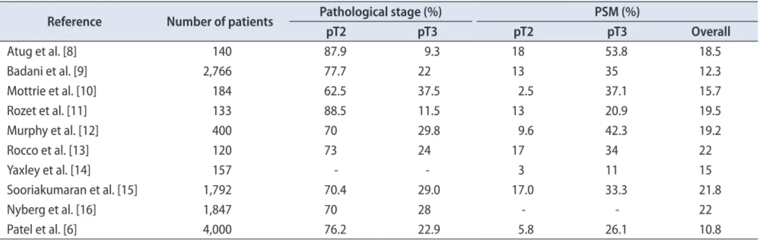
In several studies, the PSMs for pT2 and pT3 were re- ported to be approximately 8.92% and 33%, respectively. The results for the PSMs of representative studies are summa- rized in Table 1 [6,8-16]. According to our study, the overall PSM rate was 14% to 20.8%, specifically 5.8% in pT2 and 26.1% in pT3. In high-risk (D’Amico classification) patients, the overall PSM rate was 25.1%, with 8.6% in pT2, 26.6% in
pT3a, and 53.3% in pT3b [17].
Efforts to reduce PSM have led to the determination of risk factors for PSM. Our previous findings demonstrated that factors that correlated with the aggressiveness of can- cer, such as clinical/pathologic stage and tumor volume, were the most important predictors for PSM. In terms of preop- erative factors, the clinical stage was the only significant predictive factor, with higher PSM rates for T3 versus T1c (odds ratio [OR], 10.7; p<0.0001) and for T2 versus T1c. With regard to perioperative variables, pathologic stage (p<0.0001) and percentage of tumor in the surgical specimen (p=0.0022) were the only independent predictive factors for PSM [18].
In high-risk patients, Kang et al. [17] reported that the only significant predictive factors of PSMs were pathological out- comes such as the percentage of tumors in the specimen and the pathological stage (p<0.001, both).
Therefore, to reduce PSM in high volume/stage tumors, it is necessary to remove suspected tissues soundly outside the prostate capsule. Indeed, previously, all the nerve tis- sue besides the lobe was radically removed to control the margin of the ECE; this approach was known as the “all-or- none” concept, in that the entire nerve bundle was either preserved or removed. However, later studies have found that even high-risk tumors are often organ-confined tumors and that even with ECE, tumors are confined within a few millimeters. Thus, it seemed unnecessary to remove all the nerve tissue of the lobe to control the margin in the patient with ECE.
To date, it has not been possible to standardize the deci- sion-making process in terms of when to take a more or less conservative approach. Interestingly, as indicated in several papers, 85% of the ECEs are within 3 mm of the prostate capsule, and in 97.6%, within 5 mm [19]; thus, we used these statistics for our outcomes. Previously, we investigated 11,794
Table 1. Positive surgical margins (PSMs)
Reference Number of patients Pathological stage (%) PSM (%)
pT2 pT3 pT2 pT3 Overall
Atug et al. [8] 140 87.9 9.3 18 53.8 18.5
Badani et al. [9] 2,766 77.7 22 13 35 12.3
Mottrie et al. [10] 184 62.5 37.5 2.5 37.1 15.7
Rozet et al. [11] 133 88.5 11.5 13 20.9 19.5
Murphy et al. [12] 400 70 29.8 9.6 42.3 19.2
Rocco et al. [13] 120 73 24 17 34 22
Yaxley et al. [14] 157 - - 3 11 15
Sooriakumaran et al. [15] 1,792 70.4 29.0 17.0 33.3 21.8
Nyberg et al. [16] 1,847 70 28 - - 22
Patel et al. [6] 4,000 76.2 22.9 5.8 26.1 10.8
-, not mentioned.
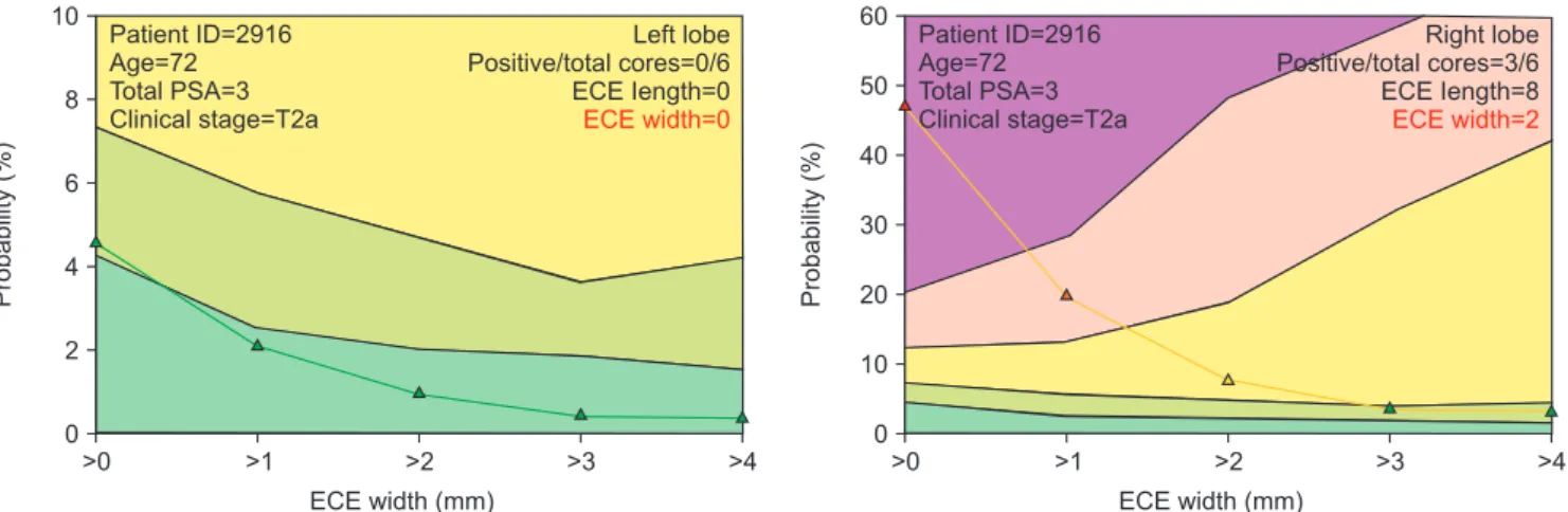
lobes of the prostate and developed a nomogram that allows the generation of a graph showing the likely level of exten- sion (Fig. 1). Therefore, we have a better chance of getting a negative margin considering 3 mm out during RARP [19].
This model is currently undergoing validation.
Additionally, we have studied the anatomy of the NVB and found vascular landmarks coming off the obturator nerve, in the form of small arteries, which assist in deter- mining its location in relation to the prostate and NVB. We presented this landmark artery as a criterion for this nerve- sparing tailoring, which was pathologically validated [20].
The main point of partial nerve-sparing is margin con- trol by sacrificing about 3 mm of nerve tissue based on the landmark artery that occupies the most medial portion of the NVB. Lateral to the plane of dissection of this artery will give the operator at least 3 mm of clearance from the prostate capsule. The anatomic grading of the proportion of NVB-saving based on the landmark artery and the grading were categorized and are described in the potency section.
CONTINENCE
Functional outcomes, such as continence and potency, were the main focus of the current study. It is not sufficient to only consider the removal of the prostate; functional out- comes that are related to the patient’s quality of life must also be considered. During a decade of evolution up until the present day, we have published every technique that we perform in our surgeries.
Excellent continence outcomes have been consistently re- ported after RARP, with the 1-year continence rate reaching
>90% in most of the large, single-center, prospective studies [2,21]. Although we previously reported a 96.4% continence rate 1 year after RARP, the early recovery of urinary conti- nence remains a challenge (Table 2) [7,22-29].
The results for early continence outcomes have been reported by various researchers and have been shown to be approximately 58.5% at 1 month and 79% at 3 months [6]. In our study, the continence rate was 67.7% at 6 weeks Table 2. Continence outcomes
Reference Number of
patients Age (y) Follow-up (mo)
Definition of
continence Technique Continence (% at n months)
1 3 6 12
Joseph et al. [23] 325 60 6 No pads No reconstruction 56 93 96 -
Zorn et al. [24] 300 59 24 No pads No reconstruction 23 47 68 90
Rocco et al. [22] 31 66 6 No pads or 1 safety pad Posterior reconstruction 84 92 - - Tewari et al. [25] 182 61 6 No pads or 1 small liner Ant/post reconstruction 83 91 97 -
Shikanov et al. [26] 380 58 24 No leak No reconstruction - 57 80 92
Patel et al. [27] 1,100 58 18 No pads Ant/post reconstruction 68 (6 wk) 85 96 97
Haglind et al. [28] 1,847 63 12 <1 pad Not mentioned - - - 79
Coughlin et al. [29] 157 35–70 24 No pads Not mentioned - - 84 90
Patela 100 58 5 No pads MAD/LPFP 78 (6 wk) 88 93 -
MAD/LPFP, minimal apical dissection and lateral prostatic fascia preservation technique applied.
a:Unpublished data.
Fig. 1. Output yielded by the graphical user interface for a 72-year-old patient with T2a clinical stage and a prostate-specific antigen (PSA) level of 3 ng/mL. The left lobe had no positive cores, while the right lobe had three positive cores, all with Gleason score >7. Produced with permission from Vipul R. Patel. ECE, extracapsular extension.
>0 >1 >2 >3
10
8
6
4
2
>4
Probability(%)
ECE width (mm) 0
Patient ID=2916 Age=72 Total PSA=3 Clinical stage=T2a
Left lobe Positive/total cores=0/6 ECE Iength=0 ECE width=0
>0 >1 >2 >3
60 50 40 30 20 10
>4
Probability(%)
ECE width (mm) 0
Patient ID=2916 Age=72 Total PSA=3 Clinical stage=T2a
Right lobe Positive/total cores=3/6 ECE Iength=8 ECE width=2
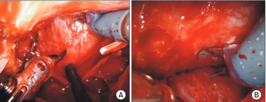
postoperatively and 85.4% at 3 months postoperatively. After a recent modification of our technique, which maximized preservation of periurethral tissue around the urethral stump avoiding the classic incision of the endopelvic fascia (Fig. 2), our 1st and 6th-week continence (no pads/d) rate was increased to 37% and 77.6%, respectively (unpublished data).
Postoperative continence recovery is affected by a num- ber of factors and possibly by both patient characteristics and surgical techniques [30]. Among the preoperative char- acteristics of patients, age, body mass index, prostate volume, and comorbidities are known to affect the postoperative continence recovery [31,32]. In addition, Shikanov et al. [33]
reported that age (OR, 0.97; p=0.002), baseline International Prostate Symptom Score (IPSS) (OR, 0.98; p=0.02), and Sexual Health Inventory for Men (SHIM) scores (OR, 1.02; p=0.005) were independent factors for postoperative continence. With regard to surgical techniques, van der Poel et al. [34] reported that they were influenced by the amount of fascia preserva- tion of the lateral aspect of the prostate, which was in line with our recent technique.
SURGICAL TECHNIQUES FOR CONTINENCE
1. Bladder neck reconstruction
The vesico-urethral anastomosis (VUA) is a critical step during RARP, and it is essential to reduce the bladder neck diameter before starting VUA under some circumstances, including cases with large prostates or large median lobes.
Before starting the bladder neck reconstruction, it is es- sential to check the position of the ureteric orifices and their distance from the edge of the bladder neck. Bilateral plication over the lateral side of the bladder is subsequently performed; the suture begins laterally and runs medially until the bladder neck size matches that of the membranous
urethra. The same suture then runs laterally back to the beginning of the suture and is tied [35].
We do not generally perform bladder neck preservation because it can be associated with PSM, especially in high- risk cancer, as it can increase the continence rate by recon- structing the bladder neck later. From March to November 2006, 279 patients underwent RARP at our institution; ap- proximately 27% (74) of these patients required bladder neck reconstruction. In this group of patients, 12.7% resumed pad- free continence immediately after the removal of the Foley catheter. The short-term pad-free continence rates at 3, 6, and 12 months after surgery were 91.8%, 97.3%, and 97.3%, respectively [35].
2. Periurethral suspension stitch
The use of a periurethral retropubic suspension stitch has been described by Walsh [36] in an open radical retropu- bic prostatectomy series, and Patel et al. [37] were the first to describe this suspension technique in RARP. We added the periurethral suspension stitch to our standard RARP tech- nique with the initial purpose of improving the hemostasis of the dorsal venous complex and facilitating dissection of the prostate apex and urethra. The continence mechanism is the anatomical support of the urethra by the suspension of the tissues ventral to the urethra on the fascia of the pubic bone [38]. We reported that the suspension stitch resulted in a significantly shorter interval to the recovery of continence (suspension group: median, 6 weeks; 95% confidence interval [CI], 6.387 to 8.288 vs. non-suspension group: median, 7 weeks;
95% CI, 7.558 to 11.612; log-rank test, p=0.02) and higher conti- nence rates 3 months after the procedure (p=0.013, 94 with- out suspension vs. 237 with suspension) [37].
3. Posterior reconstruction
The posterior reconstruction technique was first de-
Fig. 2. (A) Left. Existing method: a sus- pension stitch and incised endopelvic fascia were observed. (B) Right. Minimal apical dissection: an intact endopelvic fascia was observed.
A B
Suspension stitch
Arcus tendineus fascia pelvis
scribed by Rocco et al. [22] in an open approach. It was fur- ther investigated and modified in detail as in a review of literature on RARP [39,40] and the most recent description of the technique was implemented in our study [38]. Briefly, we performed the first layer of the reconstruction between the remaining Denonvillier’s fascia and the posterior aspect of the rhabdosphincter/posterior median raphe. The sec- ond layer of reconstruction was then performed between the bladder neck and the posterior urethra. The supposed mechanism for achieving continence is the realignment of the tissues dorsal to the bladder and urethra, which in turn provides a tension-free VUA and the ability to recreate pos- terior support [38] (Fig. 3).
Our modified technique for posterior reconstruction of the rhabdosphincter resulted in a significantly shorter in- terval to the recovery of continence and higher continence
rates in the early period after catheter removal (23%/43%
and 29%/52%; p=0.045 and p=0.016 at 1 and 4 weeks, respec- tively). A lower incidence of cystographic leaks was also observed in the posterior reconstruction group (0.4% vs. 2.1%;
p=0.036) [41,42].
4. Minimal apical dissection
Recently, we modified our techniques regarding apical dissection. After the bladder is dropped, posterior dissection and retrograde nerve-sparing are done prior to opening the endopelvic fascia. Then, the endopelvic fascia is opened closer to the prostate instead of opening it closer to the pelvic side- wall, thus leaving all other tissues behind and all ligaments in place. After finishing the NVB dissection, the dorsal ve- nous complex (DVC) is divided and sutured with running 2-0 Quill suture (Fig. 2). We compared the continence outcomes of the minimal apical dissection and lateral prostatic fascia preservation (MAD/LPFP) technique with a control group that was created by propensity-score matching from a cohort of 2,064 patients who underwent our conventional RARP (c-RALP) technique and achieved earlier continence and potency recovery. The mean time to achieve continence was 32 days in the MAD/LPFP group vs. 87 days in the c-RALP group (p<0.001), and mean time to potency was significantly shorter in the MAD/LPFP group than in the c-RALP group (37 vs. 156 days, p<0.001) (unpublished data). Continence (no pads/d) rates were 77.6% vs. 44.7% at 6 weeks and 87.9% vs.
66.7% at 3 months (MAD/LPFP vs. control group) (p<0.001, both) (Table 2, unpublished data). Similarly, de Carvalho et al. [43], using a similar technique of MAD, reported that continence was reached immediately in 85.9% of the patients and in 98.4% at 3 months postoperatively.
POTENCY OUTCOMES
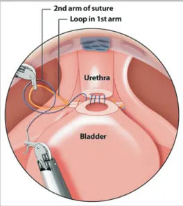
Although the proficiency of the technique (learning curve) is important to maximize the functional outcome, the Fig. 3. Posterior reconstruction (second layer suture). The two-layer
reconstruction involved the realignment of the sphincteric muscle to the Denonvillier’s fascia, followed by a second suture that fixed the posterior bladder wall to the urethra. Produced with permission from Vipul R. Patel.
Table 3. Potency outcomes of representative studies Reference Number of
patients Age (y) Follow-up (mo) Overall potency (% at n months)
3 6 12 18
Menon et al. [50] 1,142 60 - - - 70 100
Zorn et al. [24] 300 59 24 47 58 74 77
Rocco et al. [13] 120 63 12 31 43 61 -
Finley et al. [51] 62 57 > 18 32 57 77 90
Shikanov et al. [26] 380 58 24 57 63 82 -
Sooriakumaran et al. [15] 1,792 63 24 58 - 73 -
Coughlin et al. [29] 157 35–70 24 - 41 53 -
Patel et al. [27] 404 58 18 69 82 92 97
fundamentals of the technique itself could be more impor- tant. Therefore, we have focused on this aspect by continu- ously improving and modifying the surgical technique.
The surgical anatomy of the nerve-sparing radical pros- tatectomy was initially laid down by the pioneering work of Walsh and Donker [44] who documented the concept of the NVB in relation to the prostate. The preservation or return of potency post radical prostatectomy is one of the most challenging and variable parts of prostatectomy. “Nerve- sparing” means NVB preservation, which means preserva- tion of the complex of the tissues lateral to the prostate.
During this procedure, the principles of athermal and atrau- matic manipulation for nerve preservation are considered to be the most important.
In many studies, age, Charlson score, baseline Inter- national Index of Erectile Function-6 (IIEF-6) score/SHIM score, and the performance of an NVB-sparing procedure were independent factors for predicting erectile function re- covery [33,45]. Several factors affect the recovery of erectile function, including age, preoperative sexual function, and technical aspects during surgery; however, cavernosal nerve preservation is considered to be the most important factor for recovery [46-48]. Kang et al. [49] also showed that the surgeon’s subjective NVB-sparing score system could predict potency recovery. In addition, they reported that when more than a certain amount of tissue applicable to NVB-sparing grade 3 is preserved, the preservation of more nerve tissue results in incrementally shorter times to potency recovery.
In our study, by using SHIM, IIEF-6, and subjective evalu- ations, the potency recovery rates at 3, 6, 12, and 18 months were about 38.8%, 65.4%, 73.9%, and 95%, respectively (Table 3) [6,13,15,24,26,27,29,50,51].
TECHNIQUES OF NVB PRESERVATION
Our technique was performed in a retrograde manner, using the method of toggling. This process is not only about preserving the nerves, but also about manipulating the nerves carefully without using energy or traction and pre- venting the neurapraxia response.
1. Athermal retrograde approach of NVB preservation
Our techniques involve the use of an athermal technique to avoid injury of the cavernosal nerves; we believe that this technique is now performed in most institutions [52-55].
The approach to NVB-sparing can be antegrade (from the prostate base to the apex), retrograde (from the apex to the
Fig. 4. Neurovascular bundle (NVB) penetration from the Denonvillier’s fascia to the prostatic anterior aspect; the palpating landmark artery on the NVB is clearly observed.
Fig. 5. (A) The 30 degrees down view is shown. The left vas deferens is retracted with the fourth arm and the right vas is retracted by the assistant.
The dissection plane between the prostate fascia and the neurovascular bundle (NVB) is rarely seen in this view. (B) The 30 degrees up view is shown. In this view, we can easily access the proper plane for interfascial dissection. If the adhesion is not severe, we can see the already pen- etrated space between the prostatic anterior aspect and Denonvillier's fascia following separation of the NVB.
A B
base), or a combination of the two. The antegrade approach is a heritage of a pure laparoscopic procedure, which has various methods [56,57]. However, since all the procedures for NVB-sparing are performed from the inside, the elements of prostatic vasculature are not easily identified. There is also a high risk of falling into the intrafascial plane, which is not the natural plane between the prostate and NVB. This ap- proach also increases the potential risk of capsular incision or PSMs [58].
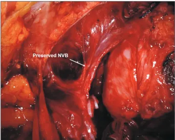
The retrograde method originates from the open ret- ropubic radical prostatectomy; the NVB approach is made from the outside, and the NVB is gently detached from the prostate. In contrast to open surgery with limited visualiza- tion, the retrograde approach during RARP allows for fine tailoring of the NVB through enhanced identification and delineation of the NVB and the surrounding tissues. Be- cause the NVB is very close to the pedicle of the base of the prostate, we believe that releasing it in a retrograde manner can prevent inadvertent clipping and the increased risk of PSMs (Fig. 4). In addition, releasing the nerve bundles before the apex is released from the base decreases the traction and positions it in a good plane [59]. A major advantage of this technique is that the NVB is released away at the mid-pros- tate, where the nerves converge to form a more condensed NVB [60].
2. Toggling technique (30-degree lens up and down in DaVinci Xi)
Since we used DaVinci Xi (Intuitive Surgical, Sunnyvale, CA, USA), we could dissect the prostate using a toggling technique, which means changing the camera from 30 de- grees down to up (Fig. 5).
Using this technique, the NVB can be released from below, achieving a good plane between the prostate fascia and the NVB. Through this avascular plane, the surgeon can release the NVB considerably higher into the prostate.
The camera can then be flipped 180 degrees downward, and
toggling can be done once again from 30 degrees up to 30 degrees down. Next, we can attend to the anterior and move into the space created posteriorly, albeit with much less bleeding (Fig. 6). Thus, this technology is most useful because understanding of the anatomy allows for better use of the technology to release the NVB and flipping of the pedicle.
3. Anatomic nerve-sparing grade
1. Grade 5 (≥95% nerve sparing): The dissection plane is at the medial side to the landmark artery, just outside the prostatic fascia between the prostate and the NVB.
Intraoperatively, we can observe a pink coloration on the prostate with an absence of fatty tissue; this is the interfascial plane (Fig. 7).
2. Grade 4 (75%): The dissection plane is between the landmark artery and the prostatic capsule across the NVB. We can observe a strip of fat over the prostate with an absence of arterial vessels. In cases with a minimal ECE, this could be an adequately safe margin.
Fig. 6. Neurovascular bundle (NVB) separation. The NVB is completely and easily saved by retrograde early release on the mid-prostate level.
Sally Shisler All Rights Reserved
0% <50%
50%
75%
95%
Sally Shisler All Rights Reserved
95% 75% 50% <50% 0%
LA
Prostate
Prostate
LA
LA
Fig. 7. Cross-section of the neurovascular bundle, represented as a histology slide (left) and a diagram (right), demon- strating our graded approach to nerve- sparing. Several degrees of partial nerve- sparing can be obtained when careful attention is given to the anatomic cues that are discussed. Produced with per- mission from Sally Shisler. LA, landmark artery.
3. Grade 3 (50%): The dissection plane is at the landmark artery’s lateral aspect. We can observe a fat strip over the prostate, with the landmark artery.
4. Grade 2 (<50%): The dissection plane is several milli- meters lateral to the artery. We can observe a thick fat strip over the prostate with embedded arteries. In this plane, the most lateral aspect of the NVB is preserved.
5. Grade 1 (0%): This represents a wide resection of the NVB. The correct plane of dissection is confirmed by the presence of the levator fascia, which is not incised.
Our study has some limitations. First, the specific data on the MAD are still being analyzed and will be reported in a future publication. Second, the 12,000 RARP procedures were performed by a single surgeon and the analysis of the learning curve was insufficient. Future data will include perioperative parameters and complication rates as well as oncologic and functional outcomes.
CONCLUSIONS
In this review, we focused on the oncologic outcomes, represented by PSM, and functional outcomes, represented by continence and potency, in terms of surgical principle and technique. This surgical journey was to improve functional outcomes while maintaining negative surgical margins.
Therefore, concepts such as NVB-saving by early retrograde release, partial nerve-saving by grading system, ECE nomo- gram, landmark artery criterion, and MAD have been intro- duced and developed.
After 12,000 cases of surgery, the surgeons can still learn and improve their skills, despite there being no huge surgi- cal advancements. All patients want the full pentafecta, but sometimes the factors that affect this pentafecta may be out of the surgeon’s control. Therefore, the surgery should aim to treat each patient with a modest attitude, acknowledging that every patient is different.
CONFLICTS OF INTEREST
The authors have nothing to disclose.
AUTHORS’ CONTRIBUTIONS
Research conception and design: Sung Gu Kang and Ji Sung Shim. Data acquisition: Fikret Onol and K. R. Seeth- aram Bhat. Drafting of the manuscript: Ji Sung Shim. Criti- cal revision of the manuscript: Sung Gu Kang. Supervision:
Vipul R. Patel. Approval of the final manuscript: all authors.
REFERENCES
1. Ficarra V, Cavalleri S, Novara G, Aragona M, Artibani W. Evi- dence from robot-assisted laparoscopic radical prostatectomy:
a systematic review. Eur Urol 2007;51:45-55; discussion 56.
2. Ficarra V, Novara G, Artibani W, Cestari A, Galfano A, Grae- fen M, et al. Retropubic, laparoscopic, and robot-assisted radi- cal prostatectomy: a systematic review and cumulative analysis of comparative studies. Eur Urol 2009;55:1037-63.
3. Porpiglia F, Morra I, Lucci Chiarissi M, Manfredi M, Mele F, Grande S, et al. Randomised controlled trial comparing lapa- roscopic and robot-assisted radical prostatectomy. Eur Urol 2013;63:606-14.
4. Willis DL, Gonzalgo ML, Brotzman M, Feng Z, Trock B, Su LM. Comparison of outcomes between pure laparoscopic vs robot-assisted laparoscopic radical prostatectomy: a study of comparative effectiveness based upon validated quality of life outcomes. BJU Int 2012;109:898-905.
5. Eastham JA, Scardino PT, Kattan MW. Predicting an optimal outcome after radical prostatectomy: the trifecta nomogram. J Urol 2008;179:2207-10; discussion 2210-1.
6. Patel VR, Abdul-Muhsin HM, Schatloff O, Coelho RF, Valero R, Ko YH, et al. Critical review of 'pentafecta' outcomes after robot-assisted laparoscopic prostatectomy in high-volume cen- tres. BJU Int 2011;108(6 Pt 2):1007-17.
7. Patel VR, Sivaraman A, Coelho RF, Chauhan S, Palmer KJ, Orvieto MA, et al. Pentafecta: a new concept for reporting outcomes of robot-assisted laparoscopic radical prostatectomy.
Eur Urol 2011;59:702-7.
8. Atug F, Castle EP, Srivastav SK, Burgess SV, Thomas R, Davis R. Positive surgical margins in robotic-assisted radical prosta- tectomy: impact of learning curve on oncologic outcomes. Eur Urol 2006;49:866-71; discussion 871-2.
9. Badani KK, Kaul S, Menon M. Evolution of robotic radical prostatectomy: assessment after 2766 procedures. Cancer 2007;110:1951-8.
10. Mottrie A, Van Migem P, De Naeyer G, Schatteman P, Carpen- tier P, Fonteyne E. Robot-assisted laparoscopic radical pros- tatectomy: oncologic and functional results of 184 cases. Eur Urol 2007;52:746-50.
11. Rozet F, Jaffe J, Braud G, Harmon J, Cathelineau X, Barret E, et al. A direct comparison of robotic assisted versus pure laparo- scopic radical prostatectomy: a single institution experience. J Urol 2007;178:478-82.
12. Murphy DG, Kerger M, Crowe H, Peters JS, Costello AJ. Oper- ative details and oncological and functional outcome of robot- ic-assisted laparoscopic radical prostatectomy: 400 cases with a minimum of 12 months follow-up. Eur Urol 2009;55:1358-66.
13. Rocco B, Matei DV, Melegari S, Ospina JC, Mazzoleni F, Errico
G, et al. Robotic vs open prostatectomy in a laparoscopically naive centre: a matched-pair analysis. BJU Int 2009;104:991-5.
14. Yaxley JW, Coughlin GD, Chambers SK, Occhipinti S, Samara- tunga H, Zajdlewicz L, et al. Robot-assisted laparoscopic pros- tatectomy versus open radical retropubic prostatectomy: early outcomes from a randomised controlled phase 3 study. Lancet 2016;388:1057-66.
15. Sooriakumaran P, Pini G, Nyberg T, Derogar M, Carlsson S, Stranne J, et al. Erectile function and oncologic outcomes fol- lowing open retropubic and robot-assisted radical prostatec- tomy: results from the laparoscopic prostatectomy robot open trial. Eur Urol 2018;73:618-27.
16. Nyberg M, Hugosson J, Wiklund P, Sjoberg D, Wilderäng U, Carlsson SV, et al.; LAPPRO group. Functional and oncologic outcomes between open and robotic radical prostatectomy at 24-month follow-up in the Swedish LAPPRO trial. Eur Urol Oncol 2018;1:353-60.
17. Kang SG, Schatloff O, Haidar AM, Samavedi S, Palmer KJ, Cheon J, et al. Overall rate, location, and predictive factors for positive surgical margins after robot-assisted laparoscopic radical prostatectomy for high-risk prostate cancer. Asian J Androl 2016;18:123-8.
18. Coelho RF, Chauhan S, Orvieto MA, Palmer KJ, Rocco B, Patel VR. Predictive factors for positive surgical margins and their locations after robot-assisted laparoscopic radical prostatec- tomy. Eur Urol 2010;57:1022-9.
19. Patel VR, Sandri M, Grasso AAC, De Lorenzis E, Palmisano F, Albo G, et al. A novel tool for predicting extracapsular exten- sion during graded partial nerve sparing in radical prostatec- tomy. BJU Int 2018;121:373-82.
20. Schatloff O, Chauhan S, Kameh D, Valero R, Ko YH, Sivara- man A, et al. Cavernosal nerve preservation during robot- assisted radical prostatectomy is a graded rather than an all-or- none phenomenon: objective demonstration by assessment of residual nerve tissue on surgical specimens. Urology 2012;79:
596-600.
21. Coelho RF, Chauhan S, Palmer KJ, Rocco B, Patel MB, Patel VR. Robotic-assisted radical prostatectomy: a review of cur- rent outcomes. BJU Int 2009;104:1428-35.
22. Rocco B, Gregori A, Stener S, Santoro L, Bozzola A, Galli S, et al. Posterior reconstruction of the rhabdosphincter allows a rapid recovery of continence after transperitoneal videolaparo- scopic radical prostatectomy. Eur Urol 2007;51:996-1003.
23. Joseph JV, Rosenbaum R, Madeb R, Erturk E, Patel HR. Ro- botic extraperitoneal radical prostatectomy: an alternative ap- proach. J Urol 2006;175(3 Pt 1):945-50; discussion 951.
24. Zorn KC, Gofrit ON, Orvieto MA, Mikhail AA, Zagaja GP, Shalhav AL. Robotic-assisted laparoscopic prostatectomy:
functional and pathologic outcomes with interfascial nerve
preservation. Eur Urol 2007;51:755-62; discussion 763.
25. Tewari A, Jhaveri J, Rao S, Yadav R, Bartsch G, Te A, et al.
Total reconstruction of the vesico-urethral junction. BJU Int 2008;101:871-7.
26. Shikanov SA, Zorn KC, Zagaja GP, Shalhav AL. Trifecta out- comes after robotic-assisted laparoscopic prostatectomy. Urol- ogy 2009;74:619-23.
27. Patel VR, Coelho RF, Chauhan S, Orvieto MA, Palmer KJ, Rocco B, et al. Continence, potency and oncological outcomes after robotic-assisted radical prostatectomy: early trifecta re- sults of a high-volume surgeon. BJU Int 2010;106:696-702.
28. Haglind E, Carlsson S, Stranne J, Wallerstedt A, Wilderäng U, Thorsteinsdottir T, et al.; LAPPRO steering committee. Uri- nary incontinence and erectile dysfunction after robotic versus open radical prostatectomy: a prospective, controlled, nonran- domised trial. Eur Urol 2015;68:216-25.
29. Coughlin GD, Yaxley JW, Chambers SK, Occhipinti S, Sama- ratunga H, Zajdlewicz L, et al. Robot-assisted laparoscopic prostatectomy versus open radical retropubic prostatectomy:
24-month outcomes from a randomised controlled study. Lan- cet Oncol 2018;19:1051-60.
30. Ficarra V, Novara G, Rosen RC, Artibani W, Carroll PR, Costello A, et al. Systematic review and meta-analysis of stud- ies reporting urinary continence recovery after robot-assisted radical prostatectomy. Eur Urol 2012;62:405-17.
31. Link BA, Nelson R, Josephson DY, Yoshida JS, Crocitto LE, Kawachi MH, et al. The impact of prostate gland weight in robot assisted laparoscopic radical prostatectomy. J Urol 2008;180:928-32.
32. Novara G, Ficarra V, D'elia C, Secco S, Cioffi A, Cavalleri S, et al. Evaluating urinary continence and preoperative predictors of urinary continence after robot assisted laparoscopic radical prostatectomy. J Urol 2010;184:1028-33.
33. Shikanov S, Desai V, Razmaria A, Zagaja GP, Shalhav AL. Ro- botic radical prostatectomy for elderly patients: probability of achieving continence and potency 1 year after surgery. J Urol 2010;183:1803-7.
34. van der Poel HG, de Blok W, Joshi N, van Muilekom E. Pres- ervation of lateral prostatic fascia is associated with urine continence after robotic-assisted prostatectomy. Eur Urol 2009;55:892-900.
35. Lin VC, Coughlin G, Savamedi S, Palmer KJ, Coelho RF, Patel VR. Modified transverse plication for bladder neck reconstruc- tion during robotic-assisted laparoscopic prostatectomy. BJU Int 2009;104:878-81.
36. Walsh PC. Anatomic radical prostatectomy: evolution of the surgical technique. J Urol 1998;160(6 Pt 2):2418-24.
37. Patel VR, Coelho RF, Palmer KJ, Rocco B. Periurethral suspen- sion stitch during robot-assisted laparoscopic radical prostatec-
tomy: description of the technique and continence outcomes.
Eur Urol 2009;56:472-8.
38. Vis AN, van der Poel HG, Ruiter AEC, Hu JC, Tewari AK, Rocco B, et al. Posterior, anterior, and periurethral surgical reconstruction of urinary continence mechanisms in robot- assisted radical prostatectomy: a description and video compi- lation of commonly performed surgical techniques. Eur Urol 2019;76:814-22.
39. Gautam G, Rocco B, Patel VR, Zorn KC. Posterior rhabdo- sphincter reconstruction during robot-assisted radical prosta- tectomy: critical analysis of techniques and outcomes. Urology 2010;76:734-41.
40. Joshi N, de Blok W, van Muilekom E, van der Poel H. Impact of posterior musculofascial reconstruction on early continence after robot-assisted laparoscopic radical prostatectomy: results of a prospective parallel group trial. Eur Urol 2010;58:84-9.
41. Freire MP, Weinberg AC, Lei Y, Soukup JR, Lipsitz SR, Prasad SM, et al. Anatomic bladder neck preservation during robotic- assisted laparoscopic radical prostatectomy: description of technique and outcomes. Eur Urol 2009;56:972-80.
42. Coelho RF, Chauhan S, Orvieto MA, Sivaraman A, Palmer KJ, Coughlin G, et al. Influence of modified posterior reconstruc- tion of the rhabdosphincter on early recovery of continence and anastomotic leakage rates after robot-assisted radical pros- tatectomy. Eur Urol 2011;59:72-80.
43. de Carvalho PA, Barbosa JABA, Guglielmetti GB, Cordeiro MD, Rocco B, Nahas WC, et al. Retrograde release of the neurovascular bundle with preservation of dorsal venous com- plex during robot-assisted radical prostatectomy: optimizing functional outcomes. Eur Urol 2018 Jul 21 [Epub]. http://doi.
org/10.1016/j.eururo.2018.07.003.
44. Walsh PC, Donker PJ. Impotence following radical prosta- tectomy: insight into etiology and prevention. 1982. J Urol 2002;167(2 Pt 2):1005-10.
45. Novara G, Ficarra V, D'Elia C, Secco S, De Gobbi A, Cavalleri S, et al. Preoperative criteria to select patients for bilateral nerve- sparing robotic-assisted radical prostatectomy. J Sex Med 2010;7(2 Pt 1):839-45.
46. Briganti A, Gallina A, Suardi N, Capitanio U, Tutolo M, Bian- chi M, et al. Predicting erectile function recovery after bilateral nerve sparing radical prostatectomy: a proposal of a novel pre- operative risk stratification. J Sex Med 2010;7:2521-31.
47. Marien T, Sankin A, Lepor H. Factors predicting preservation of erectile function in men undergoing open radical retropubic prostatectomy. J Urol 2009;181:1817-22.
48. Moskovic DJ, Alphs H, Nelson CJ, Rabbani F, Eastham J, Toui- jer K, et al. Subjective characterization of nerve sparing pre- dicts recovery of erectile function after radical prostatectomy:
defining the utility of a nerve sparing grading system. J Sex
Med 2011;8:255-60.
49. Kang SG, Schatloff O, Haidar AM, Samavedi S, Palmer KJ, Cheon J, et al. Does surgeon subjective nerve sparing score predict recovery time of erectile function following robot- assisted radical prostatectomy? J Sex Med 2015;12:1490-6.
50. Menon M, Shrivastava A, Kaul S, Badani KK, Fumo M, Bhan- dari M, et al. Vattikuti Institute prostatectomy: contemporary technique and analysis of results. Eur Urol 2007;51:648-57;
discussion 657-8.
51. Finley DS, Rodriguez E Jr, Skarecky DW, Ahlering TE. Quan- titative and qualitative analysis of the recovery of potency after radical prostatectomy: effect of unilateral vs bilateral nerve sparing. BJU Int 2009;104:1484-9.
52. Coughlin G, Dangle PP, Palmer KJ, Samevedi S, Patel VR.
Athermal early retrograde release of the neurovascular bundle during nerve-sparing robotic-assisted laparoscopic radical prostatectomy. J Robot Surg 2009;3:13-7.
53. Lei Y, Alemozaffar M, Williams SB, Hevelone N, Lipsitz SR, Plaster BA, et al. Athermal division and selective suture liga- tion of the dorsal vein complex during robot-assisted lapa- roscopic radical prostatectomy: description of technique and outcomes. Eur Urol 2011;59:235-43.
54. Takenaka A, Leung RA, Fujisawa M, Tewari AK. Anatomy of autonomic nerve component in the male pelvis: the new con- cept from a perspective for robotic nerve sparing radical pros- tatectomy. World J Urol 2006;24:136-43.
55. Tavukçu HH, Aytac O, Atug F. Nerve-sparing techniques and results in robot-assisted radical prostatectomy. Investig Clin Urol 2016;57(Suppl 2):S172-84.
56. Asimakopoulos AD, Annino F, D'Orazio A, Pereira CF, Mug- nier C, Hoepffner JL, et al. Complete periprostatic anatomy preservation during robot-assisted laparoscopic radical prosta- tectomy (RALP): the new pubovesical complex-sparing tech- nique. Eur Urol 2010;58:407-17.
57. Menon M, Shrivastava A, Bhandari M, Satyanarayana R, Siva S, Agarwal PK. Vattikuti Institute prostatectomy: technical modi- fications in 2009. Eur Urol 2009;56:89-96.
58. Curto F, Benijts J, Pansadoro A, Barmoshe S, Hoepffner JL, Mugnier C, et al. Nerve sparing laparoscopic radical prostatec- tomy: our technique. Eur Urol 2006;49:344-52.
59. Ko YH, Coelho RF, Sivaraman A, Schatloff O, Chauhan S, Abdul-Muhsin HM, et al. Retrograde versus antegrade nerve sparing during robot-assisted radical prostatectomy: which is better for achieving early functional recovery? Eur Urol 2013;63:169-77.
60. Costello AJ, Brooks M, Cole OJ. Anatomical studies of the neurovascular bundle and cavernosal nerves. BJU Int 2004;94:
1071-6.