CASE REPORT
내시경 정맥류 밴드 결찰술을 통한 항문직장 정맥류 출혈 치료 후 시행된 성공적인 경경정맥 간내문맥-정맥 단락술 및 색전술
박상우, 전충환, 최성규, 김현수, 박창환, 류종선, 박선영, 조은애, 김재규1, 김형욱1
전남대학교병원 소화기내과, 전남대학교 의과대학 영상의학교실1
Successful Transjugular Intrahepatic Portosystemic Shunt with Embolization Subsequent to Endoscopic Variceal Band Ligation for Bleeding Anorectal Varices
Sang Woo Park, Chung Hwan Jun, Sung Kyu Choi, Hyun Soo Kim, Chang Hwan Park, Jong Sun Rew, Seon Young Park, Eunae Cho, Jae Kyu Kim1 and Hyoung Ook Kim1
Division of Gastroenterology, Department of Internal Medicine, Chonnam National University Hospital, Department of Radiology, Chonnam National University Medical School1, Gwangju, Korea
Anorectal variceal bleeding is a rare occurrence; however, in such event, it could be fatal due to large size and high blood flow rate of varices. However, to date, there is no standardized treatment modality. Although endoscopic treatment can be provided, in cases of recurrent anorectal variceal bleeding, other therapeutic modalities for hemostasis are necessary. Here, we present a case of 58-year-old female patient with liver cirrhosis, who suffered from massive bleeding of anorectal varices. Endoscopic variceal band ligation was performed for primary hemostasis. Additionally, transjugular intrahepatic portosystemic shunt (TIPS) with embolization was performed to reduce the risk of rebleeding. Following the procedure, she had no further bleeding episodes, and the size of ano- rectal varices decreased, as seen on an abdomino-pelvic computed tomography. Our case illustrates the effectiveness of combined radiological intervention of TIPS with embolization after endoscopic hemostasis, for variceal obliteration and prevention of rebleeding. (Korean J Gastroenterol 2018;71:234-238)
Key Words: Varices; Treatment; Hemostasis; Portosystemic shunt; Portal hypertension
Received November 2, 2017. Revised February 8, 2018. Accepted February 8, 2018.
CC This is an open access article distributed under the terms of the Creative Commons Attribution Non-Commercial License (http://creativecommons.org/licenses/
by-nc/4.0) which permits unrestricted non-commercial use, distribution, and reproduction in any medium, provided the original work is properly cited.
Copyright © 2018. Korean Society of Gastroenterology.
교신저자: 최성규, 61469, 광주시 동구 제봉로 42, 전남대학교병원 소화기내과
Correspondence to: Sung Kyu Choi, Division of Gastroenterology, Department of Internal Medicine, Chonnam National University Hospital, 42 Jaebong-ro, Dong-gu, Gwangju 61469, Korea. Tel: +82-62-220-6215, Fax: +82-62-225-8578, E-mail: [email protected]
Financial support: None. Conflict of interest: None.
INTRODUCTION
Portal hypertension leads to the dilatation of collateral por- tosystemic veins along the gastrointestinal tract. Varices are most commonly observed in the esophagus and stomach;
however, they can also develop in the anorectal region, occur- ring in about 3.6% to 78% of patients with portal hypertension.1,2 Although anorectal variceal bleeding is rare, it can be fatal;
hence, prompt diagnosis and management are critical.
Currently, there is no definitive standardized treatment mo- dality due to insufficient data and rarity. Various modalities are used for the management of bleeding anorectal varices, including endoscopic treatment (band ligation, injection scle- rotherapy), surgical treatment, and radiologic interventions, such as transjugular intrahepatic portosystemic shunt (TIPS), balloon-occluded retrograde transvenous obliteration, and embolization.
Here, we present a case of massive anorectal variceal
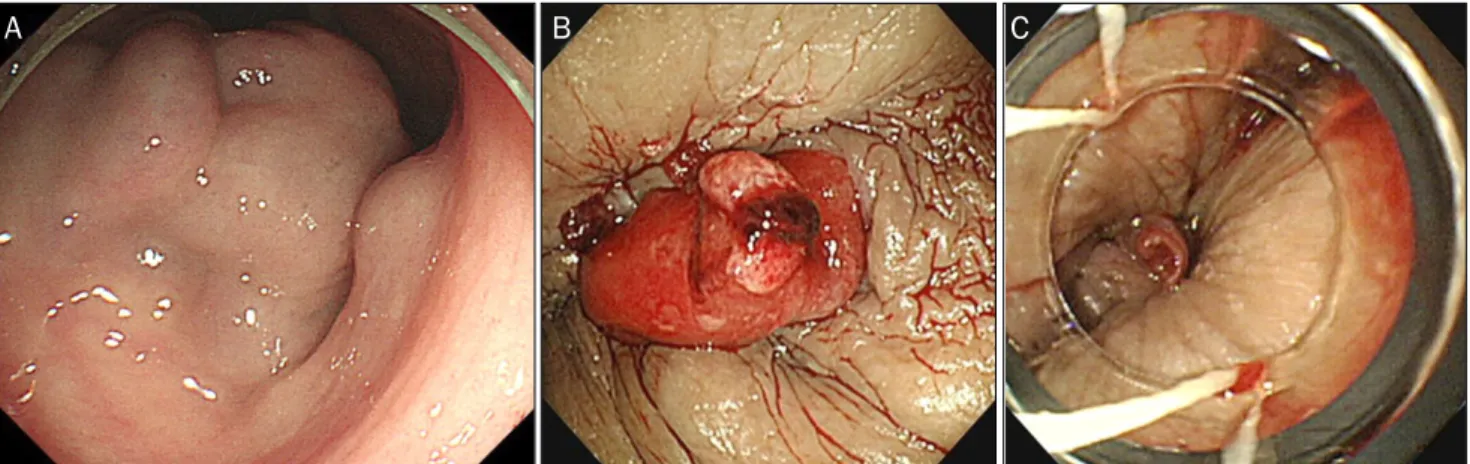
Fig. 1. Sigmoidoscopic finding. Huge anorectal varices (A) and stigmata of recent hemorrhage near the dentate line (B). Endoscopic variceal band ligation was performed to prevent further rebleeding (C).
bleeding, which was primarily controlled by endoscopic vari- ceal band ligation (EVL) followed by TIPS with embolization.
CASE REPORT
A 58-year-old female patient with liver cirrhosis associated with hepatitis B virus infection was transferred to our hospital for massive hematochezia. On arrival, she was alert and ori- ented, with an anemic conjunctiva. She showed distended abdomen with shifting dullness. Her prescribed medications were diuretics and anti-viral agent for hepatitis B virus infection. We did not prescribe her beta blockers due to pre- vious refractory ascites. Fresh blood was observed on digital rectal examination. Blood pressure was 90/60 mmHg, heart rate was 84 beats/min, and body temperature and respira- tory rate were within normal range. The initial laboratory findings were as follows: hemoglobin 6.9 g/dL, platelet 99,000/mm3, prothrombin time, international normalized ratio 1.47, AST 29 IU/L, ALT 22 IU/L, total bilirubin 0.32 mg/dL, and serum albumin 2.2 g/dL.
An emergent esophagogastroduodenoscopy showed tor- tuous esophageal varices without definitive bleeding stigma- ta, and subsequent sigmoidoscopy revealed huge anorectal varices and stigmata of recent hemorrhage near the dentate line. During examination, a sudden spurt of blood from the stigmata was observed, and EVL for the bleeding lesion was performed (Fig. 1). Following the procedure, an abdomi- no-pelvic computed tomography (CT) was performed, which revealed liver cirrhosis with splenomegaly and large amount of ascitic fluid. Despite undergoing a successful endoscopic hemostatic procedure, there were persistent, huge tortuous
anorectal varices (Fig. 2). At that time, model for end stage liver disease score was eleven and Child-Pugh-Turcotte score was nine. Hence, we decided to proceed with additional elec- tive radiologic interventional procedures. Two days after EVL, TIPS combined with embolization was performed to reduce ascites and risk of rebleeding.
Under sedation and sterile conditions, the right internal jugular vein was punctured, and the right hepatic vein was catheterized. The right portal vein was directly punctured through the right hepatic vein, using Colapinto needle (Colapinto; Cook Medical, Bloomington, IN, USA). Portal ve- nogram obtained after cannulation of the inferior mesenteric vein, showed that anorectal varices were being fed by the tor- tuous and dilated superior rectal branches arising from the inferior mesenteric vein. Embolization was performed at both ends of the superior rectal branches using 8 mm Amplatzer vascular plugs (Amplatzer™ Vascular Plug 4; St. Jude Medical, Plymouth, MN, USA) (Fig. 3). Because there were no collateral channels of varices, we did not add any additional emboliza- tion material. A follow-up portal venogram revealed an im- provement in the anorectal varices. Subsequently, two self-expandable stents, 10×80 mm and 10×60 mm each (Niti-S Vascular Stent; TaeWoong Medical, Gimpo, Korea), were deployed between the right hepatic and portal veins. The pre-treatment portosystemic pressure gradient was initially measured to be 18 mmHg, which was decreased to 10 mmHg after TIPS. The procedure was completed without proce- dure-related complications.
A follow-up abdomino-pelvic CT two weeks after the proce- dure revealed significantly improved anorectal varices with a decrease in ascites (Fig. 2). Although she was readmitted for A B C
Fig. 2. An abdomino-pelvic computed tomography (CT) showing. Huge and tortuous varices in the anorectal region and large amount of ascites in the abdomino-pelvic cavity (A, B). A follow-up abdomino-pelvic CT two weeks after TIPS with embolization showed marked improvement of anorectal varices with a decrease in ascites (C, D).
Fig. 3. Portal venogram (contrast-enhanced series) obtained after inferior mesenteric vein (IMV) cannulation. Anorectal varices being fed by the tortuous and dilated superior rectal branches arising from the IMV (A). Embolization was performed at both ends of the superior rectal branches using 8 mm Amplatzer vascular plugs (arrowheads) (B).
mental change one month after discharge, hepatic ence- phalopathy was well controlled with medication. She was then regularly followed-up for six months without any episode of bleeding or hepatic encephalopathy in the outpatient clinic.
DISCUSSION
The term, ‘ectopic varices’, is used to describe dilated por- tosystemic collateral veins due to portal hypertension, lo- cated anywhere, except in the gastro-esophageal region.
Among them, anorectal varices are discrete and dilated sub- mucosal veins located in the anorectal area, which can devel- op when portal hypertension exceeds the 10 mmHg of hep- atic venous pressure gradient.3 Anorectal variceal bleeding has been reported in about 0.5% to 5% of patients with ano- rectal varices.4 No relationship has been found between the increased incidence of anorectal variceal bleeding and the pres-
A B
C D
A B
ence of esophageal variceal bleeding, a previous treatment his- tory of esophageal varices or etiology of portal hypertension.5 Since anorectal varices can be confused with hemor- rhoids, which can coexist, they are likely to be misdiagnosed by physicians. However, anorectal variceal bleeding can be fatal, unlike hemorrhoidal bleeding; hence, it is critical to dif- ferentiate between them and make the right diagnosis to manage further bleeding.6 Hemorrhoids are purplish colored vascular cushions of the venous and arterial anastomosis.
They are present only in the anal canal, and do not extend to the rectum. On the contrary, anorectal varices are serpen- tine, submucosal varicose veins located on the rectum and anal canal, which communicate with the superior rectal veins of the portal origin to the mid and inferior rectal vein draining into the systemic circulation.7 They are bluish-grey colored veins, about 3-6 mm in diameter, compressed easily with dig- ital pressure and refilled on release. In our case, sigmoido- scopic examination showed bluish-grey tortuous dilated vein in the anorectal region and stigmata of a recent bleeding in the anal canal. The patient was a known case of portal hyper- tension and presented with massive hematochezia and shock. As a result, there was a higher likelihood of a diagnosis of anorectal variceal bleeding, rather than hemorrhoidal bleeding.
Surgical treatments, such as ligation, under-running su- tures, and portosystemic shunt, can be considered for ano- rectal variceal bleeding. However, most of these patients are in their advanced stages of liver cirrhosis. Surgical treatment in such cases is difficult due to an increase in the overall risk of surgery, including poor hepatic reserve that can result in hepatic failure.
Although various treatments for anorectal variceal bleeding, such as endoscopic (band ligation, injection sclerotherapy), surgical, and radiological (TIPS, balloon-occluded retrograde transvenous obliteration, embolization) treatments have been developed to date, there lacks a consensus on the definitive therapeutic modalities for management. Case reports or small case series have been reported, but no randomized controlled trials have been published. This is most likely due to extreme rarity. Endoscopic treatments are sometimes pre- ferred because diagnosis and treatment can be accom- plished simultaneously, and it is simple to achieve hemo- stasis of the primary bleeding lesion.8
In cases of rectal variceal bleeding, however, local ther-
apeutic modalities are more restrictive, compared with esophageal and gastric variceal bleeding. The large diameter of rectal varices makes band ligation difficult and increases the likelihood of rebleeding due to a wide defect in the varix induced by band sloughing. Large volume and high blood flow rate in rectal varices can dilute the sclerosing agent, resulting in a failure of hemostasis and incomplete obliteration.
Besides, these endoscopic treatments cannot reduce the portosystemic pressure gradient, which can cause the devel- opment of collateral vessels and ascites. Some patients present with refractory, large amount of ascites, as seen in our patient, caused by increased portosystemic pressure gradient. Hence, alternative or additional treatments are necessary to reduce portosystemic pressure gradient, which can decrease the size of varices and risk of rebleeding.
TIPS, which was first introduced by Katz et al.9, is an effec- tive and recommended procedure for intractable variceal bleeding from anorectal varices.10 Shibata et al.11 reported seven patients with ectopic variceal bleeding successfully treated with TIPS. However, Ahn et al.12 reported a patient who had rebleeding even after a successful TIPS procedure and was controlled by variceal embolization. Vangeli et al.13 re- ported five patients with rebleeding ectopic varices, despite successful TIPS resulting in a reduction of portosystemic pres- sure gradient below 12 mmHg. There are also some reports of shunt dysfunction, such as acute thrombosis of the shunt or stent thrombosis, resulting in incomplete hemostasis.10 Hence, after EVL for primary hemostasis of anorectal variceal bleeding, we performed a prophylactic combination therapy of TIPS and embolization to minimize the risk of rebleeding.
However, since TIPS is associated with severe complications, such as encephalopathy and hepatic failure that can lead to early mortality, it should be used cautiously in patients with relatively well persevered liver function.14
Variceal embolization is performed by occluding the feed- ing vein using embolization substances, including vascular plug, coils, gelfoam, thrombin, and ethanol.15,16 The advant- age of variceal embolization is that it is less invasive and suit- able for patients with advanced liver disease. Similar to endo- scopic treatments, this procedure can be used to simulta- neously diagnose and treat active bleeding. Several cases have been successfully treated with angiographic emboliza- tion without complications.17,18 Until now, there have been no randomized controlled trials and studies comparing the risk
of rebleeding between TIPS alone and TIPS combined with embolization. Vangeli et al.13 reported five cases of rebleed- ing even after successful TIPS. Another study reported a case of rebleeding after successful TIPS, which was treated well with additional embolization.12 Thus, we thought that there may be a difference in the risk of rebleeding if TIPS alone is performed without embolization. However, further studies are warranted to prove that a combinational treatment is in fact more superior than TIPS alone.
In our case, EVL was used for both the diagnosis and treat- ment, due to its ease and availability. Since the patient was in the stage of advanced liver cirrhosis, surgical treatment was not considered. An abdomino-pelvic CT after the endo- scopic procedure showed persistent, huge and tortuous ano- rectal varices, with large amount of ascites. Thus, in our case, it was essential to reduce the portosystemic pressure gradient. Embolization was also performed with TIPS to re- duce the risk of rebleeding, and no further bleeding was observed. However, more data are necessary to establish this method as a definitive therapeutic modality. We hope our case will strengthen the concept of combinational modalities when dealing with anorectal variceal bleeding.
REFERENCES
1. Rabinovitz M, Schade RR, Dindzans VJ, Belle SH, Van Thiel DH, Gavaler JS. Colonic disease in cirrhosis. An endoscopic evalua- tion in 412 patients. Gastroenterology 1990;99:195-199.
2. Wang TF, Lee FY, Tsai YT, et al. Relationship of portal pressure, anorectal varices and hemorrhoids in cirrhotic patients. J Hepatol 1992;15:170-173.
3. Maslekar S, Toh EW, Adair R, Bate JP, Botterill I. Systematic review of anorectal varices. Colorectal Dis 2013;15:e702-e710.
4. Norton ID, Andrews JC, Kamath PS. Management of ectopic varices. Hepatology 1998;28:1154-1158.
5. Chawla Y, Dilawari JB. Anorectal varices--their frequency in cir- rhotic and non-cirrhotic portal hypertension. Gut 1991;32:309-311.
6. Batoon SB, Zoneraich S. Misdiagnosed anorectal varices result- ing in a fatal event. Am J Gastroenterol 1999;94:3076-3077.
7. Takagi S, Kinouchi Y, Takahashi S, Shimosegawa T. Hemodynamics of rectal varices. J Gastroenterol 2006;41:611-612.
8. Boursier J, Oberti F, Reaud S, Person B, Maurin A, Cales P.
Bleeding from rectal varices in a patient with severe decom- pensated cirrhosis: success of endoscopic band ligation. A case report and review of the literature. Gastroenterol Clin Biol 2006;
30:783-785.
9. Katz JA, Rubin RA, Cope C, Holland G, Brass CA. Recurrent bleed- ing from anorectal varices: successful treatment with a trans- jugular intrahepatic portosystemic shunt. Am J Gastroenterol 1993;88:1104-1107.
10. Ory G, Spahr L, Megevand JM, Becker C, Hadengue A. The long-term efficacy of the intrahepatic portosystemic shunt (TIPS) for the treatment of bleeding anorectal varices in cirrhosis. A case report and review of the literature. Digestion 2001;64:261- 264.
11. Shibata D, Brophy DP, Gordon FD, Anastopoulos HT, Sentovich SM, Bleday R. Transjugular intrahepatic portosystemic shunt for treatment of bleeding ectopic varices with portal hypertension.
Dis Colon Rectum 1999;42:1581-1585.
12. Ahn SS, Kim EH, Kim MD, Lee WJ, Kim SU. Successful hemo- stasis of intractable rectal variceal bleeding using variceal embolization. World J Gastroenterology 2015;21:2558-2562.
13. Vangeli M, Patch D, Terreni N, et al. Bleeding ectopic vari- ces--treatment with transjugular intrahepatic porto-systemic shunt (TIPS) and embolisation. J Hepatol 2004;41:560-566.
14. Sakib SM, Kobayashi K, Jawed M. Potential pitfalls in trans- jugular portosystemic shunt placement for bleeding rectal varices. Case Rep Gastroenterol 2015;9:296-301.
15. Almadi MA, Almessabi A, Wong P, Ghali PM, Barkun A. Ectopic varices. Gastrointest Endosc 2011;74:380-388.
16. Helmy A, Al Kahtani K, Al Fadda M. Updates in the pathogenesis, diagnosis and management of ectopic varices. Hepatol Int 2008;2:322-334.
17. Okada M, Nakashima Y, Kishi T, et al. Percutaneous transhepatic obliteration for massive variceal rectal bleeding. Emerg Radiol 2012;19:355-358.
18. Kang DH, Park JW, Jeon EY, et al. Successful treatment of bleed- ing duodenal varix by percutaneous transsplenic embolization.
Korean J Gastroenterol 2015;66:286-290.