CASE REPORT
크론병의 드문 합병증으로 발생한 패혈성 문맥염
신아리, 이창균, 김효종, 심재준, 장재영, 동석호, 김병호, 장영운
경희대학교 의학전문대학원 내과학교실 소화기내과
Septic Pylephlebitis as a Rare Complication of Crohn’s Disease
A Ri Shin, Chang Kyun Lee, Hyo Jong Kim, Jae-Jun Shim, Jae Young Jang, Seok Ho Dong, Byung Ho Kim and Young Woon Chang Division of Gastroenterology, Department of Internal Medicine, Kyung Hee University School of Medicine, Seoul, Korea
Thrombophlebitis of the portal venous system (PVS) with superimposed bacterial infection (septic pylephlebitis) is an extremely rare complication of Crohn’s disease (CD), and therefore diagnosis of septic pylephlebitis is difficult without high clinical suspicion.
A 16-year old male patient who was diagnosed with CD 3 months earlier was admitted with recurrent fever and abdominal pain. CD activity had been well controlled with conventional medical treatment during a follow-up period. Abdominal con- trast-enhanced computed tomography showed massive thrombosis in the PVS without evidence of intra-abdominal infection, and blood cultures were positive for Streptococcus viridians. There was no evidence of deep vein thrombosis or pulmonary thromboembolism, and all laboratory tests for thrombophilia were normal. Based on these findings, the patient was diagnosed with septic pylephlebitis complicated with CD, and was successfully treated with intravenous antibiotic therapy combined with anticoagulation. This case suggests that early comprehensive evaluation is crucial for immediate diagnosis and proper treatment of septic pylephlebitis in patients with CD who present with fever and abdominal pain of unknown origin, even with stable disease activity and absence of other intra-abdominal infections. (Korean J Gastroenterol 2013;61:219-224)
Key Words: Pylephlebitis; Thromboembolism; Crohn disease; Portal venous system; Sepsis
Received July 4, 2012. Revised August 17, 2012. Accepted August 20, 2012.
CC This is an open access article distributed under the terms of the Creative Commons Attribution Non-Commercial License (http://creativecommons.org/licenses/
by-nc/3.0) which permits unrestricted non-commercial use, distribution, and reproduction in any medium, provided the original work is properly cited.
교신저자: 이창균, 130-872, 서울시 동대문구 경희대로 23, 경희대학교 의학전문대학원 내과학교실 소화기내과
Correspondence to: Chang Kyun Lee, Division of Gastroenterology, Department of Internal Medicine, Kyung Hee University Hospital, Kyung Hee University School of Medicine, 23 Kyungheedae-ro, Dongdaemun-gu, Seoul 130-872, Korea. Tel: +82-2-958-8258, Fax: +82-2-968-1848, E-mail: [email protected]
Financial support: None. Conflict of interest: None.
INTRODUCTION
Venous thromboembolism is a serious extraintestinal complication of inflammatory bowel disease (IBD) asso- ciated with substantial mortality1 and often overlooked in clinical practice. The portal venous system (PVS), which com- monly describes the portal vein and superior mesenteric vein,2 is an unusual location for venous thromboembolism in IBD. In addition, in exceptional cases a combination of throm- bophlebitis of the PVS and superimposed bacterial infection can develop, called septic pylephlebitis. Early detection and proper treatment of these cases is crucial to avoid debilitat-
ing complications such as liver abscess, venous ischemia or infarction of the bowel and portal hypertension.3
We here report a rare case of septic pylephlebitis in a 16-year-old adolescent male with Crohn’s disease (CD). To the best of our knowledge, this is the first report of septic pyle- phlebitis complicating CD in Korea.
CASE REPORT
A 16-year-old male adolescent was admitted to Kyung Hee University Medical Center for the evaluation of recurrent fe- ver and abdominal pain for 3 days. He was a non-smoker and
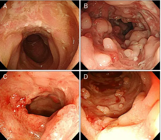
Fig. 1. Colonoscopic findings. (A, B) Multiple longitudinal active ulcers with cobblestone appearance and luminal narrowing were seen in the terminal ileum and ascending colon at first diagnosis. (C, D) Two months later, multiple active mucosal ulcer lesions were markedly improved.
was diagnosed with ileocolonic CD 3 months ago. He had no other significant medical history. The disease activity of CD was well-controlled with conventional medical treatment in- cluding azathioprine (100 mg/day), 5-aminosalicyclic acid (5-ASA) and oral prednisolone (tapered dose of 5 mg/day for weaning off) during a follow-up period. One month earlier, he was admitted with abrupt onset of fever and abdominal pain.
A physical examination revealed tenderness in the right up- per quadrant of the abdomen, and laboratory analysis showed the elevation of acute phase reactants. First, we sus- pected disease flare or intra-abdominal complications of CD such as fistula, abscess and bowel perforations. However, abdominal CT showed no evidence of specific intra-abdomi- nal complications related to CD. Colonoscopy confirmed that mucosal inflammation of CD involving the ileum and ascend- ing colon was markedly improved in comparison with the ini- tial examination (Fig. 1). The symptoms were spontaneously resolved within 2 days and the patient was discharged with azathioprine, 5-ASA and oral prednisolone. During follow- up in the outpatient clinic, he complained of intermittent vague abdominal discomfort without recurrence of fever or severe abdominal pain.
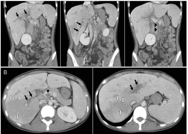
One month after discharge, however, he was re-admitted with a 3-day history of abdominal pain and fever; the same symptoms as before. He had taken oral prednisolone (5 mg/day) and azathioprine (100 mg/day). Physical examina- tion on admission revealed a blood pressure of 110/60 mmHg, heart rate of 117 beats/min, respiration rate of 22 breaths/min, and body temperature of 38.4oC. The abdo- men was soft, but tender in the right upper quadrant, with hy- poactive bowel sounds. Initial laboratory data were as fol- lows; white blood cells 16,070/mm3 (neutrophil 92.3%), he- moglobin 10.7 g/dL, platelets 220,000/mm3, erythrocyte sedimentation rate 55 mm/h, CRP 29.07 mg/dL, prothrom- bin time 15.8 seconds, total bilirubin 1.15 mg/dL. ALP 95 IU/L, AST 31 IU/L, ALT 17 IU/L, LDH 293 IU/L, total protein 5.5 g/dL, albumin 3.3 g/dL. Chest and plain abdominal radiog- raphy showed no specific abnormalities. For the evaluation of recurrent fever and abdominal pain, peripheral blood cul- tures and abdominal CT were performed. Abdominal CT showed massive thrombi in the right, middle, left and main portal vein, associated with periportal edema, and in the su- perior mesenteric vein, and showed transhepatic attenu- ation difference, which reflected decreased portal venous
Fig. 2. Contrast-enhanced abdominal computed tomography findings. Multiple thromboses with periportal edema were seen in the right, middle, left and main portal veins (black arrows). Thrombus in the main portal vein was extended to the superior mesenteric vein (arrow heads). Liver parenchyma shows a transient hepatic attenuation difference (white arrows), which exhibited low-density regions on the portal venous phase due to decreased hepatic blood flow.
blood supply to a segment of the liver due to portal vein branch occlusion and/or parenchymal hyperemia due to hepatic infection (Fig. 2). However, there was no evidence of definite intra-abdominal infectious focus such as hepatic or intestinal abscess. Doppler ultrasonography did not show evidence of deep vein thrombosis of the lower extremities. In addition, chest CT and transesophageal echocardiography revealed no evidence of pulmonary thromboembolism and intracardiac thrombus, respectively. Laboratory tests for ex- clusion of underlying coagulation abnormality were normal and included dysfibrinogenemia, lupus anticoagulant, anti- cardiolipin antibody, antiphospholipid antibody, antinuclear antibodies, deficiencies of protein C and protein S, antith- rombin 3, and genetic testing for factor V Leiden.
The patient was suspected to have septic pylephlebitis complicated with CD, and was treated with intravenous
broad-spectrum antibiotics (ceftriaxone and metronidazole) and anticoagulant medication. Anticoagulation was per- formed with intravenous heparin infusion, followed by oral anticoagulation with warfarin (target international normal- ized ratio of 2). Both azathioprine and prednisolone were dis- continued during hospitalization. Following anticoagulation and antibiotic therapy, clinical symptoms were greatly re- solved within 3 days and inflammatory parameters were nor- malized gradually. He was afebrile on the third day in hospital and had no complaint of abdominal pain on the fifth day.
Finally, Streptococcus viridans sensitive to cephalosporin was isolated from two pairs of blood cultures, and so intra- venous ceftriaxone was continued for 2 weeks until normal- ization of elevated acute phase reactants and leukocytosis.
On hospital day 14, he was discharged with azathioprine and 5-ASA. A follow-up CT scan 2 months later showed marked im-
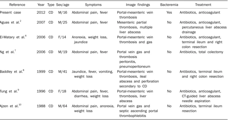
Table 1. Review of Reported Cases with Pylephlebitis or Thrombophlebitis of PVS in Patients with Inflammatory Bowel Disease
Reference Year Type Sex/age Symptoms Image findings Bacteremia Treatment
Present case 2012 CD M/16 Abdominal pain, fever Portal-mesenteric vein thrombosis
Yes Antibiotics, anticoagulant
Aguas et al.2 2007 CD M/25 Abdominal pain, fever Mesenteric partial thrombosis, multiple liver abscess
No Antibiotics, anticoagulant, percutaneous liver abscess drainage
El-Matary et al.6 2006 CD F/14 Anorexia, weight loss, jaundice
Portal-mesenteric vein thrombosis and gas
No Antibiotics, anticoagulant, terminal ileum and right colon resection
Ng et al.7 2006 CD M/19 Abdominal pain, fever Portal vein gas and thrombosis peritonitis, pneumoperitoneum
No Antibiotics, total colectomy
Baddley et al.8 1999 CD M/41 Jaundice, fever, vomiting, weight loss
Portal-mesenteric vein thrombosis, ileal abscess and perforation secondary to CD
No Antibiotics, terminal ileum and right colon resection
Tung et al.9 1996 CD F/18 Abdominal pain, fever, diarrhea, weight loss
Portal-mesenteric vein thrombosis, liver abscess
No Antibiotics, anticoagulant, CT-guided liver abscess needle aspiration Ajzen et al.10 1988 CD M/64 Abdominal pain, anorexia,
weight loss
Portal vein gas and septic ascending portal thrombophlebitis
No Antibiotics, terminal ileum resection
PVS, portal venous system; CD, Crohn’s disease; M, male; F, female.
provement of the PVS thrombosis. Warfarin was continued for the next 6 months, and no adverse events associated with anticoagulation occurred. The patient has remained well without recurrence of symptoms related to pylephlebitis and was in clinical remission of CD at 12-month follow-up.
DISCUSSION
Septic pylephlebitis or septic thrombophlebitis of the PVS is an infective suppurative thrombosis of the portal vein or any of its branches, which typically occurs when there is an ascending bacterial infection from the gastrointestinal tract into the portal vein.4 It is an extremely rare complication of in- tra-abdominal or pelvic infection, which is mostly caused by diverticulitis or appendicitis.5 However, IBD has also been re- ported to be a predisposing factor for pylephlebitis. In our re- view of the literature, several cases of septic pylephlebitis in patients with CD were found (Table 1).2,6-10 Briefly, the mean age of the patients was 30.2 years (range 14-64 years) and 71.4 % (5/7) of the patients were men. Most patients were presented by sudden abdominal pain and fever. Portal or mesenteric vein thrombosis was defined by CT findings in all cases. Median interval from diagnosis of CD to pylephlebitis or thrombophlebitis of PVS was 3.5 years (range 1-8 years).
Interestingly, two patients were diagnosed with CD at the on- set of pylephlebitis. While no microorganisms were yielded in previous studies, pathogenic organism (S. viridians) was iso- lated from peripheral blood cultures in the present case.
Antibiotics and anticoagulant were used on admission as em- pirical therapy in most cases. Four patients underwent surgi- cal procedures for the diagnosis of pylephlibitis or the man- agement of critical complications including bowel perfo- ration and infarction. To the best of our knowledge, the pres- ent case is the first report of septic pylephlebitis in a patient with CD in Korea.
The exact mechanism of pylephlebitis in IBD is not clear.
These complications can be caused by bowel inflammation or interaction with bacterial infection and the endothelium, which may precipitate the clotting cascade, which results in pylethrombosis or pylephlebitis.11 Steroid therapy affects platelet aggregation, coagulation proteins and the vascular system in ways that facilitate thrombosis. Smoking also can be attributed to thrombosis via injury in endothelial cells.12 Other risk factors include a hereditary clotting disorder, im- mobilization and prior abdominal surgery, particularly in the perioperative phase. However, about half of patients with IBD that develop a thromboembolic event have no identifiable risk factor.13 Therefore, the disease activity of IBD is an addi-
tional point to consider. Previous studies have suggested that thromboembolic complications in IBD occur during active disease or in the presence of complications such as stricture, fistula or abscess.1,14,15 Importantly, thromboembolic risk is prominently increased at the time of a flare and the relative risk at the time of a flare is higher during non-hospitalised pe- riods than during hospitalized periods.15 According to our re- view of literature (Table 1), all patients in previous studies had active disease at the onset of pylephlebitis. However, the disease activity of CD in the present case was well controlled with conventional medical treatments and was reaching re- mission state. As stated above, the patient did not have other risk factors associated with thromboembolism. In this con- text, our study suggests that clinicians should consider a po- tential risk of thromboembolism and/or pylephlebitis even in IBD patients with periods of remission and no obvious other risk factors.
The clinical features of pylephlebitis are generally non- specific. Common presenting symptoms are abdominal pain, fever and nausea/vomiting, which can be obscured by ex- acerbation of CD or intra-abdominal infections such as in- fectious colitis.2,8 Laboratory tests are also non-specific, and the most common findings are leukocytosis, anemia and ab- normal liver function tests. For these reasons, it is not thor- oughly investigated and often overlooked, which results in delayed or incorrect diagnosis. Therefore, a high clinical sus- picion is the most important for immediate diagnosis and proper treatment in IBD patients with the above non-specific symptoms and without evidence of intra-abdominal infec- tion. Abdominal ultrasound with color Doppler or CT are the most frequently used imaging techniques for detecting thrombi within the portal or mesenteric veins, and underlying foci of infection in the abdomen or pelvis.5
There is no standardized treatment for pylephlebitis asso- ciated with IBD. Treatment in these circumstances is based on a strategy of anticoagulation and broad-spectrum antibiotics. The aim of anticoagulant treatment is to reverse or prevent progression to thrombosis and treat complica- tions of established portal vein thrombosis, such as bowel is- chemia, infarction and portal hypertension.3 The optimal du- ration of anticoagulant therapy is unknown, but in general vary depending on the severity of thrombosis and bleeding risk. In the first episode, 6 months’ coverage is recom- mended, but it is extended if the risk factors have not dis-
appeared (surgery or immobilization), lifelong anticoagula- tion in patients with an inherited hypercoagulable state.16 The optimum duration of antibiotic therapy is also unclear, but a prolonged course is suggested on the assumption of dif- ficult penetration of antibiotics into the infected thrombus.
To date, there is no consensus statement that addresses the implementation of venous thromboembolism prophy- laxis in IBD patients. The current guidelines recommend that pharmacological prophylaxis for venous thromboembolism should be considered for hospitalized patients with severe disease or for non-ambulating hospitalized patients.17,18 In addition, thromboprophylactic regimens during flares of ac- tive disease should be considered especially for IBD patients with a history of venous thromboembolism, given that pa- tients with IBD are at high risk of recurrent thrombosis after a first venouos thromboembolism.19
Septic pylephlebitis is a serious condition with significantly high morbidity and mortality. However, the outcome of this condition has improved recently, because modern imaging techniques may facilitate an early diagnosis and institution of treatment. We conclude that, in IBD patients with abdomi- nal pain or fever of unknown origin, the possibility of pyle- phlebitis or thrombophlebitis of PVS should be considered re- gardless of the disease activity of CD. Awareness of such a complication allows more efficient diagnosis and proper treatment of these patients, which will result in better out- comes.
REFERENCES
1. Miehsler W, Reinisch W, Valic E, et al. Is inflammatory bowel dis- ease an independent and disease specific risk factor for throm- boembolism? Gut 2004;53:542-548.
2. Aguas M, Bastida G, Nos P, Beltrán B, Grueso JL, Grueso J. Septic thrombophlebitis of the superior mesenteric vein and multiple liver abscesses in a patient with Crohn's disease at onset. BMC Gastroenterol 2007;7:22.
3. Sherigar R, Amir KA, Bobba RK, Arsura EL, Srinivas N. Abdominal pain secondary to pylephlebitis: an uncommon disease of the portal venous system, treated with local thrombolytic therapy.
Dig Dis Sci 2005;50:983-987.
4. Taylor FW. Regional enteritis complicated by pylephlebitis and multiple liver abscesses. Am J Med 1949;7:838-840.
5. Saxena R, Adolph M, Ziegler JR, Murphy W, Rutecki GW.
Pylephlebitis: a case report and review of outcome in the anti- biotic era. Am J Gastroenterol 1996;91:1251-1253.
6. El-Matary W, Jaffray B, Scott J, Hodges S. Portal pyaemia as a pre-
senting feature of paediatric crohn disease. J Pediatr Gastroen- terol Nutr 2006;43:260-262.
7. Ng SS, Yiu RY, Lee JF, Li JC, Leung KL. Portal venous gas and thrombosis in a Chinese patient with fulminant Crohn's colitis:
a case report with literature review. World J Gastroenterol 2006;12:5582-5586.
8. Baddley JW, Singh D, Correa P, Persich NJ. Crohn's disease pre- senting as septic thrombophlebitis of the portal vein (pylephlebi- tis): case report and review of the literature. Am J Gastroenterol 1999;94:847-849.
9. Tung JY, Johnson JL, Liacouras CA. Portal-mesenteric pyle- phlebitis with hepatic abscesses in a patient with Crohn's dis- ease treated successfully with anticoagulation and antibiotics.
J Pediatr Gastroenterol Nutr 1996;23:474-478.
10. Ajzen SA, Gibney RG, Cooperberg PL, Scudamore CH, Miller RR.
Enterovenous fistula: unusual complication of Crohn disease.
Radiology 1988;166:745-746.
11. Drabick JJ, Landry FJ. Suppurative pylephlebitis. South Med J 1991;84:1396-1398.
12. Sanghavi P, Paramesh A, Dwivedi A, Markova T, Phan T.
Mesenteric arterial thrombosis as a complication of Crohn's disease. Dig Dis Sci 2001;46:2344-2346.
13. Freeman HJ. Venous thromboembolism with inflammatory bow- el disease. World J Gastroenterol 2008;14:991-993.
14. Murthy SK, Nguyen GC. Venous thromboembolism in in- flammatory bowel disease: an epidemiological review. Am J Gastroenterol 2011;106:713-718.
15. Grainge MJ, West J, Card TR. Venous thromboembolism during active disease and remission in inflammatory bowel disease: a cohort study. Lancet 2010;375:657-663.
16. Di Fabio F, Obrand D, Satin R, Gordon PH. Intra-abdominal ve- nous and arterial thromboembolism in inflammatory bowel disease. Dis Colon Rectum 2009;52:336-342.
17. Carter MJ, Lobo AJ, Travis SP; IBD Section, British Society of Gastroenterology. Guidelines for the management of in- flammatory bowel disease in adults. Gut 2004;53(Suppl 5):
V1-V16.
18. Geerts WH, Bergqvist D, Pineo GF, et al. Prevention of venous thromboembolism: American College of Chest Physicians evi- dence-based clinical practice guidelines (8th edition). Chest 2008;133(6 Suppl):381S-453S.
19. Novacek G, Weltermann A, Sobala A, et al. Inflammatory bowel disease is a risk factor for recurrent venous thromboembolism.
Gastroenterology 2010;139:779-787.