gingivalis heat shock protein peptide- specific regulatory T cells
Myung-Jin Kim1, Eui-Kyong Jeong2, Eun-Young Kwon1, Ji-Young Joo1, Ju-Youn Lee1, Jeomil Choi1,*
1Department of Periodontology, Pusan University School of Dentistry, Yangsan, Korea
2Department of Molecular Biology, Pusan University College of Natural Sciences, Yangsan, Korea
Research Article
J Periodontal Implant Sci 2014;44:235-241 http://dx.doi.org/10.5051/jpis.2014.44.5.235
Purpose: Regulatory T cells (Tregs), expressing CD4 and CD25 as well as Foxp3, are known to play a pivotal role in immunoregulatory function in autoimmune diseases, cancers, and graft rejection. Dendritic cells (DCs) are considered the major antigen-presenting cells (APCs) for initiating these T-cell immune responses, of which CD103+ DCs are derived from precursor human peripheral blood mononuclear cells (PBMCs). The aim of the present study was to evaluate the capacity of these PBMC-derived CD103+ DCs to promote the differen- tiation of antigen-specific Tregs.
Methods: Monocyte-derived DCs were induced from CD14+ monocytes from the PBMCs of 10 healthy subjects. Once the CD103+ DCs were purified, the cell population was enriched by adding retinoic acid (RA). Peptide numbers 14 and 19 of Porphyromonas gingivalis heat shock protein 60 (HSP60) were synthesized to pulse CD103+ DCs as a tool for presenting the peptide antigens to stimulate CD3+ T cells that were isolated from human PBMC. Exog- enous interleukin 2 was added as a coculture supplement. The antigen-specific T-cell lines established were phenotypically identified for their expression of CD4, CD25, or Foxp3.
Results: When PBMCs were used as APCs, they demonstrated only a marginal capacity to stimulate peptide-specific Tregs, whereas CD103+ DCs showed a potent antigen presenting capability to promote the peptide-specific Tregs, especially for peptide 14. RA enhanced the conversion of CD103+ DCs, which paralleled the antigen-specific Treg-stimulating ef- fect, though the differences failed to reach statistical significance.
Conclusions: We demonstrated that CD103+ DCs can promote antigen-specific Tregs from naive T cells, when used as APCs for an epitope peptide from P. gingivalis HSP60. RA was an effective reagent that induces mature DCs with the typical phenotypic expression of CD103 that demonstrated the functional capability to promote antigen-specific Tregs.
Keywords: Autoimmune diseases, Cells, Immunity, Periodontitis, Proteins.
Received: Jul. 26, 2014 Accepted: Sep. 24, 2014
*Correspondence:
Jeomil Choi
Department of Periodontology, Pusan University School of Dentistry, 20 Geumo-ro, Mulgeum-eup, Yangsan 626-770, Korea
E-mail: [email protected] Tel: +82-55-360-5200 Fax: +82-55-360-5194
INTRODUCTION
Regulatory T cells (Tregs), expressing CD4, CD25, and forkhead/winged helix transcription factor p3 (Foxp3), are key regulators of the immune response [1,2]. The mechanism of Treg action is unclear, but these cells are important for controlling a wide range of immune- mediated pathologies, including autoimmunity, colitis, and chronic infections [3,4]. Among the many types of Tregs reported thus far, naturally occurring CD4+ CD25+ Tregs have been the main focus of current research because accumulating evidence suggests that this pop- ulation plays a key role in the maintenance of immunological self-tolerance and the nega-
This is an Open Access article distributed under the terms of the Creative Commons Attribution Non-Commercial License (http://creativecommons.org/licenses/by-nc/3.0/).
tive control of pathological and physiological immune responses [5]. Therefore, a congenital deficiency of this population results in serious impairment of self-tolerance and immunoregulation, lead- ing to severe autoimmunity, immunopathology, and allergy in hu- mans [6]. Indeed, Tregs are deficient in patients with multiple scle- rosis, type 1 diabetes, rheumatoid arthritis, and other autoimmune diseases [7-9].
The transcriptional factor Foxp3 appears to be a master control gene for the development and function of natural CD4+ CD25+ Tregs [1]. Mutations in the Foxp3 gene cause rapidly fatal X-linked lymphoproliferative disease (scurfy) in mice [10]. Similarly, muta- tions in the human ortholog of Foxp3 result in aggressive, fatal lymphoproliferative disorders, such as immune dysregulation, poly- endocrinopathy, enteropathy and X-linked syndrome [11,12]. Fon- tenot et al. [13] reported that Foxp3 is specifically required for CD4+ CD25+ regulatory T-cell development and is sufficient to activate a suppressor function in peripheral nonregulatory CD4+ T cells.
Inflammatory periodontal diseases are associated with chronic inflammation, resulting in the destruction of the periodontal liga- ment and bone. In periodontal patients, elevated inflammatory bio- markers can contribute to the perpetuation of atherosclerotic car- diovascular disease [14]. Furthermore, the microbial components responsible for a periodontal infection can trigger the development of various autoimmune diseases [15].
The concept of molecular mimicry was recently introduced, where the antigenic components of infectious pathogens mimicking the structures of the autoantigens in human tissues can induce either effector T cells (Teffs) or Tregs depending on their nature [16-18].
Based on this concept, cellular therapeutics have attracted consid- erable interest in regulating or suppressing pathogen-triggered au- toimmune diseases or cancer [19,20]. Recently, Choi and Seymour [21] introduced the concept of a vaccine trial based on the hypoth- esis that an array of synthetic peptides from pathogenic organisms might be capable of inducing antigen-specific Tregs for suppressing the development of autoimmune diseases, such as atherosclerosis, type 1 diabetes, or rheumatoid arthritis.
Dendritic cells (DCs) are highly specialized antigen-presenting cells (APCs) with the unique capacity to establish and control the primary immune responses. In their immature state, DCs reside in the peripheral tissues, where they capture and process antigens for presentation in the context of major histocompatibility complex molecules [22]. Recently, DCs have been recognized for their role in the differentiation of naive T cells and are believed to play an important role in the generation of Treg cell responses. In mice, the DCs from the lamina propria, Peyer’s patches, or mesenteric lymph nodes (MLN) can drive the differentiation of Treg cells [23,24].
Furthermore, the DCs present in gut-associated lymphoid tissue possess several functional specializations suggesting that they might be capable of inducing regulatory-type responses [25,26]. Remark- ably, the integrin chain, CD103, was recently shown to identify DCs that are important for maintaining the proper ratio of Tregs to ef- fector cells and avoiding autoimmune colitis [27]. Recent evidence
suggests that only a subset of MLN DCs, which express CD103, are efficient in inducing gut-homing receptors on responding T cells and Foxp3+ T-cell differentiation [23,24]. Another study reported that CD103-deficient mice were unable to mediate the suppression of colitis by wild-type regulatory T cells, suggesting that the CD103+ DCs in mice are essential for the suppression of colitis [28].
Therefore, this study was performed to develop a strategy for promoting the conversion of T cells into peptide-specific CD4+CD25+ Foxp3+ Tregs by introducing the peptides of the heat shock protein (HSP)60 from Porphyromonas gingivalis, a major periodontal patho- gen, into the CD103+ DCs derived from human peripheral blood mononuclear cells (PBMCs).
MATERIALS AND METHODS
Preparation of APCs from PBMCs and the generation of DCs
PBMCs were isolated from 10 healthy subjects by density gradi- ent centrifugation using a Ficoll-Paque Plus (Amersham Bioscienc- es, Saclay, France). For use as APCs, the PBMCs were treated with mitomycin (50 µg/mL) at 37°C for 20 minutes.
Monocyte-derived DCs (MoDCs) were generated from CD14+ monocytes. Briefly, monocytes were purified by positive selection with anti-CD14 coupled to magnetic beads (Miltenyi, Bergisch Gladbach, Germany). To obtain immature DCs, the CD14+ cells were incubated for 6 days in complete medium (Rosewell Park Memorial Institute 1640, 100 U/mL penicillin, 100 g/mL streptomycin, and 10% fetal bovine serum) containing 50 ng/mL of granulocyte- monocyte colony-stimulating factor (R&D Systems, Minneapolis, MN, USA) and 20 ng/mL of interleukin (IL) 4 (R&D Systems), and with or without 1µM all-trans retinoic acid (RA; Sigma-Aldrich Co., St. Louis, MO, USA). The morphology of the DCs was examined by optical microscopy. The study was approved by the Institutional Review Board of Pusan National University Hospital. Informed con- sent for the surgical procedure was obtained from all patients.
Isolation of CD3+ T cells
Human CD3+ T cells were isolated from PBMCs by immunomag- netic selection using anti-CD3 coupled to magnetic beads. The pu- rity of the CD3+ T cells was greater than 95%, as assessed by flow cytometry.
Synthetic peptide
Peptide numbers 14 and 19 (each consisting of 20 amino acids) from the entire HSP60 sequences of P. gingivalis as published and peptide number 19 of Streptococcus sanguinis HSP60, respective- ly, were synthesized by Fmoc solid-phase peptide synthesis using an ASP48S (Peptron Inc., Daejeon, Korea) (Table 1). The peptides were purified by reverse phase high-performance liquid chroma- tography (HPLC) using a Vydac Everest C18 column (Grace Vydac, Hesperia, CA, USA). Elution was carried out with a water/acetoni- trile linear gradient (10%–75% (v/v) of acetonitrile) containing
0.1% (v/v) trifluoroacetic acid.
Coculture of peptides and DCs with CD3+ T cells
The PBMCs and MoDCs were washed extensively with Dulbecco’s phosphate buffered saline (PBS; Invitrogen, Grand Island, NY, USA), and cocultured in complete medium with T cells (2×10⁵/well) at a 1:10 ratio in 96-well round-bottom plates (Corning Costar, Corning, NY, USA) and exogenous IL-2 (50 U/mL; eBioscience Inc., San Diego, CA, USA) in the presence of each synthetic peptide (10 g/mL). The
coculture was maintained for 2 weeks with alternating stimulating and resting cycles. For the control experiment, PBS was added to T cells for coculture with PBMCs and MoDCs.
Flow cytometry analysis
Cultured T cells were incubated with fluorescently-labeled anti- bodies to CD4 and CD25, and with CD103, for 20 minutes at 4°C.
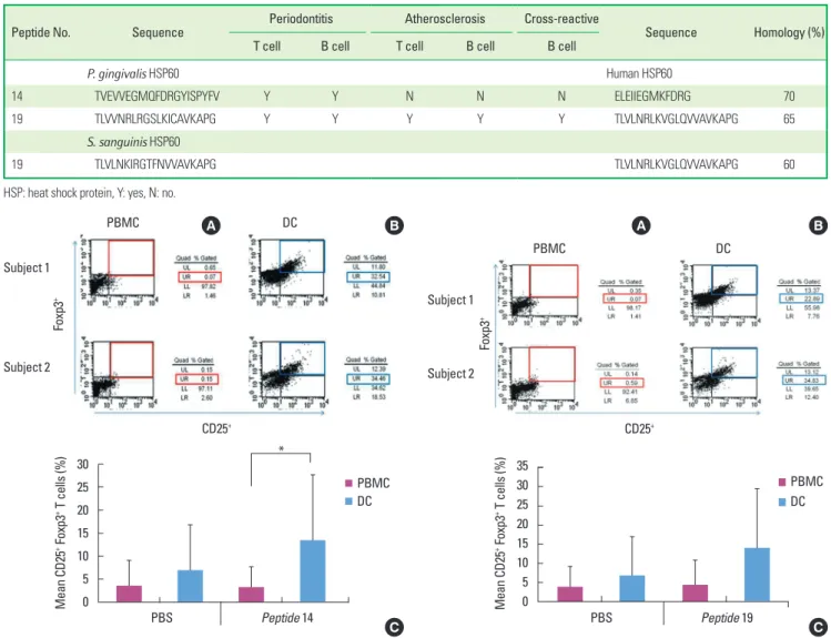
After staining, the cells were washed, fixed, and permeabilized us- ing a Cytofix/Cytoperm solution (BD Biosciences, San Jose, CA, USA) Table 1. One-letter amino acid sequences of the peptide numbers 19 and 14 from Porphyromonas gingivalis HSP60 and Streptococcus sanguinis HSP60.
Peptide No. Sequence Periodontitis Atherosclerosis Cross-reactive
Sequence Homology (%)
T cell B cell T cell B cell B cell
P. gingivalis HSP60 Human HSP60
14 TVEVVEGMQFDRGYISPYFV Y Y N N N ELEIIEGMKFDRG 70
19 TLVVNRLRGSLKICAVKAPG Y Y Y Y Y TLVLNRLKVGLQVVAVKAPG 65
S. sanguinis HSP60
19 TLVLNKIRGTFNVVAVKAPG TLVLNRLKVGLQVVAVKAPG 60
HSP: heat shock protein, Y: yes, N: no.
Figure 1. Fluorescence activated cell sorter profiles of peptide 14-specific regulatory T cells (Tregs) (CD4+, CD25+, and Foxp3+) in peripheral blood mononuclear cells (PBMCs) and dendritic cells (DCs) from two representative subjects (A, B) demonstrating the capability of DCs to enhance Tregs from 0.07% to 32.54% in subject 1 and from 0.15% to 34.46% in subject 2. The bar graph shows the mean percentage of peptide 14-specific Tregs of 10 sub- jects (C), evidencing a statistically significant difference between PMBCs and DCs (*P <0.05). Phosphate buffered saline (PBS) was used as a control anti- gen. UL: upper left panel, UR: upper right panel, LL: lower left panel, LR: low- er right panel.
C Mean CD25+ Foxp3+ T cells (%)
30 25 20 15 10 5
0 PBS Peptide 14
*
A B
PBMC DC
Subject 1
Subject 2
Foxp3+
CD25+
PBMC DC
Figure 2. Fluorescence activated cell sorter profiles of peptide 19-specific regulatory T cells (Tregs) (CD4+, CD25+, and Foxp3+) in peripheral blood mononuclear cells (PBMCs) and dendritic cells (DCs) from two representative subjects (A, B) demonstrating the capability of DCs to enhance Tregs from 0.07% to 22.89% in subject 1 and from 0.59% to 34.83% in subject 2, re- spectively. The bar graph shows the mean percentage of peptide 19-specific Tregs of 10 subjects (C). The difference between PMBCs and DCs was not sta- tistically significant. Phosphate buffered saline (PBS) was used as a control antigen. UL: upper left panel, UR: upper right panel, LL: lower left panel, LR:
lower right panel.
Mean CD25+ Foxp3+ T cells (%) 35 30 25 20 15 10 5
0 PBS Peptide 19
DC PBMC
Subject 1
Subject 2
Foxp3+
CD25+
A B
C PBMC DC
at room temperature for 20 minutes, rewashed with Perm Wash Buffer (BD Biosciences), and stained at the intracellular level with fluorescent-labeled antibodies to Foxp3 for 30 minutes at 4°C. The stained cells were acquired with a FACSCalibur flow cytometer and analyzed using CELLQuest software (BD Biosciences). All antibodies were purchased from eBioscience.
Statistical analysis
A Student t-test was applied wherever applicable to identify the differences between two variables.
RESULTS
Promoting peptide-specific Tregs by conventional PBMCs and DCs as APCs
In case for peptide 14 of P. gingivalis HSP60, the mean proportion of Tregs were 13.5%±14.2% when the DCs were used as APCs, while it was 3.3%±4.3% when PBMCs were used for APCs in ten subjects (Fig. 1C). The mean percentage of peptide 14-specific Tregs of 10 subjects was statistically significant difference between PMBCs and DCs (P<0.05). In the case of peptide number 19 from P. gingivalis HSP60, the values were 14.1%±15.3% when DCs was used as APCs, while it was 4.2%±6.6% (Fig. 2C) when PBMCs were used for APCs.
The mean percentage of peptide 19-specific Tregs of 10 subjects was not statistically significant difference between PMBCs and DCs.
When DCs were pulsed with peptide number 19 from S. sanguinis
HSP60 in one subject, the value increased from 0.05% to 17.02%
when PBMCs were used as the APCs (Fig. 3).
Effect of RA on the phenotype and expression of CD103+ DCs
RA has been reported to stimulate the expression of CD103 by DCs when added to the culture medium after culturing the DCs for 6 days. As shown in Fig. 4, the morphology of the DCs exhibited the typical extension of dendritic processes characteristic of maturing DCs. The CD103+ cells were increased from 4.28% to 32.67%. This characteristic phenotype expression occurred in parallel with its functional capability of inducing Tregs. After adding RA, the per- centage of induced Tregs increased from 21.12% to 22.89% in sub- ject 1, and from 27.73% to 34.46% in subject 2 for peptide number 19, respectively (Fig. 5). In case of peptide 14, it increased from 23.70% to 32.54% in subject 1 and from 27.57% to 34.83% in sub- ject 2 (Fig. 6). There were no statistical differences in Tregs stimulat- ed by DCs and RA-stimulated DCs for both peptide 19 and 14.
DISCUSSION
The tolerizing effect conferred by antigen-specific Tregs has been of interest to both immunologists and clinicians in their at-
0.05
17.02
CD25+ Foxp3+ cells (%) 20
15
10
5
0 Subject 1
PBMC DC
Foxp3+
PBMC DC
CD25+
A B
C Figure 3. Stimulation of peptide-specific regulatory T cells (Tregs) by periph- eral blood mononuclear cells (PBMCs) and dendritic cells (DCs). (A) Peptide number 19 from Streptococcus sanguinis heat shock protein 60 has shown a minimal capability (0.05%) to convert T cells into Tregs when PBMCs are used as antigen-presenting cells. (B) DCs were used for presenting peptide anti- gens, which enhanced induction of Tregs (17.02%). (C) The bar graph shows the percentage of peptide-specific Tregs among T cells in one subject. UL: up- per left panel, UR: upper right panel, LL: lower left panel, LR: lower right panel.
GM-CSF + IL-4
CD103+ CD103+
GM-CSF + IL-4+1μM RA
CD103+ DC (%) 35 30 25 20 15 10 5 0
DC RA treated DC
A B
C Figure 4. Comparison of effect of retinoic acid treatment with nontreatment on the phenotype expression of dendritic cells (DCs). (A) The morphology of DCs has demonstrated the typical extension of dendritic processes character- istic of maturing DCs when retinoic acid (RA) has been added to the culture medium. (B) The percentage of CD103+ cells was increased from 4.28% to 32.67%. (C) A bar graph summarizing the fluorescence activated cell sorter results is shown. GM-CSF: granulocyte-monocyte colony-stimulating factor, IL: interleukin, UL: upper left panel, UR: upper right panel, LL: lower left pan- el, LR: lower right panel.
tempts to suppress pathogen-triggered autoimmune diseases. The emerging concept that a periodontal infection might predispose a patient to the development of cardiovascular diseases has prompt- ed the suggestion that HSP60 of P. gingivalis might play a role in modulating the immunopathologic process by either triggering or suppressing the Teffs through mobilizing Tregs [29]. Van Eden et al.
[30] reported that HSP60 can be a target for molecular mimicry, and identifying a candidate T-cell epitope capable of inducing im- munological tolerance might provide an opportunity for antigen- tailored therapy against infectious diseases or infection-triggered autoimmune disease, such as atherosclerosis.
Strategies for in vitro or in vivo augmenting (or expanding) the Tregs for use in immunotherapy against autoimmune diseases have been devised [31]. One of these has been to pulse the immuno- dominant epitope of the pathogenic antigens into CD103+ imma- ture DCs, as APCs, which have a tolerizing effect on pathogenic Teffs [27]. In this experiment, the DCs pulsed with peptide number 19 or 14 profoundly stimulated the development of Tregs, regard-
less of the peptide number used. This is consistent with another study that reported on the capability of expanding CD4+CD25+ T cells [32].
Lohr et al. [33] claimed that although there may not be special conditions for antigen stimulation in the development of effect and regulatory cell populations, different subsets of DCs might in- duce these T-cell populations. transforming growth factor-β to- gether with a subpopulation of CD103+ DCs was reported to pro- mote the differentiation of Teffs to Foxp3+ Tregs [23].
In this study, peptide number 19 from P. gingivalis HSP60 exhibit- ed a minimal ability to induce Tregs when PBMCs were used as APCs, which was confirmed in previous observations [34]. In the present experiment, peptide number 14 from P. gingivalis HSP60 exhibited a comparable capability to induce Tregs when PBMCs were used as APCs to peptide number 19. The peptide has been identified as an immunodominant epitope both for T and B cells in periodontitis pa- tients, while not for atherosclerosis patients, despite the high se- quence homology shared with their human counterpart (unpub- Figure 5. Comparison of effect of retinoic acid (RA) treatment with nontreat-
ment on regulatory T cells (Tregs) induction by peptide number 19 from Por- phyromonas gingivalis heat shock protein 60 (HSP60). (A) T cells were stained for CD4, CD25, and Foxp3 and analyzed by fluorescence activated cell sorter.
The percentage of peptide-specific Tregs induced increased from 21.12% to 22.89% (subject 1 in C), and it increased from 27.73% to 34.46% (subject 2 in C) for peptide number 19 from P. gingivalis HSP60 by the addition of RA.
(B) The bar graph shows the percentage of peptide-specific Tregs among T cells in two subjects. The difference between cases with and without RA failed to reach statistical significance. UL: upper left panel, UR: upper right panel, LL: lower left panel, LR: lower right panel.
CD25+ Foxp3+ cells (%) 40 35 30 25 20 15 10 5 0
DC RA treated DC
Subject 1 Subject 2 CD25+
Foxp3+
CD3+ T cells + DCs
Peptied 19
Peptied 19 Peptied 19
Peptied 19
CD3+ T cells + RA treated-DCs
A B
C
Figure 6. Comparison of the effect of retinoic acid (RA) treatment and non- treatment on regulatory T cells (Tregs) induction by peptide number 14 from Porphyromonas gingivalis heat shock protein 60 (HSP60). (A) T cells were stained for CD4, CD25, and Foxp3 and analyzed by fluorescence activated cell sorter. The percentage of peptide-specific Tregs induced increased from 23.70% to 32.54% (subject 1 in C), and it increased from 27.57% to 34.83%
(subject 2 in C) for peptide number 14 from P. gingivalis HSP60 by the addi- tion of RA. (B) The bar graph shows the percentage of peptide-specific Tregs among T cells in two subjects. The difference between cases with and without RA failed to reach statistical significance. UL: upper left panel, UR: upper right panel, LL: lower left panel, LR: lower right panel.
CD25+
Foxp3+
Peptied 14
Peptied 14 Peptied 14
Peptied 14
CD3+ T cells + DCs A CD3+ T cells + RA treated-DCs B
CD25+ Foxp3+ cells (%) 40 35 30 25 20 15 10 5 0
DC RA treated DC
Subject 1 Subject 2 C
lished observations). Therefore, this peptide has the potential for development as a vaccine to suppress autoimmunity without elic- iting cross-reactive immune responses with human autoantigens.
DCs have the ability to enhance the induction of Tregs by both peptide numbers 19 from P. gingivalis HSP60 and S. ganguinis HSP60 and peptide number 14 from P. gingivalis HSP60. However, the promotion of Tregs by the DCs was not greater for peptide 14 than by PMBCs. However, it was not the case for peptide number 19. Thus, CD103+ DCs seem to promote the antigen-specific Tregs in an antigen-dependent manner when compared with those ob- served by PMBC. This contrasts with our most recent observation that CD103+ DCs promoted Tregs in an antigen nondependent manner in an experimental mouse model [35].
One of the aims of this study was to determine whether peptide number 19 from S. sanguinis HSP60 can induce Tregs because com- mensal or probiotic bacteria induce Tregs to suppress autoimmune diseases or intestinal colitis [36]. Nevertheless, this phenomenon was not observed, presumably due to its inability to induce regula- tory T cells, as observed in the case of P. gingivalis. Peptide number 19 cross-reacts with its human counterpart and HSPs from other bacteria. Future studies should examine the enhanced induction of Tregs by peptide number 14 from S. sanguinis HSP60.
In the present study, the development of Tregs was enhanced when the epitope peptides were introduced into DCs as APCs. This effect appeared to be anticipated consistently regardless of the epitope peptide sequences, despite slight differences by sequence.
With the additional strategy of expanding the number of Tregs in vitro, this promising result would provide a venue for prospective cell-based immunotherapy in modulating infection-induced auto- immune diseases. Antigen-specific Tregs, once induced by a specific antigen, exhibit an antigen-nonspecific immune suppressive effect.
Nevertheless, the importance of antigen- or organ-specific Treg therapy is being increasingly emphasized [1].
In the intestines, CD103+ DCs have the unique ability to induce adaptive Tregs because of their ability to produce RA, which is need- ed to stimulate naïve T cells to differentiate into Foxp3-expressing Tregs [4]. This phenomenon was confirmed in the present experi- ment. RA stimulates the development of mature DCs with the en- hanced expression of CD103+ DCs with the typical extension of den- dritic processes characteristic of mature DCs, which can again drive the naïve T cells into Tregs [37,38]. The enhanced expression of CD103 was associated with its functional capability of stimulating antigen-specific Tregs.
Overall, within the context of this experiment, CD103+ DCs have the potential to stimulate peptide-specific Tregs, which can be uti- lized further in the customized cell-based vaccine therapy against infection-induced autoimmune diseases, such as atherosclerosis and rheumatoid arthritis.
In conclusion, CD103+ DCs can stimulate the differentiation of antigen-specific Tregs from naïve T cells, when used as APCs for an epitope peptide from a periodontal pathogenic organism. RA is an effective reagent that induces mature DCs with the typical pheno-
typic expression of CD103, and enhanced their functional ability to stimulate Tregs.
CONFLICT OF INTEREST
No potential conflict of interest relevant to this article was re- ported.
ACKNOWLEDGEMENTS
This work was supported by the National Research Foundation of Korea (NRF) grant funded by the Korean Government (MEST) (NRF-2011-0015501), and a 2013 Clinical Research Grant from Pu- san National University Dental Hospital.
ORCID
Myung-Jin Kim http://orcid.org/0000-0003-0528-6848 Eui-Kyong Jeong http://orcid.org/0000-0001-9866-9842 Eun-Young Kwon http://orcid.org/0000-0001-9555-0360 Ji-Young Joo http://orcid.org/0000-0002-4050-5797 Ju-Youn Lee http://orcid.org/0000-0002-0772-033X Jeomil Choi http://orcid.org/0000-0002-7491-6711
REFERENCES
1. Tang Q, Bluestone JA. The Foxp3+ regulatory T cell: a jack of all trades, master of regulation. Nat Immunol 2008;9:239-44.
2. Vignali DA, Collison LW, Workman CJ. How regulatory T cells work.
Nat Rev Immunol 2008;8:523-32.
3. Guilliams M, Oldenhove G, Noel W, Herin M, Brys L, Loi P, et al.
African trypanosomiasis: naturally occurring regulatory T cells fa- vor trypanotolerance by limiting pathology associated with sus- tained type 1 inflammation. J Immunol 2007;179:2748-57.
4. Groux H, O'Garra A, Bigler M, Rouleau M, Antonenko S, de Vries JE, et al. A CD4+ T-cell subset inhibits antigen-specific T-cell re- sponses and prevents colitis. Nature 1997;389:737-42.
5. Fehervari Z, Sakaguchi S. CD4+ Tregs and immune control. J Clin Invest 2004;114:1209-17.
6. Gambineri E, Torgerson TR, Ochs HD. Immune dysregulation, poly- endocrinopathy, enteropathy, and X-linked inheritance (IPEX), a syndrome of systemic autoimmunity caused by mutations of FOXP3, a critical regulator of T-cell homeostasis. Curr Opin Rheu- matol 2003;15:430-5.
7. Viglietta V, Baecher-Allan C, Weiner HL, Hafler DA. Loss of func- tional suppression by CD4+CD25+ regulatory T cells in patients with multiple sclerosis. J Exp Med 2004;199:971-9.
8. Lindley S, Dayan CM, Bishop A, Roep BO, Peakman M, Tree TI. De- fective suppressor function in CD4(+)CD25(+) T-cells from pa- tients with type 1 diabetes. Diabetes 2005;54:92-9.
9. Ehrenstein MR, Evans JG, Singh A, Moore S, Warnes G, Isenberg DA, et al. Compromised function of regulatory T cells in rheuma-
toid arthritis and reversal by anti-TNFalpha therapy. J Exp Med 2004;200:277-85.
10. Brunkow ME, Jeffery EW, Hjerrild KA, Paeper B, Clark LB, Yasayko SA, et al. Disruption of a new forkhead/winged-helix protein, scur- fin, results in the fatal lymphoproliferative disorder of the scurfy mouse. Nat Genet 2001;27:68-73.
11. Bennett CL, Christie J, Ramsdell F, Brunkow ME, Ferguson PJ, Whi- tesell L, et al. The immune dysregulation, polyendocrinopathy, en- teropathy, X-linked syndrome (IPEX) is caused by mutations of FOXP3. Nat Genet 2001;27:20-1.
12. Wildin RS, Ramsdell F, Peake J, Faravelli F, Casanova JL, Buist N, et al. X-linked neonatal diabetes mellitus, enteropathy and endocri- nopathy syndrome is the human equivalent of mouse scurfy. Nat Genet 2001;27:18-20.
13. Fontenot JD, Gavin MA, Rudensky AY. Foxp3 programs the devel- opment and function of CD4+CD25+ regulatory T cells. Nat Im- munol 2003;4:330-6.
14. Friedewald VE, Kornman KS, Beck JD, Genco R, Goldfine A, Libby P, et al. The American Journal of Cardiology and Journal of Peri- odontology editors' consensus: periodontitis and atherosclerotic cardiovascular disease. J Periodontol 2009;80:1021-32.
15. Kebschull M, Demmer RT, Papapanou PN. "Gum bug, leave my heart alone!": epidemiologic and mechanistic evidence linking periodontal infections and atherosclerosis. J Dent Res 2010;89:
879-902.
16. Wucherpfennig KW. Structural basis of molecular mimicry. J Au- toimmun 2001;16:293-302.
17. Rajaiah R, Moudgil KD. Heat-shock proteins can promote as well as regulate autoimmunity. Autoimmun Rev 2009;8:388-93.
18. Wucherpfennig KW. Mechanisms for the induction of autoimmu- nity by infectious agents. J Clin Invest 2001;108:1097-104.
19. Delogu LG, Deidda S, Delitala G, Manetti R. Infectious diseases and autoimmunity. J Infect Dev Ctries 2011;5:679-87.
20. Vanneman M, Dranoff G. Combining immunotherapy and targeted therapies in cancer treatment. Nat Rev Cancer 2012;12:237-51.
21. Choi JI, Seymour GJ. Vaccines against periodontitis: a forward- looking review. J Periodontal Implant Sci 2010;40:153-63.
22. Banchereau J, Briere F, Caux C, Davoust J, Lebecque S, Liu YJ, et al. Immunobiology of dendritic cells. Annu Rev Immunol 2000;
18:767-811.
23. Coombes JL, Siddiqui KR, Arancibia-Carcamo CV, Hall J, Sun CM, Belkaid Y, et al. A functionally specialized population of mucosal CD103+ DCs induces Foxp3+ regulatory T cells via a TGF-beta and retinoic acid-dependent mechanism. J Exp Med 2007;204:
1757-64.
24. Sun CM, Hall JA, Blank RB, Bouladoux N, Oukka M, Mora JR, et al.
Small intestine lamina propria dendritic cells promote de novo generation of Foxp3 T reg cells via retinoic acid. J Exp Med 2007;
204:1775-85.
25. Chirdo FG, Millington OR, Beacock-Sharp H, Mowat AM. Immu- nomodulatory dendritic cells in intestinal lamina propria. Eur J Immunol 2005;35:1831-40.
26. Johansson C, Kelsall BL. Phenotype and function of intestinal den- dritic cells. Semin Immunol 2005;17:284-94.
27. Izcue A, Coombes JL, Powrie F. Regulatory T cells suppress systemic and mucosal immune activation to control intestinal inflamma- tion. Immunol Rev 2006;212:256-71.
28. Annacker O, Coombes JL, Malmstrom V, Uhlig HH, Bourne T, Jo- hansson-Lindbom B, et al. Essential role for CD103 in the T cell- mediated regulation of experimental colitis. J Exp Med 2005;202:
1051-61.
29. Belkaid Y. Regulatory T cells and infection: a dangerous necessity.
Nat Rev Immunol 2007;7:875-88.
30. Van Eden W, Wick G, Albani S, Cohen I. Stress, heat shock pro- teins, and autoimmunity: how immune responses to heat shock proteins are to be used for the control of chronic inflammatory diseases. Ann N Y Acad Sci 2007;1113:217-37.
31. Mills KH, McGuirk P. Antigen-specific regulatory T cells: their in- duction and role in infection. Semin Immunol 2004;16:107-17.
32. Yamazaki S, Iyoda T, Tarbell K, Olson K, Velinzon K, Inaba K, et al.
Direct expansion of functional CD25+ CD4+ regulatory T cells by antigen-processing dendritic cells. J Exp Med 2003;198:235-47.
33. Lohr J, Knoechel B, Abbas AK. Regulatory T cells in the periphery.
Immunol Rev 2006;212:149-62.
34. Choi J, Lee SY, Kim K, Choi BK. Identification of immunoreactive epitopes of the Porphyromonas gingivalis heat shock protein in periodontitis and atherosclerosis. J Periodontal Res 2011;46:240-5.
35. Jeong E, Kim K, Kim JH, Cha GS, Kim SJ, Kang HS, et al. Porphy- romonas gingivalis HSP60 peptides have distinct roles in the de- velopment of atherosclerosis. Mol Immunol Forthcoming 2014.
36. Forsythe P, Bienenstock J. Immunomodulation by commensal and probiotic bacteria. Immunol Invest 2010;39:429-48.
37. Hand T, Belkaid Y. Microbial control of regulatory and effector T cell responses in the gut. Curr Opin Immunol 2010;22:63-72.
38. del Rio ML, Bernhardt G, Rodriguez-Barbosa JI, Forster R. Devel- opment and functional specialization of CD103+ dendritic cells.
Immunol Rev 2010;234:268-81.