대한소화기학회지 2005;46:404-408
서 론
혈관육종은 혈관 내피세포에서 기원한 악성종양의 하나 로서 연부조직 육종의 1-2%를 차지한다.1 호발부위는 주로 피부와 피하조직이나,2 간, 비장, 심장, 부신과 같은 내부 장 기에도 혈관육종이 발생할 수 있다.3,4 특히 소장에 발생한 원발 혈관육종은 극히 드물다.5-7 상피세포양 혈관육종은 샘 암종, 림프종, 악성 흑색종과 감별이 어려워 정확한 진단을 위해서는 면역조직화학염색이 필수이며,8 광범위한 절제와 항암치료에도 불구하고 전체 예후는 불량하다.5,9
저자들은 반복적인 혈변으로 내원한 환자에서 장중첩증이
의심되어 개복술 후 소장의 혈관육종을 진단한 예를 경험하 여 문헌고찰과 함께 보고한다.
증 례
54세 남자가 수일간 복통과 내원 당일 발생한 흑색변으로 응급실을 방문하였다. 환자는 내원 1달 전 흑색변을 보여 시행한 상부위장관 내시경검사에서 십이지장 제2부에 발적 과 함께 중심부에 혈괴가 붙어 있는 병변이 발견되었고, 출 혈 병변으로 생각해 밴드결찰술을 시행했다(Fig. 1). 당시 대 장내시경검사와 복부 전산화단층촬영에서 특별한 소견은
반복적인 위장관 출혈로 발현한 소장의 원발 혈관육종 1예
부산대학교 의과대학 내과학교실, 병리학교실*
류동엽․황상연․이동원․김태오․박도윤*․김광하․허 정․강대환․송근암․조 몽
A Case of Primary Angiosarcoma of Small Intestine Presenting as Recurrent Gastrointestinal Bleeding
Dong Yup Ryu, M.D., Sang Youn Hwang, M.D., Dong Won Lee, M.D., Tae Oh Kim, M.D., Do Youn Park, M.D.*, Gwang Ha Kim, M.D., Jeong Heo, M.D., Dae Hwan Kang, M.D.,
Geun Am Song, M.D., and Mong Cho, M.D.
Departments of Internal Medicine and Pathology*, Pusan National University College of Medicine, Busan, Korea
Angiosarcoma is a rare malignant tumor which occurs
frequentlyin the skin and soft subcutis. Moreover, primary gastrointestinal angiosarcomas are very rare. This tumor manifests as non-specific symptoms such as gastroin- testinal bleeding, abdominal pain and nausea. The diagnosis is often made at an advanced stage. Surgery, chemo- therapy and radiotherapy are the mainstay of treatment. However, the prognosis is very poor. We report a case of primary angiosarcoma of the small intestine presenting as recurrent gastrointestinal bleeding. A 54-year-old man was admitted with recurrent gastrointestinal bleeding. An abdominal CT scan revealed an ileo-ileal intussusception.
Segmental resection was performed with ileo-ileal anastomosis. The ileal mass was diagnosed as angiosarcoma on immunohistochemical stain. He received 3 cycles of chemotherapy, but died 5 months after the diagnosis.
(Korean J Gastroenterol 2005;46:404-408)
ꠏꠏꠏꠏꠏꠏꠏꠏꠏꠏꠏꠏꠏꠏꠏꠏꠏꠏꠏꠏꠏꠏꠏꠏꠏꠏꠏꠏꠏꠏꠏꠏꠏꠏꠏꠏꠏꠏꠏꠏꠏꠏꠏꠏꠏꠏꠏꠏꠏꠏꠏꠏꠏꠏꠏꠏꠏꠏꠏꠏꠏꠏꠏꠏꠏꠏꠏꠏꠏꠏꠏꠏꠏꠏꠏꠏꠏꠏꠏꠏꠏꠏꠏꠏꠏꠏꠏꠏꠏꠏꠏꠏꠏꠏꠏꠏꠏꠏꠏꠏꠏꠏꠏꠏꠏꠏꠏꠏꠏꠏꠏꠏꠏ
Key Words: Angiosarcoma; Small intestine
ꠏꠏꠏꠏꠏꠏꠏꠏꠏꠏꠏꠏꠏꠏꠏꠏꠏꠏꠏꠏꠏꠏꠏꠏꠏꠏꠏꠏꠏꠏꠏꠏꠏꠏ 접수: 2005년 3월 10일, 승인: 2005년 9월 20일
연락처: 송근암, 602-739, 부산광역시 서구 아미동 1가 10번지 부산대학교병원 소화기병센터
Tel: (051) 240-7869, Fax: (051) 244-8180 E-mail: gasong@pusan.ac.kr
ꠏꠏꠏꠏꠏꠏꠏꠏꠏꠏꠏꠏꠏꠏꠏꠏꠏꠏꠏꠏꠏꠏꠏꠏꠏꠏꠏꠏꠏꠏꠏꠏꠏꠏ Correspondence to: Geun Am Song, M.D.
Division of Gastroenterology and Medicine, Pusan National Uni- versity Hospital, 1-10 Ami-dong, Seo-gu, Busan 602-739, Korea Tel: +82-51-240-7869, Fax: +82-51-244-8180
E-mail: gasong@pusan.ac.kr
류동엽 외 9인. 반복적인 위장관 출혈로 발현한 소장의 원발 혈관육종 1예 405
없었으며, 수혈 등 보존치료 후 호전되어 퇴원하였다. 퇴원 2주 후부터 간헐적인 복통이 있었고, 내원 당일 흑색변을 보여 응급실을 방문하였다.
입원 당시 급성 병색을 보였으며, 혈압 130/70 mmHg, 맥 박 78회/분, 호흡수 20회/분, 체온 36.6oC였다. 두․경부 진찰 에서 결막은 창백하였고 공막에 황달은 없었으며, 경부 림 프절은 촉지되지 않았다. 흉부 청진에서 호흡음은 깨끗하였 고, 심음은 규칙적이었으며 십잡음은 들리지 않았다. 복부 진찰에서 장음이 거의 들리지 않았고 타진 시 복부 전반에 걸쳐 압통이 있었으나 반발통은 없었다. 간이나 비장비대는 관찰되지 않았고, 복부 종괴는 만져지지 않았다.
혈액 검사에서 백혈구 6,500/mm3, 혈색소 6.1 g/dL, 헤마토 크리트 19.0%, 혈소판 377,000/mm3이었고, 총 단백 6.3 g/dL, 알부민 2.0 g/dL, AST/ALT 64/61 IU/L, ALP/LDH 254/479 IU/L, 총 빌리루빈 0.7 mg/dL, 아밀라아제/리파아제 72/17 IU/L, BUN/크레아티닌 18/1.1 mg/dL이었다.
상부위장관 내시경검사에서 이전에 밴드결찰을 시행한 부위에서 치유기 궤양이 관찰되었고, 그 외에 출혈을 일으 킬 만한 병변은 없었다. 치유기 궤양에서 시행한 조직검사 는 만성염증 소견이었다. 대장내시경검사에서 돌막창자판 막을 통해 다수의 핏덩이가 흘러내려 소장 출혈을 의심했 다. 복부 전산화단층촬영에서 공장-공장 장중첩증이 의심되 었고(Fig. 2), 우측 부신비대(3.7×1.3 cm)가 관찰되었으며, 시험 개복술을 시행하였다.
개복 시 장중첩증은 없었으며 트라이츠 인대 50 cm 하방 공장에 내강을 거의 막고 있는 흑갈색의 종괴가 관찰되어, 공장 부분절제술과 문합술, 장간막 림프절 절제술을 시행하 였다.
절제된 장에 육안으로 각각 6×3.5 cm, 3.5×3 cm 크기의 흑갈색 종괴가 관찰되었고, 종괴 표면에는 궤양이 동반되어 있었다(Fig. 3). 조직 소견은 종양 일부에서는 비전형 내피세 포가 서로 교통하는 혈관구조를 형성했으며, 다른 부분에서 는 상피세포와 닮은 종양세포가 고형 성장의 형태를 보였다 (Fig. 4). 10개의 고배율 시야에서 10개 이상의 유사분열이 관찰되었으며, 비정형 유사분열도 보였다. 면역조직화학염 색에서 vimentin, 제8인자(factor VIII), CD34에 양성(Fig. 5), cytokeratin, S-100, leukocyte common antigen (LCA), c-kit, HMB-45, smooth muscle actin (SMA), desmin, myoglobin에는 음성으로 혈관육종으로 진단했다.
수술 후 촬영한 뼈 스캔에서 양측 무릎과 엉덩이 관절에 대칭으로 섭취증가를 보였으나, 전이를 시사하는 소견은 아 니었다. 환자는 3개월 동안 3차례에 걸쳐 doxorubicin, ifos- Fig. 1. Endoscopic finding. A well-demarcated hyperemic mucosa
with blood clot is seen at the second portion of duodenum.
Fig. 2. Abdominal CT finding. A mass-like lesion with lobulated contour (arrow) is noticed. The intussusceptum is seen within the intussuscipiens.
Fig. 3. Gross finding. Two ulcerative masses with hemorrhage are seen.
406 The Korean Journal of Gastroenterology: Vol. 46, No. 5, 2005
famide, dacarbazine을 이용한 항암화학요법을 시행 받았다.
항암치료 3회 후 촬영한 복부 전산화단층촬영에서 간에 저 음영 종괴가 다발로 관찰되었고, 우측 부신이 4×3.6 cm 크 기로 커져 있어 전이가 의심되었다. 이후 추적관찰이 이루 어지지 않았고, 환자는 수술 5개월 후 파종혈관내응고증으 로 사망하였다.
고 찰
소장은 전체 위장관의 약 75%에 해당하나, 위장관 종양 의 1% 정도만 소장에 발생한다. 소장에 가장 흔히 발생하는
악성종양은 유암종, 샘암종, 평활근육종이다.10-12
혈관육종은 혈관 내피세포에서 기원한 악성종양의 하나 로서 연부조직 육종의 1-2% 정도를 차지한다.1 주로 피부와 피하조직에 발생하고,2 간, 비장, 심장과 같은 내부 장기에 도 발생한다.3,4 하지만 소장이 원발부위가 되는 경우는 매 우 드물다.5-7 99예의 혈관육종 환자를 대상으로 한 연구에 서 소장이 원발 병소가 된 예는 없었으며,3 이번 환자처럼 소장에서 출혈을 보였던 49명의 환자를 대상으로 한 12년간 연구에서는 1예가 혈관육종으로 진단되었다.13
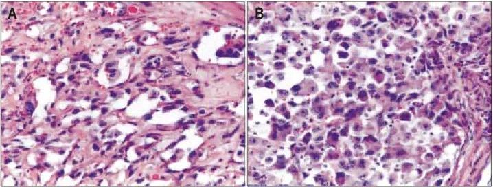
혈관육종의 발생원인은 불분명하지만, 몇 가지 유발인자 가 있다. Vinyl chloride, thorotrast, arsenic와 같은 물질을 오 Fig. 4. Microscopic findings. (A) Irregular but well-developed vascular channels lined by atyical endothelial cells are seen (H&E stain, ×400).
(B) Clusters of large epithelioid cells with vesicular nuclei and a central nucleolus are seen (H&E stain, ×400).
A B
Fig. 5. Immunohistochemical stain.
The tumor cells shows diffuse positivity for factor VIII (A) and CD 34 (B) immunostaining, res- pectively (×400).
A B
Ryu DY, et al. A Case of Primary Angiosarcoma of Small Intestine Presenting as Recurrent Gastrointestinal Bleeding 407
랫동안 취급한 사람에서 간에 혈관육종이 발생할 수 있
다.14,15 이전에 방사선을 치료받은 환자에서 발생한 다발 혈
관육종이 보고되어 있으며,16 장기간 이물질 잔류,17 림프부 종 또한 혈관육종의 발생과 관련이 있다. 하지만 이번 증례 에서 이러한 유발인자는 없었다.
임상증상은 대체로 첫 증상이 출혈로 나타나는 경우가 많 으나,5 복통, 구토, 어지러움, 빈혈 등의 비특이 증상을 보이 기도 하여 종종 진단이 지연된다.18,19
조직소견으로 혈관육종은 크게 혈관을 형성하는 형태와 고형 성장의 형태를 보인다.8 혈관을 형성하는 경우 비전형 내피세포가 서로 교통하는 혈관 통로를 구성하고, 고형 성 장 형태인 경우 세포는 방추형 또는 풍부한 호산 세포질을 가지는 다각형 상피세포 모양을 보인다.18 특히 혈관육종이 상피세포양 형태를 보일 경우 전이암종, 림프종, 악성 흑색 종과 감별이 어렵다. 따라서 정확한 진단을 위해서는 면역 조직화학 검사가 필수이다.18 가장 특징적인 염색소견은 CD31, CD34, 제8인자와 같은 혈관 내피세포 표지자에 양성 소견을 보이는 것이다.5,7,8 이번 증례는 vimentin, CD34, 제8 인자에 대해서만 양성소견을 보여 종양이 중배엽에서 기원 한 혈관육종으로 확진했다.
대체로 진단 후 수개월 이내에 사망해 예후는 불량하다.
치료는 수술 절제, 항암화학요법, 방사선 치료를 시행하며, 광범위 절제에 이은 항암치료를 시행받은 환자들은 2년 생 존율이 29%로서 그 밖의 치료를 받은 환자들의 2년 생존율 인 9%보다 예후가 향상된다.5,9 특히 완전히 절제되지 않았 거나 전이된 연조직 육종의 치료로 doxorubicin, dacarbazine, ifosfamide를 병합하면 종양의 반응률을 높이고, 병의 진행 을 늦출 수 있다.20 하지만 환자 대부분은 2년 이상 생존하 지 못하며, 평균 생존기간은 2개월에서 6개월 정도이다.5 고 령(나이 50세 이상), 종양 크기(5 cm 이상), 후복강의 원발병 소, 유사분열의 정도(고배율에서 10개 이상) 등이 불량한 예 후와 관련된 인자이다.4,9 이번 증례는 진단 당시 나이가 50 세 이상, 종양의 크기가 6 cm, 유사분열이 10개의 고배율 시 야에서 10개 이상으로써 불량한 예후를 예측할 수 있었다.
혈관육종 환자의 85%에서 폐, 뼈, 간, 부신 등에 전이될 수 있으며,9 이번 증례는 두 번째 내원 시 복부 전산화단층 촬영에서 우측 부신이 커져 있었기 때문에 이미 전이된 상 태에서 혈관육종이 진단되었다고 생각한다. 완전 절제되지 않은 종양은 항암화학요법과 방사선 치료에 잘 반응하지 않 는다.5 따라서 수술을 통해 소장의 병변과 장간막 림프절은 제거되었지만, 우측 부신에 전이병소가 남아 있어 항암치료 에도 불구하고 간전이가 추가로 발생하는 등 상태가 악화되 어 사망한 것으로 생각한다.
소장의 혈관육종은 다발로 발생하기도 한다. 복통과 흑색 변으로 내원하여 내시경검사에서 특이 소견이 없었으나, 소
장촬영에서 국소 협착과 점막변화가 발견되어 수술 후 소장 의 다발 혈관육종이 진단된 예,18 빈혈로 내원하여 시험 개 복술을 통해 십이지장과 소장의 다발 혈관육종이 진단된 예 가 있다.19 이번 증례의 경우 첫 내원 시 발견된 십이지장 병변은 소장 종괴와 육안에서 유사하였다. 따라서 비록 조 직검사를 통해 확인되지는 않았지만, 십이지장 병변 또한 혈관육종이었을 가능성이 있으며, 십이지장 병변을 밴드 결 찰술로 치료할 당시 소장에도 이미 혈관육종이 존재하였을 가능성을 배제할 수 없다.
위장관 출혈은 임상에서 흔히 접할 수 있지만, 드물게 이 번 증례처럼 혈관육종이 원인이 될 수 있다. 특히 출혈의 원 인이 불분명할 경우 항상 소장에 발생하는 악성종양을 감별 진단에 포함시켜야 조기 진단을 통한 치료가 가능할 것으로 생각한다.
참고문헌
1. Weiss SW, Goldblum JR. Enzinger and Weiss's soft tissue tumors. 4th ed. St. Louis: Mosby, 2001:917-954.
2. Maddox JC, Evans HL. Angiosarcoma of skin and soft tissue:
a study of forty-four cases. Cancer 1981;48:1907-1921.
3. Naka N, Ohsawa M, Tomita Y, Kanno H, Uchida A, Aozasa K. Angiosarcoma in Japan. A review of 99 cases. Cancer 1995;75:989-996.
4. Meis-Kindblom JM, Kindblom LG. Angiosarcoma of soft tissue: a study of 80 cases. Am J Surg Pathol 1998;22:683- 697.
5. Allison KH, Yoder BJ, Bronner MP, Goldblum JR, Rubin BP. Angiosarcoma involving the gastrointestinal tract: a series of primary and metastatic cases. Am J Surg Pathol 2004;28:
298-307.
6. Chami TN, Ratner LE, Henneberry J, Smith DP, Hill G, Katz PO. Angiosarcoma of the small intestine: a case report and literature review. Am J Gastroenterol 1994;89:797-800.
7. Taxy JB, Battifora H. Angiosarcoma of the gastrointestinal tract. A report of three cases. Cancer 1988;62:210-216.
8. Alles JU, Bosslet K. Immunocytochemistry of angiosarcomas.
A study of 19 cases with special emphasis on the appli- cability of endothelial cell specific markers to routinely pre- pared tissues. Am J Clin Pathol 1988;89:463-471.
9. Naka N, Ohsawa M, Tomita Y, et al. Prognostic factors in angiosarcoma: a multivariate analysis of 55 cases. J Surg Oncol 1996;61:170-176.
10. Barclay TH, Schapira DV. Malignant tumors of the small intestine. Cancer 1983;51:878-881.
11. Wilson JM, Melvin DB, Gray GF, Thorbjarnarson B. Primary malignancies of the small bowel: a report of 96 cases and
408 대한소화기학회지: 제46권 제5호, 2005
review of the literature. Ann Surg 1974;180:175-179.
12. Awrich AE, Irish CE, Vetto RM, Fletcher WS. A twenty-five year experience with primary malignant tumors of the small intestine. Surg Gynecol Obstet 1980;151:9-14.
13. Lau WY, Yuen WK, Chu KW, Poon GP, Li AK. Obscure bleeding in the gastrointestinal tract originating in the small intestine. Surg Gynecol Obstet 1992;174:119-124.
14. Ito Y, Kojiro M, Nakashima T, Mori T. Pathomorphologic characteristics of 102 cases of thorotrast-related hepatocellular carcinoma, cholangiocarcinoma, and hepatic angiosarcoma.
Cancer 1988;62:1153-1162.
15. Makk L, Creech JL, Whelan JG Jr, Johnson MN. Liver damage and angiosarcoma in vinyl chloride workers. A sys- tematic detection program. JAMA 1974;230:64-68.
16. Wolov RB, Sato N, Azumi N, Lack EE. Intra-abdominal
“angiosarcomatosis” report of two cases after pelvic irradi-
ation. Cancer 1991;67:2275-2279.
17. Ben-Izhak O, Kerner H, Brenner B, Lichtig C. Angiosarcoma of the colon developing in a capsule of a foreign body.
Report of a case with associated hemorrhagic diathesis. Am J Clin Pathol 1992;97:416-420.
18. Delvaux V, Sciot R, Neuville B, et al. Multifocal epithelioid angiosarcoma of the small intestine. Virchows Arch 2000;
437:90-94.
19. Ordonez NG, del Junco GW, Ayala AG, Ahmed N. Angio- sarcoma of the small intestine: an immunoperoxidase study.
Am J Gastroenterol 1983;78:218-221.
20. Antman K, Crowley J, Balcerzak SP, et al. An intergroup phase III randomized study of doxorubicin and dacarbazine with or without ifosfamide and mesna in advanced soft tissue and bone sarcomas. J Clin Oncol 1993;11:1276-1285.