Journal of Bacteriology and Virology 2015. Vol. 45, No. 4 p.328 – 338 http://dx.doi.org/10.4167/jbv.2015.45.4.328
Recharacterization of Morphological and Genetic Feature of Getah Virus Isolated from South Korea
Seung Heon Lee, Dong-Kun Yang*, Ha-Hyun Kim, Hyun-Ye Jo, Sung-Suk Choi, Jung-Won Park, Kang-Seuk Choi and In-Soo Cho
Viral Disease Division, Animal and Plant Quarantine Agency, Gyeonggi-do, Korea
Three QIAG93 strains, QIAG9301, QIAG9302 and QIAG9303 that have been identified as Getah virus (GETV) are analyzed in this study. The morphological features of three virus isolates were observed by using electron microscopy, suggesting that the QIAG9301, QIAG9302 and QIAG9303 isolate can be classified as tentative member of Alphavirus species in the Semliki Forest complex. The full length of the structural polyprotein gene of each QIAG93 isolate (QIAG9301, QIAG9302 and QIAG9303) was determined that are identical in size, comprising 3759 nucleotides that encoded 1253 amino acids. The sequence analysis of the structural polyprotein gene, including the C, E3, E1, 6K and E2 domain, showed that each QIAG93 isolate shares >98.9% sequence identity. The phylogenetic analysis and evolutionary distance (ED) estimation based on the structural polyprotein gene sequence showed that the QIAG9301 isolate is closely related to GETV South Korea strain (99.9% sequence identity and ED value 0.001) and Chinese GETV YN0540 strain (99.3% sequence identity ED value 0.007) than other Alphavirus species analyzed in this study. Both QIAG9032 and QIAG9303 isolate exhibited genetically close relationship with Mongolian GETV LEIV17741MPR strain (at least 99.3%
sequence identity and mean ED value 0.0065). Therefore, our findings will be valuable for molecular epidemiological analyses of GETV in Korea and contribute to a further study on pathogenicity of three QIAG93 isolates in animals.
Key Words: Getah virus, Genetic epidemiology
INTRODUCTION
Getah virus (GETV) is an Alphavirus (formerly, group A arthropod-borne virus) that was isolated for the first time in 1955 from mosquitoes (Culex gelidus) in Malaysia (1, 2).
GETV has since been frequently isolated from mosquitoes in several countries (3). GETV infection has been found in many species of vertebrates (4) and is particularly known
as a pathogen of horse and pig. GETV infection of horse is a mild, self-limiting illness characterized by fever, hind-limb edema and stiffness (5~8). A few cases of fetal death have been reported following natural or experimental infection of pigs. Other clinical signs reported in experimentally infected pigs include transient fever, anorexia and mild depression and diarrhea (9, 10). In humans, although neutralizing anti- bodies have been identified, GETV infection is considered subclinical (11).
328
Received: September 18, 2015/ Revised: November 11, 2015/ Accepted: November 18, 2015
*Corresponding author: Dong-Kun Yang. Viral Disease Division, Animal and Plant Quarantine Agency, 175 Anyang-ro, Anyang-si, Gueonggi-do 14089, Korea.
Phone: +82-31-467-1783, Fax: +82-31-467-1797, e-mail: [email protected]
**This work was supported financially by a grant (M-1543083-20150150-1) from Animal, and Plant Quarantine Agency, Ministry of Agriculture, Food and Rural Affairs (MAFRA), Republic of Korea.
○CCThis is an Open Access article distributed under the terms of the Creative Commons Attribution Non-Commercial License (http://creativecommons.org/license/by-nc/3.0/).
Original Article
Based upon serologic criteria, GETV is classified into the Semliki Forest antigenic complex of the genus Alphavirus in the family Togaviridae (12). The Semliki Forest complex includes Ross River (RRV), Sagiyama (SAGV), Semliki Forest, Middleburg, Chikungunya (CHIKV), Barmah Forest, Getah, Bebaru, Mayaro, Una, and O'nyong-nyong viruses (13). Among them, the closet phylogenic relatives of GETV are RRV and SAGV (14, 15). Serological evidence suggests that GETV is widely distributed from Eurasia to Australasia (16~20). To date, 31 strains of GETV have been isolated worldwide, and their genetic information has been collected and annotated by several web sites such as the Virus Patho- gen Resource (ViPR; http://www.viprbrc.org). In addition, their nucleotide sequences, including 8 complete genome sequences, 15 E2 sequences, and 13 partial nsP1 sequences, have been deposited in GenBank (21).
The Alphavirus genome comprises a linear, positive-sense, single-stranded RNA approximately 11 kb in length. The genome is composed of a 5' terminal cap, four nonstructural protein genes (nsP1 to nsP4), five structural protein genes (C, E3, E2, 6K and E1 protein), and a 3' poly A tail (22).
Virus replication occurs within the cytoplasm and virions are formed by budding of nucleocapsids through the plasma membrane, in which the virus-encoded envelope glycopro- teins E2 and E1 are embedded. These two glycoproteins are the targets of numerous serologic reactions and tests, including neutralization and hemagglutination inhibition.
Based upon antigenic cross-reactivities, Alphaviruses can be classified into eight complexes (23). In addition, these cross-reactivities reflect the level of conservation of amino acid sequences in structural proteins, including the capsid protein and E1 glycoprotein, while antibodies directed against the E2 glycoprotein are virus-specific because it contains the neutralizing epitopes.
GETV is maintained in nature by means of a zoonotic transmission cycle involving nonhuman primate/vertebrate hosts and primary arthropod vectors (2). Mosquitoes (mainly Culex and Aedes species) are the most important arthropod vectors of GETV infection. Pigs and horses act mainly as amplifying hosts for GETV in this cycle (24, 25). A recent sero-surveillance study in Korea reported GETV seroposi-
tivity rates in Thoroughbred horses of 12.4% and 12.2% in 2013 and 2014, respectively (26). These rates were signifi- cantly lower than those of horses in 1986 (41.5%). These findings suggest that natural GETV infection is occurring in Korea, despite the fact that the incidence of GETV in horses has gradually decreased due to an aggressive mos- quito eradication program. Therefore, rapid identification and accurate characterization of GETV are needed for ongoing surveillance and development of an effective vaccine.
The QIAG9301, QIAG9302 and QIAG9303 isolates analyzed in this study were first identified in 1993 from blood samples of pigs raised in Gyeongnam and Jeonnam Provinces, South Korea. They were subsequently identified as GETVs based on serological cross-reactivity. However, these viruses had not been extensively characterized. In the present study, the morphology and morphogenesis of the QIAG93 isolates after infection of Vero cells were char- acterized using electron microscopy. In addition, to increase our understanding of the evolutionary history and mech- anisms of viral emergence, nucleotide sequence analyses of the structural polyprotein gene of QIAG93 isolates were performed, and phylogenetic trees constructed using the sequence data obtained.
MATERIALS AND METHODS Viruses and cells
Virus seed stocks containing the QIAG9301, QIAG9302 and QIAG9303 isolates were obtained from the Animal and Plant Quarantine Agency, Ministry of Agriculture, Food and Rural Affairs (MAFRA), Republic of Korea. African green monkey kidney cells, Vero cells, (CCL-81) were pur- chased from the American Type Culture Collection (ATCC, Manassas, VA). Vero cells were routinely cultivated in alpha MEM medium (α-MEM, Gibco, Grand Island, NY) con- taining 1% antibiotic-antimycotic (Gibco, Carlsbad, CA) and supplemented with 10% fetal bovine serum (FBS, Gibco, Carlsbad, CA). The cells were maintained at 37℃ in a 5%
CO2 incubator.
Cell culture and viral infection
Viruses were propagated in Vero cells. In brief, confluent monolayers of cells were washed twice in phosphate-buffered saline (PBS; pH 7.2) and then inoculated with each QIAG93 isolate (QIAG9301, QIAG9302 and QIAG9303). One hour after being plated at 37℃ for viral infection, maintenance medium was added without removing the viral inoculum, and the cultures were incubated at 37℃ until a GETV- specific cytopathic effect (CPE) was observed. The cells were then disrupted by freeze-thawing three times, the culture fluid was centrifuged at 3,000 × g for 30 min to remove cell debris, and the released virus was harvested from clarified supernatants. The supernatant was stored at -70℃ until use.
Virus concentration and purification
Cells and cell debris were removed from the viral suspen- sion by filtration using membrane filters of 0.2 μm pore size. Viruses were concentrated as described previously (27, 28). Polyethylene glycol 8000 (PEG; Sigma-Aldrich) and NaCl were added to the viral suspension to final concen- trations of 10 and 0.5 M, respectively. After incubation at 4℃
for at least 20 h, pellets were precipitated by centrifugation at 2,000 × g for 10 min. The white phase pellets containing crystallized viruses were re-suspended in GTNE buffer, pH 7.6, containing 200 mM Glycine, 100 mM Tris-Cl, 100 mM NaCl and 1 mM Ethylenediaminetetraacetic acid (EDTA).
The viruses were purified on a sucrose gradient, the sucrose solutions for which were produced using GTNE buffer. The re-suspended viruses were layered on a 20 to 60% (wt/wt) sucrose gradient and centrifuged at 100,000 × g for 2 h.
The bands were collected carefully, centrifuged at 2,000 × g for 10 min to clarify the virus, and then stored at -70℃
until use.
Electron microscopy
Vero cells were seeded onto a 25-cm2 flask at a density sufficient for growth to result in monolayer formation (3 × 10⁶ cells). Two days after being plated at 37℃, Vero cells were infected with the QIAG9301, QIAG9302 or QIAG9303 isolate. At 72 h post-infection, the cells were harvested by
scraping with a rubber policeman. After low-speed centri- fugation at 200 × g for 10 min, cell pellets were fixed with 2.5% glutaraldehyde in PBS without antibiotics at 4℃ for 2 h. After dehydration in a graded series of ethanol and propylene oxide, the cells were embedded into spur resin.
Ultrathin sections were made and stained with uranyl acetate and lead citrate, and then observed under a Hitachi 7100 electron microscope (Hitachi, Nakakami, Japan).
Viral RNA preparation and reverse transcription
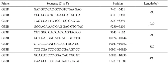
Viral genomic RNA was extracted from 200 μl of virus- infected Vero cell culture supernatant using a viral RNA extraction kit (Bioneer, Daejeon, Korea) according to the manufacturer's instructions. To synthesize cDNA from the extracted RNA, reverse transcription was conducted using specific primers with a Qiagen one-step RT-PCR kit (Qiagen, Valencia, CA, USA). The PCR products were purified using the QIAEX II Gel Extraction kit (Qiagen) and cloned into a pGEM-T easy vector (Promega, Madison, WI, USA) prior to sequencing. Oligonucleotide primers (Table 1) were designed from the complete sequences of the GETV South Korea strain (GenBank Accession No. AY702913). These primers were used for amplification of structural protein genes (the C, E3, E1, 6K and E2 polyprotein) and sequencing.
Sequence analysis and phylogenic comparison
An individual clone containing an amplified gene was then used as a template in cycle sequencing with an ABI PRISM Big Dye Terminator Cycle Sequencing Kit, version 1.1 (Applied Biosystems, Foster City, CA, USA). The sense and antisense strands were analyzed using an automated Applied Biosystems 377 DNA sequencer according to the manufacturer's instructions. Nucleotide sequence alignments were generated by Clustal X version 1.81 (29, 30). Analysis of nucleotide sequence identities was performed using DNA STAR software (DNA STAR. Inc., Madison, WI, USA).
Phylogenic analyses were conducted by the neighbor-joining method using MEGA version 5.05 (http://www.megasoft- ware.net/). The bootstrap method with 1,000 replicates was applied to determine the reliability of the inferred phylo- genetic tree. Distance analyses were conducted using the
Kimura two-parameter formula to correct for multiple sub- stitutions of equivalent nucleotides. Previously reported Alphavirus strains used in this study were listed in Table 2 and their complete sequences were deposited in GenBank.
RESULTS Virus identification
After propagation of the viruses, the infectious titer of each isolate was calculated as a tissue culture infectious dose 50 (TCID50). The titers were 1 × 107.5 TCID50/ ml for QIAG9301, 1 × 10⁷ TCID50/ ml for QIAG9302 and 1
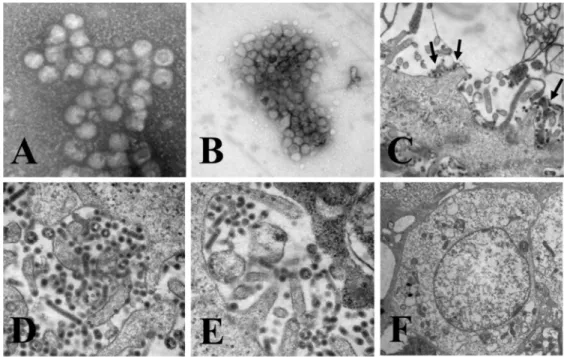
× 107 TCID50/ ml for QIAG9303. The morphology and morphogenesis of QIAG93 isolates were evaluated using negative staining electron microscopy. In this study, the QIAG9301 isolate was used because in preliminary experi- ments the morphological features of the three isolates were similar. Mature QIAG9301 virus particles were observed in
specimens prepared by direct or sucrose gradient centri- fugation (Figs. 1A and 1B). At 72 h post infection, virus particles were observed in both the cell culture fluid (Fig.
1C) and within cytoplasmic vacuoles (Fig. 1D) of Vero cells with the QIAG9301 isolate. These particles consisted of an internal nucleoid enclosed in a membrane that was surrounded by an envelope with projections. The diameter of these spherical particles, including the projection border, was 50~70 nm.
As shown in Fig. 1D, QIAG9301-infected cells showed substantial cytopathic changes, including vacuolization and condensation of the cytoplasm. One of the most striking features of infected cells was a lightly stained, circular in- terior region surrounding mitochondria, where many virus particles were present. Another feature was a single layer of mature virus particles, circular or sometimes tubular in shape, attached directly to the vacuolar membrane (data not shown). However, no virus-like particles and no cytopathic
Figure 1. Electron microscopy of QIAG93 isolates. QIAG9301 was used as a representative of the three isolates. Virus-like particles in specimens prepared by direct (A) or sucrose gradient centrifugation (B). General morphological characteristics of viruses and cytopathic changes in Vero cells after virus infection for 72 h (C, D and E). Mock-infected cells were used for comparison (F). Inset C, black solid arrows indicate virus-like particles.
changes were observed in mock-infected cells (Fig. 1F). In addition, developing virus particles were observed at the cell membrane, indicative of the terminal stage of the GETV replication cycle.
PCR Amplification
Viral RNA was extracted from cell culture supernatants, and cDNA of the structural polyprotein gene was synthesized
using each individual RNA extract as a template. The sequential series of overlapping genes was amplified by PCR using five primer sets (Table 1). The amplified DNA fragments were cloned into a plasmid vector for sequencing.
The primers GE1 to GE5 resulted in amplification of pro- ducts of 990, 1,030, 990, 880 and 490 bp, respectively (Fig.
2). The amplified DNA fragments appeared as single bands after electrophoresis and ethidium bromide staining, sug-
Table 1. Oligonucleotide primers used for RT-PCR of GETV, designed from the complete genome sequences of GETV South Korea (GenBank Accession No. AY702913)
Primer Sequence (5' to 3') Position Length (bp)
GE1F GAT GTC CAC GCT GTC TAA GAG 7401~7421
990
GE1R CGC GGG CTC TGA GCA TGG GA 8371~8390
GE2F TGG CCA TTG TCC TGG GAG GG 8221~8240
1030
GE2R GGG ACA AAC GAG GAG GTG TAC 9230~9250
GE3F CGT GGG CAC CAC CAG TAG CG 9143~9162
990
GE3R GGT GAT GGC ACG ACT GTC TTG 10124~10144
GE4F CTC CCC GAT GAC CCT ACA GC 10043~10062
880
GE4R TCG CGA TCC CGC CGA AGT CC 10901~10920
GE5F GGA CAT CCC GGA CAC CGC GT 10811~10830
490
GE5R CAA GCC TCC CGG AAT GCG GC 11281~11300
Figure 2. Amplification of the structural polyprotein gene of the GETV QIAG93 isolate with a specific primer set (see Table 1). Lanes 1 to 5, PCR products amplified using the respective GE1, GE2, GE3, GE4, and GE5 primer sets. Lane M, 100 bp DNA ladder. Use of the GE1 to GE5 primer sets resulted in amplification of products with sizes of 990, 1,030, 990, 880 and 490 bp, respectively.
gesting that the three isolates were homogenous with respect to the structural polyprotein gene. In addition, the full length of the structural polyprotein gene of each QIAG93 isolate (QIAG9301, QIAG9302 and QIAG9303) was identical in size, comprising 3759 nucleotides that encoded 1253 amino acids.
Sequence analysis
Multiple sequence alignment was conducted using the nucleotide sequences of the structural polyprotein genes to identify sequence similarities between QIAG93 isolates and other Alphavirus strains (Table 2). As shown in Table 3, the nucleotide sequence identities among the QIAG93 isolates ranged from 98.9% to 99.7%. The QIAG93 isolates shared 98.4~99.4% nucleotide sequence identities with a reference GETV strain, GETV South Korea (AY702913).
The nucleotide sequence identities of QIAG93 isolates with Chinese (HB0234, M1 and YN0540), Japanese (Kochi/01/
2005), Russian (LEIV 16275 Mag), Mongolian (LEIV 17741 MPR) and Malaysian (MM2021) GETV strains were 98.4~
99.8%, 98.2~98.5%, 97.9~98.1%, 99.1~99.4% and 95.4~
95.5%, respectively. A sequence alignment was also per- formed to identify nucleotide sequence homology between QIAG93 isolates and other Alphavirus strains within the Semliki Forest antigenic complex-such as SAGV, RRV and CHIKV (Table 2). The QIAG93 isolates showed 97.4~
97.6%, 98.6~98.8%, and 62.6~62.8% nucleotide sequence identities with SAGV (AB032553), RRV M1 (EF011023), and CHIKV 0611aTw (FJ807896), respectively.
Phylogenetic analysis
Phylogenetic relationships between QIAG93 isolates and other Alphavirus species were analyzed using the structural polyprotein gene and a variety of computational approaches.
The phylogenetic relationships among virus strains were depicted as a neighbor-joining tree (Fig. 3). A similar phy- logeny was found by maximum-parsimony analysis. In the neighbor-joining tree, monophyletic sister groups that had bootstrap values > 70% were chosen. To better understand the phylogenetic relationships among virus strains, the GETV strains were classified into three sister groups rooted at GETV MM2021. However, in our phylogeny, the Russian Table 2. Detailed information of the Alphaviruses analyzed in the present study
Taxonomy Isolates Geographic origin Host GeneBank accession
GETV QIAG9301 South Korea Swine KR081238
GETV QIAG9302 South Korea Swine KR081239
GETV QIAG9303 South Korea Swine KR081240
GETV South Korea South Korea Swine AY702913
GETV HB0234 China Mosquito EU015062
GETV M1 China Mosquito EU015061
GETV YN0540 China Mosquito EU015063
GETV Kochi/01/2005 Japan Swine AB859822
GETV LEIV 16275 Mag Russia Mosquito EF631998
GETV LEIV 17741 MPR Mongolia Mosquito EF631999
GETV MM2021 Malaysia Unknown AF339484
SAGV Unknown Japan Unknown AB032553
RRV M1 China Unknown EF011023
CHIKV 0611aTw Singapore Human FJ807896
Table 3. Nucleotide sequence comparison (above the diagonal) and estimation of evolutionary distance (below the diagonal) based on comparisons of the structural polyprotein gene of QIAG93 isolates with that of other Alphaviruses belonging to the Semliki Forest complex
Virus strains 1 2 3 4 5 6 7 8 9 10 11 12 13 14
1. GETV QIAG9301 99 98.9 99.9 99.1 98.4 99.3 98.2 97.9 99.1 95.4 97.4 98.5 62.8 2. GETV QIAG9302 0.010 99.7 99 98.6 98.7 98.8 98.5 98.2 99.4 95.5 97.6 98.8 62.7 3. GETV QIAG9303 0.011 0.003 98.9 98.5 98.5 98.7 98.4 98.1 99.3 95.4 97.5 98.6 62.6 4. GETV South Korea 0.001 0.010 0.011 99.2 98.4 99.4 98.2 98 99.2 95.4 97.4 98.5 62.9
5. GETV HB0234 0.009 0.014 0.016 0.008 98 99 97.8 97.6 98.7 95.1 97 98 62.8
6. GETV M1 0.017 0.013 0.015 0.016 0.021 98.1 97.8 98.2 98.7 95.7 97.6 99.8 62.8 7. GETV YN0540 0.007 0.012 0.013 0.006 0.010 0.018 98 97.7 99 95.2 97.2 98.2 62.9 8. GETV Kochi/01/2005 0.018 0.015 0.016 0.018 0.023 0.022 0.020 97.4 98.7 94.9 97 97.9 62.7 9. GETV LEVI16275Mag 0.021 0.018 0.019 0.021 0.025 0.018 0.023 0.027 98.3 95.5 97.6 98.3 62.6 10. GETV LEIV17741MPR 0.009 0.006 0.007 0.008 0.013 0.013 0.011 0.013 0.017 95.6 97.7 98.8 62.8 11. GETV MM2021 0.048 0.047 0.048 0.048 0.052 0.045 0.051 0.054 0.047 0.046 95.6 95.8 62.4 12. SAGV 0.027 0.024 0.025 0.027 0.031 0.025 0.028 0.031 0.025 0.024 0.045 97.6 62.6 13. RRV Alpahavirus M1 0.016 0.013 0.014 0.016 0.020 0.002 0.018 0.022 0.017 0.012 0.044 0.024 62.9 14. CHIKV 0611aTw 0.751 0.755 0.760 0.751 0.753 0.754 0.753 0.758 0.762 0.754 0.769 0.763 0.751
Figure 3. Phylogenetic relationships between QIAG93 isolates and other Alphaviruses within the Semliki Forest antigenic complex.
Phylogenies were assessed on the structural polyprotein gene using neighbor-joining methods. A bootstrap method with 1,000 replicates was applied in this analysis to evaluate the reliability of the inferred phylogenetic tree. In the neighbor-joining tree, the monophyletic sister groups that had bootstrap values > 70% were chosen. The GETV strains were classified into three sister groups rooted at GETV MM2021.
GETV LEVI 1672 Mag strain was not included in one of the sister groups because it was in a different branch. The three sister groups were named clades A to C. Clade A included the QIAG9302 and QIAG9303 isolates, GETV Kochi/01/2005 and GETV LEIV 17741 MPR. Clade B included GETV HB0234, GETV YN0540, QIAG9301 and GETV South Korea strains. Clade C included GETV M1 and the RRV Alphavirus M1 strain. A further feature of the neighbor-joining tree was that the SAGV strain was grouped into a distinct branch of GETV strains, which was also rooted from the GETV MM2021 strain. The CHIKV 0611aTw strain appeared as an out-group in this analysis.
To evaluate the evolutionary relationships between QIAG93 isolates and other virus strains, evolutionary dis- tances (EDs) were calculated using MEGA software, version 5.05. As shown in Table 3, in clade B, the ED between the QIAG9302 and QIAG9303 isolates was 0.003, indicating that these two isolates were more closely related to each other than to other GETV strains, i.e., GETV Kochi/01/2005 (ED 0.015 with QIAG9301 and 0.016 with QIAG9303) and GETV LEIV 17741 MPR (ED 0.006 with QIAG9301 and 0.007 with QIAG9303). In clade B, QIAG9301 showed EDs of 0.001, 0.007 and 0.009 with the GETV South Korea, GETV YN0540 and GETV HB0234 strains, respectively.
Therefore, QIAG9301 was evolutionarily closely related to the GETV South Korea strain.
DISCUSSION
Virus taxonomy is a useful approach not only for identifying newly discovered virus species but also, and more importantly, for enhancing our understanding of known viruses. Classification of viruses into hierarchical taxa, in- cluding species, genera and families, is based mainly on morphology, nucleic acid sequences, and the pattern of viral replication. Other biological, physicochemical and serological properties are also important in identification and classifi- cation of individual virus strains within any given taxon (31).
In the present study, we characterized QIAG9301, QIAG- 9302 and QIAG9303 using various taxonomic approaches to achieve more precise classification and subgrouping.
Application of electron microscopy in this study facilitated visualization of QIAG93 virus particles and their classifi- cation based on their morphologies and morphogenesis. The general morphological characteristics and unique cytoplasmic structures within cells infected by Semliki Forest virus (SFV) and related members of the Alphavirus group have been demonstrated by electron microscopy (32, 33). Negatively stained QIAG93 isolate particles in our electron micrographs were roughly circular in shape with a diameter of 50~70 nm (Fig. 1A and 1B). The virus particles consisted of an outer membrane, the envelope, and an inner core consisting of a nucleoid coated by an outer membrane with projections.
These features are similar to the morphological features of SFV (32). SFV is closely related to CHIKV, GETV, and Mayaro Alphaviruses (34). It is thus suggested that the QIAG93 isolates can be classified as tentative members of the Alphavirus family in the Semliki Forest complex.
Since viruses have relatively simple genomes and high multiplication rates, they are subject to more rapid evolu- tionary changes. Even a single point mutation can lead to dramatic changes in virus phenotype, which may positively or negatively affect progeny and alter the direction of viral evolution (35, 36). In general, the percentage of sequence divergence is correlated inversely with serologic cross- reactivity (12, 15). To assess the evolutionary history and mechanisms of the emergence of QIAG93 isolates, a sequence analysis of the structural polyprotein gene, in- cluding the C, E3, E1, 6K and E2 domains, was conducted (Table 2). The sequence analysis showed that the QIAG9301 isolate shares >99% sequence identity with the QIAG9302 isolate, and the GETV South Korea, GETV HB0234, GETV YN0540 and GETV LEIV17741MPR strains. Among them, the GETV South Korea and Chinese GETV YN0540 strain showed the highest sequence similarity with the QIAG9301 isolate. The QIAG9302 isolate exhibited high sequence similarity with the QIAG9301 (99.7%) and QIAG9303 (99.7%) isolates. In addition, the GETV South Korea and Mongolian GETV LEIV17741MPR strains also exhibited
>99% sequence identity with the QIAG9302 isolate. Other than the QIAG9301 and QIAG9302 isolates, only the Mon- golian GETV_LEIV17741MPR strain showed high sequence
similarity with the QIAG9303 isolate (99.3%). Viruses within a given antigenic complex are genetically more closely related than viruses in different complexes (15). Therefore, to determine the extent of relatedness of the QIAG93 isolates to other Alphavirus species, the SAGV, RRV Alphavirus M1 and CHIKV 0611aTw strains were subjected to sequence analysis (Table 2). The QIAG93 isolates shared 97.4% and 98.6% sequence similarities with SAGV and the RRV Alphavirus M1 strain, respectively. In contrast, the CHIKV 0611aTw strain exhibited 37.6% nucleotide sequence simi- larity with the QIAG93 isolates, despite the fact that they are the closest serological relatives of GETV.
Phylogenetic trees constructed by sequence analyses and evolutionary distance (ED) estimations based on the struc- tural polyprotein gene sequences showed that the QIAG- 9301 isolate (clade B), and QIAG9302 and GETV QIAG- 9303 isolates (clade A) are classified into different sister groups (Fig. 3). In clade B, the QIAG9301 isolate was located in close proximity to GETV South Korea and the GETV HB0234 and GETV YN0540 strains, forming a distinguishable branch. In clade A, the GETV QIAG9302 isolate was located in close proximity to the QIAG9303 isolate and the GETV Kochi/01/2005 and GETV LEIV 17741 MPR strains, also forming a distinguishable branch.
These results suggest that the QIAG9301 isolate is more closely related to GETV South Korea (ED value 0.001) and GETV YN0540 (ED value 0.007) than are the QIAG9302 and QIAG9303 isolates. Both QIAG9302 and QIAG9303 are genetically closely related to the Mongolian GETV LEIV17741MPR strain, as suggested by their very short evolutionary distances (mean ED value, 0.0065). As shown in Table 2, the GETV isolates in clades A and B exhibited greater evolutionary distances from the original Malaysian GETV strain (range, 0.044~0.054). In contrast, the evolu- tionary distances among them ranged from 0.001 to 0.018.
Taken together, our findings suggest that the GETV strains in clades A and B have been evolving slowly and circulating extensively in Northeast Asia since their transmission from Malaysia. In addition, the evolutionary mechanism of GETV strains is more closely correlated with their geographical separation than epidemiological features, such as infection
outcome and year of isolation.
Taken together, our findings suggest that the QIAG93 isolates are considered to be GETV strains because their morphological and genetic features are in agreement with previously reported biological features of GETV. Therefore, our findings will be valuable for molecular epidemiological analyses of GETV in Korea and contribute to further study of the pathogenicity of QIAG93 isolates in animals. Fur- thermore, use of our data to facilitate development of a vaccine to prevent GETV infection would benefit the horse and pig industries.
REFERENCES
1)Berge TO. International catalogue of arboviruses, in- cluding certain other viruses of vertebrates. Washington D. C.: US Department of Health, Education and Welfare, Public Health Service; 1975. p.278.
2) Go YY, Balasuriya UB, Lee CK. Zoonotic encephalitides caused by arboviruses: transmission and epidemiology of alphaviruses and flaviviruses. Clin Exp Vaccine Res 2014;3:58-77.
3) Elisvberg BL, Buescher EL. Getah (MM 2021). Arbo- virus catalogue card No.47, Arbovirus Information Exchange, 1963.
4)Doherty RL, Gorman BM, Whitehead RH, Carley JG.
Studies of arthropod-borne virus infections in Queens- land. V. Survey of antibodies to Group A arboviruses in man and other animals. Aust J Expt Biol Sci 1966;44:
365-77.
5) Brown CM, Timoney PJ. Getah virus infection of Indian horses. Trop Anim Health Prod 1988;30:241-52.
6)Kamada M, Ando Y, Fukunaga Y, Kumanomido T, Imagawa H, Wada R, et al. Equine Getah virus infection:
Isolation of the virus from racehorses during a enzootic in Japan. Am J Trop Med Hyg 1980;29:984-8.
7)Sentsui H, Kono Y. An epidemic of Getah virus in- fection among racehorses: isolation of the virus. Res Vet Sci 1980;29:157-61.
8)Timoney PJ. Getah virus infection. In: Infectious Dis- eases of Livestock. Coetzer JAW and Tustin RC (eds).
2nd ed. Cape Town: Oxford University Press; 2004.
p.1023-26.
9)Kumanomido T, Wada R, Kanemaru T, Kamada M, Hirasawa K, Akiyama Y. Clinical and virological obser- vations on swine experimentally infected with Getah virus. Vet Microbiol 1988;16:295-301.
10) Matsuyama T, Nakamura T, Isahai K, Oya A, Kobayashi M. Haruna virus, a group A arbovirus isolated from swine in Japan. Gunma J Med Sci 1967;16:131-4.
11)Marchette NJ, Rudnick A, Garcia R. Alphaviruses in Peninsular Malaysia: II. Serological evidence of human infection. Southeast Asian J Trop Med Public Health 1980;11:14-23.
12)Calisher CH, Karabatsos N. Arbovirus serogroups:
definition and geographic distribution. In The Arbo- viruses: Epidemiology and Ecology. Monath TP (ed).
vol. I, Boca Raton, FL: CRC Press; 1988. p.19-57.
13) Weaver SC, Dalgarno L, Frey TK, Huang HV, Kinney RM, Rice CM, et al. Family Togaviridae. In: Virus Taxonomy: Seventh Report of the International Com- mittee on Taxonomy of Viruses. van Regenmortel MV, Fauguet CM, Bishop DHI, Carstens EB, Estes MK, Lemon SM, et al (eds). San Diego: Academic Press;
2000. p.879-89.
14) Calisher CH, Shope RE, Brandt W, Casals J, Karabatsos N, Murphy FA, et al. Proposed antigenic classification of registered arboviruses I. Togaviridae, Alphavirus.
Intervirology 1980;14:229-32.
15)Powers AM, Brault AC, Shirako Y, Strauss EG, Kang W, Strauss JH, et al. Evolutionary relationships and systematics of the alphaviruses. J Virol 2001;75:10118 -31.
16) Bryant JE, Crabtree MB, Nam VS, Yen NT, Duc HM, Miller BR. Isolation of arboviruses from mosquitoes collected in northern Vietnam. Am J Trop Med Hyg 2005;
73:470-3.
17) Chang CY, Huang CC, Huang TS, Deng MC, Jong MH, Wang FI. Isolation and characterization of a Sagiyama virus from domestic pigs. J Vet Diagn Invest 2006;18:
156-61.
18) Kanamitsu M, Taniguchi K, Urasawa S, Ogata T, Wada Y, Wada Y, et al. Geographic distribution of arbovirus antibodies in indigenous human populations in the Indo- Australian archipelago. Am J Trop Med Hyg 1979;28:
351-63.
19)Norder H, Lundström JO, Kozuch O, Magnius LO.
Genetic relatedness of Sindbis virus strains from Europe, Middle East, and Africa. Virology 1996;222:440-5.
20) Turell MJ, O'Guinn ML, Wasieloski LP Jr, Dohm DJ, Lee WJ, Cho HW, et al. Isolation of Japanese enceph- alitis and Getah viruses from mosquitoes (Diptera:
Culicidae) collected near Camp Greaves, Gyonggi Pro- vince, Republic of Korea, 2000. J Med Entomol 2003;
40:580-4.
21) Tajima S, Kotaki A, Yagasaki K, Taniwaki T, Moi ML, Nakayama E, et al. Identification and amplification of Japanese encephalitis virus and Getah virus propagated from a single porcine serum sample: a case of co- infection. Arch Virol 2014;159:2969-75.
22)Pfeffer M, Kinney RM, Kaaden OR. The Alphavirus 3'-nontranslated region: size heterogeneity and arrange- ment of repeated sequence elements. Virology 1998;240:
100-8.
23)Griffin DE. Alphaviruses, In: Knipe DM, Howley PM (Eds). Fields' Virology, 4th ed. New York: Lippincott, Williams and Wilkins, 2001. p.917-62.
24) Calisher CH, Walton TE. Getah virus infections. In: Virus Infections of Equines. Studdert MK (ed). Amsterdam:
Elsevier; 1996. p.157-65.
25) Fukunaga Y, Kumanomido T, Kamada M. Getah virus as an equine pathogen. Vet Clin North Am Equine Pract 2000;16:605-17.
26) Jo HY, Yang DK, Kim HH, Choi SS, Kang KS, Yang SJ, et al. Sero-surveillance of Getah virus among Thor- oughbred horses in Korea. J Bacteriol Virol 2015;45:
235-41.
27) Colombet J, Sime-Ngando T. Use of PEG, Polyethylene glycol, to characterize the diversity of environmental viruses. Microb Ecol 2012;58:728-36.
28) Vajda BP. Concentration and purification of viruses and bacteriophages with polyethylene glycol. Folia Microbiol (Praha) 1978;23:88-96.
29) Jeanmougin F, Thompson JD, Gouy M, Higgins DG, Gibson TJ. Multiple sequence alignment with Clustal X. Trends Biochem Sci 1998;23:403-5.
30) Thompson JD, Gibson TJ, Plewniak F, Jeanmougin F, Higgins DG. The CLUSTAL_X windows interface:
flexible strategies for multiple sequence alignment aided by quality analysis tools. Nucleic Acids Res 1997;25:
4876-82.
31)Hyypiä T, Hovi T, Knowles NJ, Stanway G. Classifi- cation of enteroviruses based on molecular and biological properties. J Gen Virol 1997;78:1-11.
32)Acheson NH, Tamm I. Replication of Semliki Forest virus: an electron microscopic study. Virology 1967;32:
128-43.
33) Grimley PM, Berezesky IK, Friedman RM. Cytoplasmic structures associated with an arbovirus infection: loci of viral ribonucleic acid synthesis. J Virol 1968;2:1326 -38.
34)Fazakerley JK. Pathogenesis of Semliki Forest virus
encephalitis. J Neurovirol 2002;8:66-74.
35) Dietzgen RG, Kuzmin IV. Taxonomy of rhabdoviruses.
In: Rhabdoviruses: Molecular Taxonomy, Evolution, Genomics, Ecology, Host-Vector Interactions, Cyto- pathology and Control. Dietzgen RG, Kuzmin IV (eds).
Norfolk: Caister Academic Press; 2012. p.13-22.
36)Dietzschold B, Wunner WH, Wiktor TJ, Lopes AD, Lafon M, Smith CL, et al. Characterization of an antigenic determinant of the glycoprotein that correlates with pathogenicity of rabies virus. Proc Natl Acad Sci U S A 1983;80:70-4.