INTRODUCTION
It is important to maintain bone health as the bone plays a role in mechanical support, calcium storage, and RBC production.
Accordingly, a variety of methods have been investigated for the restoration of bone defects in the oral and maxillofa- cial regions.1
Biomaterials containing additional bone growth factors or BMP (bone morphogenic protein), including autogenous graft bone, or ceramics, are currently being used for the restoration of bone defects.2 Various substitutes are also being used, including mineralized frozen dried allograft, demineralized frozen dried allograft, coralline calcium carbonate, polylactide- polyglycolide copolymer, synthetic polymers, calcium sulfate, bovine bone, and hydroxyapatite.3
Bio-Oss�(Geistlich, Wolhusen, Switzerland), an inorganic material produced from bovine bone treated with ethylenediamine and sterilized to remove organic materials such as protein, fol- lowed by sterilization, consists of calcium-deficient carbon- ate apatite. It has been used to fill any bone defect in the cran- iofacial and periodontal regions for a long time. It was report- ed that Bio-Oss�has not only superior biocompatibility but also can be sufficiently used as a scaffold for osteogenesis and osteogenic cells.1,4,5However, it is known, that Bio-Oss�has poor osteoinduction. In addition, it was reported, that Bio-Oss� shows lower proliferation and differentiation of osteoblasts com- pared to the other substitutes.6,7
Three important factors have been proposed as prerequisites for the stable treatment of the bone defect in the recent den- tal implanting: soluble molecular signals, response cells with
The effect of immobilization of heparin and bone morphogenic protein-2 to bovine bone substitute
on osteoblast-like cell ’ ’ s function
Jung-Bo Huh1a, DDS, MSD, Sung-Eun Kim2a, PhD, Se-Kyung Song3, DDS, MSD, Mi-Jung Yun1, DDS, MSD, Ji-Suk Shim3, DDS, MSD, Jeong-Yol Lee3, DDS, PhD, Sang-Wan Shin3*, DDS, PhD
1Department of Prosthodontics, School of Dentistry, Pusan National University, Yangsan,
2Department of Orthopedic Surgery & Rare Diseases Institute, Korea University Medical Center, Guro Hospital, Seoul,
3Department of Prosthodontics, Institute for Clinical Dental Research, Korea University Guro Hospital, Seoul, Korea
PURPOSE. This study was performed to investigate the ability of recombinant human-bone morphogenic protein-2 immobilized on a heparin-grafted bone substrate to enhance the osteoblastic functions. MATERIALS AND METHODS. The Bio-Oss�, not coated with any material, was used as a control group. In rhBMP-2-Bio-Oss�group, rhBMP-2 was coated with Bio-Oss�using only deep and dry methods (50 ng/mL, 24 h). In heparinized rhBMP-2-Bio-Oss�group, dopamine was anchored to the surface of Bio-Oss�, and coated with heparin. rhBMP- 2 was immobilized onto the heparinized- Bio-Oss�surface. The release kinetics of the rhBMP-2-Bio-Oss�and heparinized rhBMP-2-Bio-Oss�were analyzed using an enzyme-linked immunosorbent assay. The biological activities of the MG63 cells on the three groups were investigated via cytotoxicity assay, cell proliferation assay, alkaline phosphatase (ALP) measurement, and calcium deposition determination. Statistical comparisons were carried out by one-way ANOVA test. Differences were considered statistically significant at *P<.05 and **P<.001.
RESULTS. The heparinized rhBMP-2-Bio-Oss�showed more sustained release compared to the rhBMP-2-Bio-Oss�over an extended time.
In the measurement of the ALP activity, the heparinized group showed a significantly higher ALP activity when compared with the non-heparinized groups (P<.05). The MG63 cells cultivated in the group with rhBMP-2 showed increased calcium deposition, and the MG63 cells from the heparinized group increased more than those that were cultivated in the non-heparinized groups. CONCLUSION. Heparin increased the rhBMP-2 release amount and made sustained release possible, and heparinized Bio-Oss�with rhBMP-2 successfully improved the osteoblastic functions.
[J Adv Prosthodont 2011;3:145-51]
KEY WORDS: Heparin; rhBMP-2; Osteoblast-like cell; Bovine bone
Corresponding author: Sang-Wan Shin
Institute for Clinical Dental Research, Korea University Hospital 97 Gurodong-Gil, Guro-Gu, Seoul, 152-703, Korea
Tel. 82 2 2626 1922: e-mail, [email protected]
Received July 12, 2011 / Last Revison August 7, 2011 / Accepted August 9, 2011
ⓒ 2011 The Korean Academy of Prosthodontics
This is an Open Access article distributed under the terms of the Creative Commons Attribution Non-Commercial License (http://creativecommons.org/licenses/by- nc/3.0) which permits unrestricted non-commercial use, distribution, and reproduction in any medium, provided the original work is properly cited.
aThese authors contributed equally to this work.
corresponding cell surface receptors, and the extracellular matrix.8,9The molecular mechanism of such regeneration is caused by osteogenic proteins that belong to the TGF-β (transforming growth factor β) subgroup. Among those pro- teins, BMP has particularly drawn attention as a potent mate- rial inducing bone differentiation.10There are more than 20 BMP subgroups, among which BMP-2 has been proven via preclinical and clinical studies to be useful for therapeutic purposes regarding to bone graft in dentistry.11,12BMP-2 promotes the proliferation of osteoblasts from mesenchymal stem cells by regulating the essential factors for the osteoinduction-regen- erating bone structure, and enhances osteogenesis by assisting the biosynthesis of the bone matrix.13Sykaras et al.14report- ed that BMP-2 was effective in titanium implant osseointegration.
Despite the successful use of BMP-2 in the enhancement of bone regeneration, BMP-2 has the disadvantages of high cost, need of large amount (1 mg BMP-2/mL defect), and short half- life in the body.15 To overcome the aforementioned prob- lems, studies on the continuous and local release of BMP-2 using collagen gels, sponges, scaffolds, hyaluronic acid, and fibrin gels have been conducted.16-23 Such methods have various problems, however, such as failure in regulating the release amount, early and short-term release.24
In this study, heparin was used to regulate rhBMP-2 release.
Heparin, which is a highly sulfationized linear natural poly- saccharide, has been known to have a binding affinity with var- ious growth factors, such as the vascular endothelial growth factor (VEGF), basic fibroblast growth factor (bFGF), and trans- forming growth factor-β(TGF-β).25 Furthermore, heparin has been reported to regulate the release of such growth fac- tors.26,27Many studies reported that the binding of BMP-2 with heparin on a titanium surface resulted in the continuous release of BMP-2 which resulted in an anti-inflammatory response and the reinforced function of the osteoblast-like cells.28-31 A study reported that the bone deposition and the amount of bone growth after six weeks implantation were higher in the BMP-2-coated implants than in the non-BMP-2-coated implants in the bone defects in rabbits. This effect was clear- ly reinforced by the surface modification of heparin.28The sur- face modification of heparin by a collagen matrix enabled the long-term release of BMP-2 in a biomimetic model. In addi- tion, the binding of rhBMP-2 and heparin on the thin apatite surface of the titanium which was suspended in the simulat- ed body fluid after the base and heat treatments seemed to stim- ulate both the ALP activity and the expression of OCN mRNA in the osteoblast-like cells.29
Accordingly, in this study, the surface of the Bio-Oss� bone substitute was modified with heparin and then was coated with rhBMP-2. Subsequently, a method that promotes osteoinduction for bone formation was explored, and its validity was assessed on a cellular level.
MATERIALS AND METHODS
Immobilization of rhBMP-2 on the heparinized Bio-Oss��
The Bio-Oss�, not coated with any material, was used as a control group. In rhBMP-2-Bio-Oss� group, rhBMP-2 (Cowellmwdi Co., Pusan, South Korea) was coated with Bio-Oss�using deep and dry methods (50 ng/mL, 24 h). In heparinized rhBMP-2-Bio-Oss�group, to immobilize rhBMP- 2 on the surface of the Bio-Oss�, the Bio-Oss�surface was coat- ed with heparinized dopamine in advance. 2 mg/mL heparinized dopamine was dissolved in a 10 mM Tris-HCl (pH 8.0) buffer, and then 100 mg Bio-Oss�was put into the Tris-HCl buffer solution, followed by reaction under blocked light for 24 h. After the reaction, the Bio-Oss�was washed with distilled water, freeze-dried, and then 50 ng/mL rhBMP-2 was immo- bilized on the heparinized Bio-Oss�. The heparinized Bio-Oss� (100 mg) was then put into a 0.1M MES (pH 5.6) buffer, and rhBMP-2 (50 ng/mL) was added to the buffer, followed by reac- tion at room temperature for 24 h.
Assessment of the morphological characteristics of Bio-Oss��
The Bio-Oss�, rhBMP-2-Bio-Oss�, and heparinized rhBMP- 2-Bio-Oss� were morphologically analyzed using a scan- ning electron microscope (SEM; S2300, Hitachi, Tokyo, Japan). The samples were coated with Pt using a sputter- coater (Eiko IB, Tokyo, Japan). The SEM was accelerated with 10 kV power before use.
Release kinetics of rhBMP-2
Each sample of rhBMP-2-Bio-Oss�and heparinized rhBMP- 2-Bio-Oss� was put into an E-tube containing 1 mL PBS (pH 7.4) 0.02% sodium azide. Then the samples were react- ed in an agitating incubator with 100 rpm, at 37℃. The supernant was collected in a specified interval and was replaced with a new buffer. The amount of BMP-2 in the col- lected supernatant was measured using an ELISA development kit (Pepro Tech, Rocky Hill, NJ, USA) in a microplate read- er, at 495 nm.
Cell culture
Aliquots of human-derived MG63 osteoblast-like cells (KCLB, Seoul, South Korea) in Dulbecco’s Modified Eagle’s Medium (DMEM), with added 10% fetal bovine serum (FBS, Gibco BRL, Grand Island, NY, USA), 100 U/mL penicillin, and 100 μg/mL streptomycin were loaded onto a 100-mm cul- ture plate. They were cultured at 37℃ and 100% humidity while continuously supplying 95% air and 5% CO2. For the induc-
tion of osteogenic differentiation, they were cultured in DMEM (osteogenic medium) containing 100 U/mL peni- cillin, 100 μg/mL streptomycin, and 10% FBS, with addition of 100 nM dexamethasone, 100 μM ascorbic acid, and 10 mM β-glycerolphosphate.
Assessment of cytotoxicity
MG63 osteoblast-like cells were loaded onto a 24-tran- swell culture plate containing Bio-Oss�, rhBMP-2-Bio-Oss�, and heparinized rhBMP-2-Bio-Oss�, respectively, to have an aliquot of 5×10⁴cells in each plate. After culturing at 37℃ for 24 and 48 h, respectively, the plate was treated with CCK-8 (Dojindo, Tokyo, Japan) reagent for 1 h to assess the cytotox- icity of the samples. The culture media that was treated with the reagent was transferred into a 96-well plate, and its absorbance was measured with a microplate reader at 450 nm.
Each group was cultured three times, according to time.
Measurement of cell proliferation
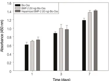
MG63 osteoblast-like cells were loaded onto a 24-tran- swell culture plate containing Bio-Oss�, rhBMP-2-Bio-Oss�, and heparinized rhBMP-2-Bio-Oss�, respectively, to have an aliquot of 5×10⁴cells in each plate. After culturing at 37℃ for 1, 3, and 7 days, respectively, the plate was treated with CCK-8 (Dojindo, Tokyo, Japan) reagent for 1 h to assess the cell proliferation of the samples. The culture media that was treated with the reagent was transferred into a 96-well plate, and its absorbance was measured with a microplate reader at 450 nm. Each group was cultured three times, according to time.
Measurement of alkaline phosphatase activity
MG63 osteoblast-like cells were loaded onto a 24-tran- swell culture plate containing Bio-Oss�, rhBMP-2-Bio-Oss�, and heparinized rhBMP-2-Bio-Oss�, respectively, to have an aliquot of 5×10⁴cells in each plate. Then the plate was cul- tured in DMEM culture media containing 100 U/mL penicillin, 100 μg/mL streptomycin, and 10% FBS, to which 100 nM dex- amethasone, 100 μM ascorbic acid, and 10 mM β-glyc- erolphosphate were added for 7, 14, and 21 days, respectively.
After culturing, the media was removed, and the cells were sep- arated with trypsin-EDTA, followed by collection via cen- trifugation. The supernatant was removed, after which 0.2 mL RIPA buffer solution was added. The solution was suspend- ed using a sonicator, and the cells were dissolved at 4℃.
The dissolved cells were centrifuged, and p-nitrophenyl phos- phate (p-NPP) solution was then added to the supernatant, fol- lowed by reaction for 30 min. Then the reaction was halted by adding 1N NaOH. The hydrolysis of p-NPP was measured with a microplate reader at 410 nm, and p-nitrophenol (p-NP)
was used as a standard value. The protein concentration was measured using a Bradford protein assay reagent, and bovine serum albumin was used as a standard. The ALP activity was denoted as μM/min/μg protein. In each experiment, the activ- ity was calculated as a percentage over the negative con- trol. Each experiment was conducted three times.
Measurement of calcium accumulation
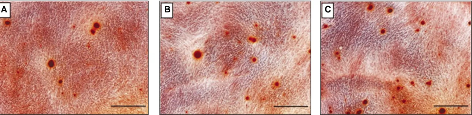
Human-derived MG63 osteoblast-like cells were loaded onto a 6-transwell culture plate containing Bio-Oss�, rhBMP- 2-Bio-Oss�, and heparinized rhBMP-2-Bio-Oss�, respec- tively, to have an aliquot of 1×10⁵cells in each plate, followed by culturing for 21 days. After 21-day culturing, the media was removed, and the cells were washed with PBS. The cells were fixed in ice-cold 70% ethanol at -20℃ for 1 h. After remov- ing the ethanol, the cells were stained with Alizarin red S (pH 4.2) at room temperature for 10 min. Alizarin red S was then removed, and the cells were washed with distilled water three times. The stained parts were photographed using an opti- cal microscope (Olympus, Tokyo, Japan) for comparative analysis.
Statistical analysis
All the data were denoted as mean±SD. Statistical analy- sis was conducted using t test and one-way analysis of variance (ANOVA, Systat Software, Inc.) to determine the signifi- cance levels of the cytotoxicity, cell proliferation, and ALP activ- ity for each sample (*P<.05, **P<.001).
RESULTS
Observation of the surface morphology of the Bio-Oss��
The surfaces of the Bio-Oss�, rhBMP-2-Bio-Oss�, and heparinized rhBMP-2-Bio-Oss�were compared by SEM to observe their microstructures (Fig. 1). No difference in surface morphology was found.
Release kinetics of recombinant human-bone morpho- genetic protein-2 (rhBMP-2)
When the release kinetics of rhBMP-2 were observed in the rhBMP-2-Bio-Oss� and heparinized rhBMP-2-Bio-Oss�, rhBMP-2 was rapidly released in the rhBMP-2-Bio-Oss� group at an early stage. Meanwhile, more rhBMP-2 was released in the heparinized rhBMP-2-Bio-Oss�group than in the rhBMP-2-Bio-Oss�group. Furthermore, rhBMP-2 was con- tinuously released during the two-week period, and approxi- mately 20% of the original amount of rhBMP-2 seemed to be released after the two-week period, showing a tendency of con-
tinuous release. Therefore, rhBMP-2 was slowly and contin- uously released in the heparinized rhBMP-2-Bio-Oss�with the passage of time (Fig. 2).
Assessment of cytotoxicity and cell proliferation
When the cytotoxicity of the three Bio-Oss�groups was assessed, no cytotoxicity was found in the three groups when compared to the control group, where only MG63 cells were cultured for 24 and 48 h (Fig. 3). When MG63 cells were cul- tured in the Bio-Oss�, rhBMP-2-Bio-Oss�, and heparinized rhBMP-2-Bio-Oss�for 1, 3, and 7 days, and the cell proliferation was observed, no significant difference was found (Fig. 4).
Measurement of alkaline phosphatase activity
When the ALP activity, which is a marker of the differentiation of osteoblast-like cells, was measured, the activity increased
Fig. 1. SEM images of the (A) Bio-Oss�, (B) rhBMP-2 (50 ng/ml)-Bio-Oss�, and (C) heparinized rhBMP-2 (50 ng/ml)-Bio-Oss�.
A B C
Fig. 2. Release kinetics of the rhBMP-2-(50 ng)-Bio-Oss�and heparinized rhBMP-2-(50 ng)-Bio-Oss�.
Fig. 3. Cytotoxicity of the osteoblast-like cells (MG63 cells) grown on the Bio-Oss�, rhBMP-2-(50 ng)-Bio-Oss�, and heparinized rhBMP-2- (50 ng)-Bio-Oss�.
Fig. 4. Proliferation of the osteoblast-like cells (MG63 cells) grown on the Bio-Oss�, rhBMP-2-(50 ng)-Bio-Oss�, and heparinized rhBmp-2- (50 ng)-Bio-Oss�.
50
40
30
20
10
0
BMP-2 cumulative release (ng/mL)
0 5 10 15 20 25 30 Time (day)
Heparinized BMP-2 (50 ng)-Bio-Oss BMP-2 (50 ng)-Bio-Oss
140 120 100 80 60 40 20 0
Cell viability (% of control)
24 48 Time (hours)
Control Bio-Oss
BMP-2 (50 ng)-Bio-Oss Heparinized BMP-2 (50 ng)-Bio-Oss
1.6 1.4 1.2 1.0 0.8 0.6 0.4 0.2 0
Absorbance (450 nm)
1 3 7 Time (days)
Bio-Oss
BMP-2 (50 ng)-Bio-Oss Heparinized BMP-2 (50 ng)-Bio-Oss
in the MG63 osteoblast-like cells which were cultured in the Bio-Oss�groups with rhBMP-2 (*P<.05) on day 7 after culture. The ALP activity increased more in the MG63 osteoblast-like cells cultured in the heparinized rhBMP-2-Bio- Oss�than in those cultured in the control and non-heparinized groups, which showed a significant difference on day 14 after culture (**P<.001). In addition, the ALP activity reached its peak in all three groups on day 14 after culture, and decreased in all three groups on day 21 after culture when com- pared to those of 14 (Fig. 5).
Assessment of calcium accumulation (Alizarin red S)
The accumulation of calcium, an inorganic material that was deposited on the bone during ossification, was observed on day
31 after culture, calcium accumulation increased in the MG63 osteoblast-like cells cultured in the Bio-Oss� groups with rhBMP-2. Furthermore, calcium accumulation increased more in the MG63 osteoblast-like cells that were cultured in the heparinized group than in those that were cultured in the control and non-heparinized groups (Fig. 6). Therefore, it was confirmed that the heparinized substitute had a superior osteogenic ability.
DISCUSSION
When autogenous graft bone or other bone substitutes are used for bone transplantation, the new bone is formed through osteoconduction, osteogenesis, and osteoinduction.
Osteoconduction is defined as the induction of bone formation in which bone substitutes act as a scaffold for bone formation.
Osteogenesis is caused by bone-forming cells and a matrix direct- ly provided from bone substitutes. Osteoinduction refers to the induction of osteoblast differentiation undifferentiated mes- enchymal cells after the chemotaxis of substitutes on the undifferentiated mesenchymal cells in the host. BMP is asso- ciated with osteoinduction.32
Bio-Oss�has been reported not only to have superior bio- compatibility but also to be sufficiently used as a scaffold of osteogenesis and osteogenic cells, but it is known to have poor osteoinduction.1,4,5Accordingly, the use of rhBMP-2 for over- coming the aforementioned problem has been investigated.
As a growth factor with the best osteoinduction ability, rhBMP-2 has been intensively investigated, but its application to clinical practice was restricted due to its short half-life, rapid dissolution by body fluid, large-amount requirement, and high production cost. In this study, rhBMP-2 from E. coli (Escherichia coli) was used. RhBMP-2 can be produced on a large scale, unlike the BMP produced from the CHO cell used in previous studies. Therefore, rhBMP-2 has the advan- tages of cost-saving and mass production. Bessho et al.33 reported that when the aforementioned two rhBMP-2 were used,
Fig. 6. Alizarin-red-S staining of the osteoblast-like cells (MG63 cells) grown on the Bio-Oss�(A), rhBMP-2-(50 ng)-Bio-Oss�(B), and heparinized rhBMP-2 (50 ng)-Bio-Oss�(C) after 21-day incubation (scale bar = 20 μm).
A B C
Fig. 5. ALP activity of the osteoblast-like cells (MG63 cells) grown on the Bio-Oss�, rhBMP-2-(50 ng)-Bio-Oss�, and heparinized rhBMP-2- (50 ng)-Bio-Oss�after 7-, 14-, and 21-day incubation.
40
30
20
10
0
ALP activity (μM/min/μg protein)
7 14 21 Time (days)
Bio-Oss
BMP-2 (50 ng)-Bio-Oss Heparinized BMP-2 (50 ng)-Bio-Oss
lower bone density was obtained from the rhBMP-2 pro- duced from E. coli, and fatty marrow was formed. The efficacy and safety of the E. coli-derived rhBMP-2 (Cowellmwdi Co., Pusan, South Korea) used in this study, has been proven in many studies.34,35
For the establishment of an effective rhBMP-2 application system and an appropriate rhBMP-2 release amount for a sufficient period, the surface of Bio-Oss�was modified with heparin in this study, followed by the immobilization of rhBMP-2 on the surface. rhBMP-2 was successfully released from the heparinized rhBMP-2-Bio-Oss� for an extended period. The continuous release of growth factors, including var- ious types of matrices, from heparin has been reported in many studies.27,36,37Lin et al.37reported that in an in vivo experiment, the ALP activity and calcified-tissue rate increased in the cells attached to the demineralized bone matrix, where heparin was crossly bound for the binding of rhBMP-2. From the results of the aforementioned studies, heparin can be considered as a suitable for the continuous release of growth factors material.
In this study, cell proliferation increased more in the MG63 cells cultured in the rhBMP-2-immobilized groups than in those cultured in the groups without rhBMP-2, but the increase was not significant. On the contrary, Park et al.38reported that cell proliferation significantly increased in the osteoblast- like cells cultured in the nanofibrous chitosan membrane with immobilized rhBMP-2 than in those cultured in the nanofibrous chitosan membrane without rhBMP-2. Therefore, it seems that the effect of rhBMP-2 on the proliferation of osteoblast-like cells is unclear.
ALP activity and calcium accumulation have been widely used as markers of the early and late differentiation of osteoblast- like cells.39,40ALP activity was measured on days 7, 14, and 21 after culture. No significant difference in cell proliferation was found between the groups with immobilized rhBMP-2 and those without immobilized rhBMP-2. Meanwhile, the ALP activi- ty was significantly higher in the groups with immobilized rhBMP-2 than in those without immobilized rhBMP-2 during the various culturing periods. Therefore, rhBMP-2 stimulat- ed the differentiation of osteoblast-like cells. The ALP activ- ity in all the matrices, however, was reduced slightly more on day 21 than on day 14. This means that the ALP activity reached the peak prior to the actual initiation of calcification.41 Furthermore, the aforementioned result showed that the ALP activity gradually decreased beyond day 14, whereas the calcium accumulation gradually increased. When the calcium accumulation was measured on day 21 after culture, it was increased in the MG63 osteoblast-like cells that were cultured in the groups with immobilized rhBMP-2, and increased more in the MG63 osteoblast-like cells that were cultured in the heparinized group than in those that were cultured in the non-heparinized group. The aforementioned results showed that the heparinized rhBMP-2-Bio-Oss�stimulated matrix formation
and enhanced the functions of the osteoblast-like cells.
In this study, the reactions were investigated on a cellular lev- el. Therefore, the results of this study should be validated through animal experiments and clinical studies. Furthermore, considering the current unclear rhBMP-2 concentration standard, further studies are required to determine the optimal concentration and amount of rhBMP-2 in combined use with Bio-Oss�, via the application of rhBMP-2 with various concentrations.
CONCLUSION
When Bio-Oss� was treated with heparin and rhBMP-2 was then immobilized on the heparinized surface, Bio-Oss� showed successful functional improvement. Heparin increased the rhBMP-2 release amount and allowed sustained release. The heparinized rhBMP-2-Bio-Oss�successfully improved the osteoblastic functions.
REFERENCES
1. Amerio P, Vianale G, Reale M, Muraro R, Tulli A, Piattelli A.
The effect of deproteinized bovine bone on osteoblast growth fac- tors and proinflammatory cytokine production. Clin Oral Implants Res 2010;21:650-5.
2. Del Fabbro M, Rosano G, Taschieri S. Implant survival rates af- ter maxillary sinus augmentation. Eur J Oral Sci 2008;116:497- 506.
3. Mangano C, Scarano A, Iezzi G, Orsini G, Perrotti V, Mangano F, Montini S, Piccirilli M, Piattelli A. Maxillary sinus augmentation using an engineered porous hydroxyapatite: a clinical, histological, and transmission electron microscopy study in man. J Oral Implantol 2006;32:122-31.
4. Esposito M, Grusovin MG, Kwan S, Worthington HV, Coulthard P. Interventions for replacing missing teeth: bone augmentation techniques for dental implant treatment. Cochrane Database Syst Rev 2008;3:CD003607.
5. Fulmer NL, Bussard GM, Gampper TJ, Edlich RF. Anorganic bovine bone and analogs of bone mineral as implants for cran- iofacial surgery: a literature review. J Long Term Eff Med Implants 1998;8:69-78.
6. Ku¨bler A, Neugebauer J, Oh JH, Scheer M, Zo¨ller JE. Growth and proliferation of human osteoblasts on different bone graft substitutes: an in vitro study. Implant Dent 2004;13:171-9.
7. Turhani D, Weissenbo¨ck M, Watzinger E, Yerit K, Cvikl B, Ewers R, Thurnher D. In vitro study of adherent mandibular os- teoblast-like cells on carrier materials. Int J Oral Maxillofac Surg 2005;34:543-50.
8. Reddi AH. Symbiosis of biotechnology and biomaterials: ap- plications in tissue engineering of bone and cartilage. J Cell Biochem 1994;56:192-5.
9. Reddi AH. Bone morphogenetic proteins, bone marrow stromal cells, and mesenchymal stem cells. Maureen Owen revisited. Clin Orthop Relat Res 1995;313:115-9.
10. Duneas N, Crooks J, Ripamonti U. Transforming growth factor- beta 1: induction of bone morphogenetic protein genes ex- pression during endochondral bone formation in the baboon, and synergistic interaction with osteogenic protein-1 (BMP-7).
Growth Factors 1998;15:259-77.
11. Hall J, Sorensen RG, Wozney JM, Wikesjo¨UM. Bone forma- tion at rhBMP-2-coated titanium implants in the rat ectopic mod- el. J Clin Periodontol 2007;34:444-51.
12. Chen D, Zhao M, Mundy GR. Bone morphogenetic proteins.
Growth Factors 2004;22:233-41.
13. Bessa PC, Balmayor ER, Azevedo HS, Nu¨rnberger S, Casal M, van Griensven M, Reis RL, Redl H. Silk fibroin microparticles as carriers for delivery of human recombinant BMPs. Physical characterization and drug release. J Tissue Eng Regen Med 2010;
4:349-55.
14. Sykaras N, Iacopino AM, Triplett RG, Marker VA. Effect of re- combinant human bone morphogenetic protein-2 on the os- seointegration of dental implants: a biomechanics study. Clin Oral Investig 2004;8:196-205.
15. Sellers RS, Zhang R, Glasson SS, Kim HD, Peluso D, D'Augusta DA, Beckwith K, Morris EA. Repair of articular cartilage defects one year after treatment with recombinant human bone mor- phogenetic protein-2 (rhBMP-2). J Bone Joint Surg Am 2000;82:
151-60.
16. Bax BE, Wozney JM, Ashhurst DE. Bone morphogenetic pro- tein-2 increases the rate of callus formation after fracture of the rabbit tibia. Calcif Tissue Int 1999;65:83-9.
17. Sheehan JP, Kallmes DF, Sheehan JM, Jane JA Jr, Fergus AH, diPierro CG, Simmons NE, Makel DD, Helm GA. Molecular methods of enhancing lumbar spine fusion. Neurosurgery 1996;
39:548-54.
18. King GN, King N, Cruchley AT, Wozney JM, Hughes FJ.
Recombinant human bone morphogenetic protein-2 promotes wound healing in rat periodontal fenestration defects. J Dent Res 1997;76:1460-70.
19. Sellers RS, Peluso D, Morris EA. The effect of recombinant hu- man bone morphogenetic protein-2 (rhBMP-2) on the healing of full-thickness defects of articular cartilage. J Bone Joint Surg Am 1997;79:1452-63.
20. Zellin G, Linde A. Importance of delivery systems for growth- stimulatory factors in combination with osteopromotive mem- branes. An experimental study using rhBMP-2 in rat mandibu- lar defects. J Biomed Mater Res 1997;35:181-90.
21. Kim J, Park Y, Tae G, Lee KB, Hwang CM, Hwang SJ, Kim IS, Noh I, Sun K. Characterization of low-molecular-weight hyaluronic acid-based hydrogel and differential stem cell responses in the hydrogel microenvironments. J Biomed Mater Res A 2009;
88:967-75.
22. Kim J, Kim IS, Cho TH, Lee KB, Hwang SJ, Tae G, Noh I, Lee SH, Park Y, Sun K. Bone regeneration using hyaluronic acid- based hydrogel with bone morphogenic protein-2 and human mes- enchymal stem cells. Biomaterials 2007;28:1830-7.
23. Lee TC, Ho JT, Hung KS, Chen WF, Chung YH, Yang YL. Bone morphogenetic protein gene therapy using a fibrin scaffold for a rabbit spinal-fusion experiment. Neurosurgery 2006;58:373- 80; discussion 373-80.
24. Jeon O, Song SJ, Yang HS, Bhang SH, Kang SW, Sung MA, Lee JH, Kim BS. Long-term delivery enhances in vivo osteogenic efficacy of bone morphogenetic protein-2 compared to short-term delivery. Biochem Biophys Res Commun 2008;369:774-80.
25. Sasisekharan R, Ernst S, Venkataraman G. On the regulation of fibroblast growth factor activity by heparin-like glycosamino- glycans. Angiogenesis 1997;1:45-54.
26. Perets A, Baruch Y, Weisbuch F, Shoshany G, Neufeld G, Cohen S. Enhancing the vascularization of three-dimensional porous alginate scaffolds by incorporating controlled release basic fi- broblast growth factor microspheres. J Biomed Mater Res A 2003;65:489-97.
27. Ishibe T, Goto T, Kodama T, Miyazaki T, Kobayashi S, Takahashi T. Bone formation on apatite-coated titanium with in- corporated BMP-2/heparin in vivo. Oral Surg Oral Med Oral Pathol Oral Radiol Endod 2009;108:867-75.
28. von Walter M, Herren C, Gensior TJ, Steffens GC, Hermanns- Sachweh B, Jahnen-Dechent W, Ru¨ger M, Erli HJ. Biomimetic modification of the TiO(2)/glass composite Ecopore with he- parinized collagen and the osteoinductive factor BMP-2. Acta Biomater 2008;4:997-1004.
29. Kodama T, Goto T, Miyazaki T, Takahashi T. Bone formation on apatite-coated titanium incorporated with bone morpho- genetic protein and heparin. Int J Oral Maxillofac Implants 2008;23:1013-9.
30. Ishibe T, Goto T, Kodama T, Miyazaki T, Kobayashi S, Takahashi T. Bone formation on apatite-coated titanium with in- corporated BMP-2/heparin in vivo. Oral Surg Oral Med Oral Pathol Oral Radiol Endod 2009;108:867-75.
31. Kim SE, Song SH, Yun YP, Choi BJ, Kwon IK, Bae MS, Moon HJ, Kwon YD. The effect of immobilization of heparin and bone morphogenic protein-2 (BMP-2) to titanium sur- faces on inflammation and osteoblast function. Biomaterials 2011;32:366-73.
32. Tadjoedin ES, de Lange GL, Bronckers AL, Lyaruu DM, Burger EH. Deproteinized cancellous bovine bone (Bio-Oss) as bone substitute for sinus floor elevation. A retrospective, his- tomorphometrical study of five cases. J Clin Periodontol 2003;
30:261-70.
33. Bessho K, Konishi Y, Kaihara S, Fujimura K, Okubo Y, Iizuka T. Bone induction by Escherichia coli -derived recombinant hu- man bone morphogenetic protein-2 compared with Chinese hamster ovary cell-derived recombinant human bone morpho- genetic protein-2. Br J Oral Maxillofac Surg 2000;38:645-9.
34. Tokuhara Y, Wakitani S, Imai Y, Kawaguchi A, Fukunaga K, Kim M, Kadoya Y, Takaoka K. Repair of experimentally in- duced large osteochondral defects in rabbit knee with various con- centrations of Escherichia coli-derived recombinant human bone morphogenetic protein-2. Int Orthop 2010;34:761-7.
35. Lee JH, Kim CS, Choi KH, Jung UW, Yun JH, Choi SH, Cho KS. The induction of bone formation in rat calvarial defects and subcutaneous tissues by recombinant human BMP-2, produced in Escherichia coli. Biomaterials 2010;31:3512-9.
36. Ho YC, Mi FL, Sung HW, Kuo PL. Heparin-functionalized chi- tosan-alginate scaffolds for controlled release of growth factor.
Int J Pharm 2009;376:69-75.
37. Lin H, Zhao Y, Sun W, Chen B, Zhang J, Zhao W, Xiao Z, Dai J. The effect of crosslinking heparin to demineralized bone matrix on mechanical strength and specific binding to human bone morphogenetic protein-2. Biomaterials 2008;29:1189-97.
38. Park YJ, Kim KH, Lee JY, Ku Y, Lee SJ, Min BM, Chung CP.
Immobilization of bone morphogenetic protein-2 on a nanofi- brous chitosan membrane for enhanced guided bone regenera- tion. Biotechnol Appl Biochem 2006;43:17-24.
39. Turksen K, Bhargava U, Moe HK, Aubin JE. Isolation of monoclonal antibodies recognizing rat bone-associated molecules in vitro and in vivo. J Histochem Cytochem 1992;40:1339-52.
40. van den Beucken JJ, Walboomers XF, Boerman OC, Vos MR, Sommerdijk NA, Hayakawa T, Fukushima T, Okahata Y, Nolte RJ, Jansen JA. Functionalization of multilayered DNA- coatings with bone morphogenetic protein 2. J Control Release 2006;113:63-72.
41. Bancroft GN, Sikavitsas VI, van den Dolder J, Sheffield TL, Ambrose CG, Jansen JA, Mikos AG. Fluid flow increases mineralized matrix deposition in 3D perfusion culture of mar- row stromal osteoblasts in a dose-dependent manner. Proc Natl Acad Sci USA 2002;99:12600-5.