46
Left ventricular systolic function can be worsened as a result of long standing pressure overload in patients with aortic ste- nosis (AS). Two-dimensional speckle tracking echocardiogra- phy can detect decreased global longitudinal strain (GLS) even in patients with preserved left ventricular ejection fraction.1) Transcatheter aortic valve implantation (TAVI) is a newly in- troduced therapy for elderly patients with severe AS with high perioperative risk.2) Because the TAVI is free from an addi- tional cardiac injury during cardioplegia or myocardial inci- sion, the comparison of strain values before and after the proce- dure can demonstrate the effect of increased pressure gradient on myocardial function more accurately. We want to show the acute effect of the decompression on the GLS in a patient un- derwent the TAVI procedure.
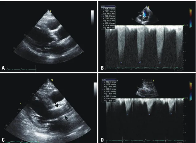
A 87 years old man was admitted to our hospital due to re- current chest discomfort and syncope during exercise. Stan- dard transthoracic echocardiography with Doppler analysis was done with Vivid E9 and M4S transducer (GE Vingmed, Horton, Norway). The baseline echocardiogram showed se- vere degenerative AS with preserved left ventricular systolic function (57%) (Fig. 1A and B) and his coronary angiography was normal. Because of the increased perioperative risk, the
patient underwent TAVI procedure with a 31 mm sized Core- Valve (Medtronic, Minneapolis, MN, USA). After the proce- dure, the patient showed marked improvement of functional capacity. The follow-up echocardiogram showed well de- ployed prosthetic valve in the aortic valve area with normal left ventricular systolic function (Fig. 1C) and the increased pressure gradient was lowered up to 14 mmHg from 74 mmHg of the baseline echocardiogram (Fig. 1D). Longitudi- nal strain analysis was performed from the apical long axis, four- and two-chamber views with 60 frames per second using offline software (EchoPacPC, GE Vingmed, Horten, Norway).
Global longitudinal peak systolic strain of the left ventricle was generated averaging peak systolic strain values of the three apical views and bull’s eye view was used to demonstrate seg- mental strain values. The baseline GLS was -14.4% (Fig. 2A) and follow-up GLS was increased up to -16.5% at one week after the procedure (Fig. 2B). This acute improvement of GLS may be originated from the effect of the pressure overload. To demonstrate the effect of increased pressure gradient on the GLS objectively, we need more patients with severe AS under- going TAVI procedure.
pISSN 1975-4612/ eISSN 2005-9655 Copyright © 2014 Korean Society of Echocardiography www.kse-jcu.org http://dx.doi.org/10.4250/jcu.2014.22.1.46
IMAGES IN CARDIOVASCULAR ULTRASOUND J Cardiovasc Ultrasound 2014;22(1):46-47
Similar Morphology, but Different Function: Acute Improvement
of Myocardial Longitudinal Strain after Percutaneous Transcatheter Aortic Valve Implantation Therapy in a Severe Aortic Stenosis Patient
Jae-Hwan Lee, MD, PhD, Jae-Hyeong Park, MD, PhD, Si Wan Choi, MD, PhD, Jin-Ok Jeong, MD, PhD, and In-Whan Seong, MD, PhD
Department of Cardiology, Internal Medicine, School of Medicine, Chungnam National University, Chungnam National University Hospital, Daejeon, Korea
KEY WORDS: Aortic stenosis · Transcatheter aortic valve implantation · Strain echocardiography.
• Received: January 12, 2014 • Revised: February 23, 2014 • Accepted: February 23, 2014
• Address for Correspondence: Jae-Hyeong Park, Department of Cardiology, Internal Medicine, School of Medicine, Chungnam National University, Chungnam National University Hospital, 282 Munhwa-ro, Jung-gu, Daejeon 301-721, Korea
Tel: +82-42-280-8237, Fax: +82-42-280-8238, E-mail: [email protected]
• This is an Open Access article distributed under the terms of the Creative Commons Attribution Non-Commercial License (http://creativecommons.org/licenses/by-nc/3.0) which permits unrestricted non-commercial use, distribution, and reproduction in any medium, provided the original work is properly cited.
Acute Change of GLSLV after TAVI | Jae-Hwan Lee, et al.
47 References
1. Delgado V, Tops LF, van Bommel RJ, van der Kley F, Marsan NA, Klautz RJ, Versteegh MI, Holman ER, Schalij MJ, Bax JJ. Strain analysis in patients with severe aortic stenosis and preserved left ventricular ejection fraction undergoing surgical valve replacement. Eur Heart J
2009;30:3037-47.
2. Cribier A, Eltchaninoff H, Bash A, Borenstein N, Tron C, Bauer F, Derumeaux G, Anselme F, Laborde F, Leon MB. Percutaneous trans- catheter implantation of an aortic valve prosthesis for calcific aortic stenosis:
first human case description. Circulation 2002;106:3006-8.
Fig. 1. The baseline parasternal long axis view demonstrates severe calcified aortic valve (A) with increased transaortic valve maximal velocity up to 4.3 m/sec (B). After the procedure, prosthetic valve is inserted in to the aortic valve area (C, arrows) and transvalvular maximal velocity is measured up to 1.9 m/sec (D).
Fig. 2. Bull’s eye diagram shows clearly visible improvement in global longitudinal strain after the transfemoral aortic valve intervention [before (A), and one week after (B) the procedure].
C
A A
D
B B