INTRODUCTION
Breast cancer is a leading cause of cancer-related death in woman worldwide. Mammography and other diagnostic tools permit earlier detection, while adjuvant chemotherapies extend survival. Death from breast cancer occurs primarily through distant metastasis. Nearly 30% of patients with axillary node- negative breast cancer and almost 50% of those with axillary node-positive breast cancer will eventually experience tumor recurrence following surgical resection with adjuvant chemo- therapy due to occult micrometastatic cells in their blood [1].
Metastasis stems from minimal residual disease (MRD) and
the presence of single disseminated or circulating tumor cells (CTC) that are undetectable even by high resolution imaging technologies [2,3].
Disseminated tumor cells (DTC) are known to predict un- favorable outcomes in breast cancer patients, but the detection of DTC in the bone marrow is cumbersome, painful and inva- sive [4,5]. Many clinicians are turning to CTC research because CTC detection is well tolerated, minimally invasive and easily reproduced. The value of peripheral blood CTC in prognosis was first evaluated in patients with metastatic breast cancer and more recently evaluated in the general breast cancer pop- ulation [6-10].
Cytokeratin-20 (CK-20) presents a valid marker for CTC in peripheral blood because it is not expressed in normal healthy hematologic tissues or in benign breast disease, including in- flammatory breast disease therefore, CK-20 is used as a mark- er of hematogenous micrometastasis and a reverse transcrip- tase polymerase chain reaction (RT-PCR) target for the detec- tion of breast CTC in patients with breast cancer [11-13].
Breast cancer can be classified in intrinsic subtypes accord-
Circulating Tumor Cells Detected by RT-PCR for CK-20 before Surgery Indicate Worse Prognostic Impact in Triple-Negative and HER2 Subtype Breast Cancer
Seong Bae Hwang, Jeoung Won Bae, Hye Yoon Lee, Hoon Yub Kim
Division of Breast-Endocrine Surgery, Department of Surgery, Korea University Anam Hospital, Seoul, Korea
Purpose: Circulating tumor cells (CTC) clearly correlate with unfa- vorable outcomes for patients with metastatic breast cancer, but the long-term prognostic implications of CTC for molecular sub- types of operable breast cancer are not yet known. We explored the relationships between previously established prognostic fac- tors and CTC in operable breast cancer, and the significance of CTC by breast cancer molecular subtype. Methods: We retro- spectively evaluated 166 patients with operable breast cancer (stage I-IIIA) diagnosed from April 1997 to May 2003. CTC were detected using cytokeratin- 20 (CK-20) mRNA expression in peripheral blood samples that were collected just prior to surgery under general anesthesia. Clinicopathological characteristics of the cancer were analyzed according to CTC status. Metastasis- free survival (MFS) and overall survival (OS) were analyzed accord- ing to CTC status and breast cancer molecular subtype. Results:
CK-20 mRNA-positive CTC was detected in 37 of 166 patients (22.3%) and was not correlated with any previous clinical factors in univariate analysis (p>0.05). After a median follow-up of 100 months, the patients with CK-20 mRNA-positive CTC had less favorable outcomes in terms of MFS and OS than those without detectable CTC (log-rank p<0.05). Among molecular subtypes of operable breast cancer, the patients with CK-20 mRNA-posi- tive CTC had shorter MFS and OS in triple negative and human epidermal growth factor 2 (HER2) breast cancer subtype (log-rank, p<0.05). Conclusion: CK-20 mRNA-positive CTC may lend insight into tumor progression as a prognostic indicator especially in the triple negative and HER2 subtypes of operable breast cancer.
Key Words: Breast neoplasms, Circulating tumor cell, Molecular subtype, Survival
Correspondence: Jeoung Won Bae
Division of Breast-Endocrine Surgery, Department of Surgery, Korea University Anam Hospital, 73 Inchon-ro, Seongbuk-gu, Seoul 136-705, Korea Tel: +82-2-920-5305, Fax: +82-2-928-1631
E-mail: [email protected]
Prognosis and response prediction were presented as poster in the Global Breast Cancer Conference 2011.
Received: September 26, 2011 Accepted: January 15, 2012
Cancer
ORIGINAL ARTICLE
ing to distinct gene profile [14,15]. This molecular classifica- tion includes four main subtypes of breast cancer: luminal A subtype, luminal B subtype, human epidermal growth factor receptor 2 (HER2) subtype, and basal-like tumors [16]. Breast cancers have different clinical features according to their intrin- sic subtypes, accordingly, they may have a different prognostic CTC value according to own subtype [17]. CTC can exhibit different characteristics compared to primary tumors [18].
Until now, few studies have addressed the prognostic signifi- cance of CTC according to breast cancer molecular subtype, particularly with regard to long-term follow-up.
In this study, we analyzed the relationships between CK-20 mRNA-positive CTC in the peripheral blood and previously recognized factors in operable breast cancer, and we evaluated the different prognostic significance of CK-20 mRNA-positive CTC according to the 4 breast cancer molecular subtypes.
METHODS
Materials
Peripheral blood samples were obtained from breast cancer patients who underwent surgery at Korea University Anam Hospital from April 1997 to May 2003. Each patient provided informed consent prior to the blood collection. The samples were collected as described to study the sensitivity of CK-20 mRNA expression for detecting CTC and the relationship be- tween the established prognostic parameters and CK-20 mRNA- positive CTC in breast cancer [12,19]. Based on retrospective reviews of medical records, patients were selected for this study according to the following inclusion criteria: 1) female gender;
2) diagnosis of invasive ductal carcinoma; 3) stage I-IIIA accord- ing to the American Joint Committee on Cancer staging sys- tem (6th edition) defined as an operable breast cancer; 4) data available to determine estrogen receptor (ER), progesterone receptor (PR), and HER2 status; and 5) no previous surgery or other treatments for the primary tumor. Accordingly, 166 patients met the criteria and were entered into this study.
All patients included in this study received treatments judged appropriate for the stage and hormone receptor status of their tumors. Surgical procedures included breast-conserving sur- gery and modified radical mastectomy. Breast-conserving sur- gery included lumpectomy, quadrantectomy and wedge resec- tion with axillary lymph node dissection. All patients who underwent breast-conserving surgery received radiation ther- apy and all ER+ patients received tamoxifen therapy (20 mg daily for 5 years). Patients with HER2-positive tumor did not receive adjuvant treatment with trastuzumab (Herceptin®; Genentech, San Francisco, USA).
Sample collection and RT-PCR amplification of CK-20 mRNA The blood samples (10 mL) were collected just prior to sur- gery under general anesthesia from the ante-cubital vein after several milliliters of blood were drained to avoid contamina- tion with skin epithelial cells. The blood samples were collect- ed in ethylenediaminetetraacetic acid-treated tubes, placed on ice, and transported immediately to the laboratory for RNA isolation. RT-PCR amplification of CK-20 mRNA was per- formed as previously described [12,19].
Immunohistochemistry for ER, PR, HER2, and molecular subtypes
ER (DAKO, Glostrup, Denmark) and PR (DAKO) status were recorded as positive if >1% of the tumor nuclei were immunoreactive. Otherwise, ER and PR were recorded as negative. For HER2 status (Labvision, Fremont, USA), 0 and 1+ were considered negative and 3+ was considered positive by immunohistochemistry (IHC). Fluorescence in situ hybrid- ization (FISH) was not performed for 2+ HER2 status by IHC, and HER2 2+ patients were excluded from the study. Molecu- lar subtypes were assigned according to ER, PR, or HER2 sta- tus: luminal A (ER+ and/or PR+, HER2-), luminal B (ER+
and/or PR+, HER2+), triple negative (ER-, PR-, HER2-), and HER2 (ER-, PR-, HER2+).
Statistical analysis
The relationships between CK-20 mRNA-positive CTC and clinical factors were evaluated using Fisher’s exact test and Pearson’s chi-square test. Metastasis-free survival (MFS) and overall survival (OS) according to molecular subtypes were estimated using the Kaplan-Meier log-rank method. To evalu- ate independent prognostic factor and relative risk, multivari- ate analyses were performed using the Cox proportional haz- ards model and logistic regression. All statistical tests were performed at the 5% significance level (p<0.05). All statistical analyses were performed using SPSS version 18.0 for Windows (SPSS Inc., Chicago, USA).
RESULTS
Patients characteristics
The characteristics of the 166 patients are presented in Table 1. The patients’ mean age was 49.5±10.4 years (range, 26-78 years). The median follow-up time was 100.6 months (range, 0.4-163.9 months). The CK-20 mRNA-positive CTC was de- tected in 37 (22.3%) of 166 patients. According to breast can- cer subtype, 73 patients were identified as luminal A (44.0%), 40 as luminal B (24.1%), 36 as triple negative (21.7%), and 17 as HER2 (10.2%).
Table 1. Characteristics in patients with operable breast cancer Characteristics Overall
No. (%)
CTC negative No. (%)
CTC positive No. (%) p-value Age (yr)
<35 13 (7.8) 8 (6.2) 5 (13.5) 0.303
≥35 to ≤50 78 (47.0) 63 (48.8) 15 (40.5)
50< 75 (45.2) 58 (45.0) 17 (46.0)
T stage
1 66 (39.8) 54 (41.9) 12 (32.4) 0.313
2 83 (50.0) 64 (49.6) 19 (51.4)
3 17 (10.2) 11 (8.5) 6 (16.2)
N stage
0 84 (50.6) 70 (54.3) 14 (37.8) 0.209
1 51 (30.7) 37 (28.7) 14 (37.8)
2 31 (18.7) 22 (17.0) 9 (24.4)
TNM stage
I 43 (25.9) 37 (28.7) 6 (16.2) 0.071
IIA 58 (34.9) 44 (34.1) 14 (37.8)
IIB 29 (17.5) 25 (19.4) 4 (10.8)
IIIA 36 (21.7) 23 (17.8) 13 (35.2)
Lymphatic invasion
No 136 (81.9) 108 (83.7) 28 (75.7) 0.262
Yes 30 (18.1) 21 (16.3) 9 (24.3)
Vascular invasion
No 160 (96.4) 125 (96.9) 35 (94.6) 0.508
Yes 6 (3.6) 4 (3.1) 2 (5.4)
p53
Negative 92 (55.4) 72 (55.8) 20 (54.1) 0.078
Positive 74 (44.6) 57 (44.2) 17 (45.9) ER
Negative 63 (38.0) 48 (37.2) 15 (40.5) 0.713
Positive 103 (62.0) 81 (62.8) 22 (59.5)
Characteristics Overall No. (%)
CTC negative No. (%)
CTC positive No. (%) p-value PR
Negative 59 (35.5) 43 (33.3) 16 (43.2) 0.267
Positive 107 (64.5) 86 (66.7) 21 (56.8) HER2
Negative (0-1+) 109 (65.7) 84 (65.1) 25 (67.6) 0.782 Positive (3+) 57 (34.3) 45 (34.9) 12 (32.4)
Subtypes
Luminal A 73 (44.0) 57 (44.2) 16 (43.2) 0.756
Luminal B 40 (24.1) 33 (25.6) 7 (18.9)
TNBC 36 (21.7) 27 (20.9) 9 (24.3)
HER2 17 (10.2) 12 (9.3) 5 (13.5)
Surgery
MRM 108 (65.1) 81 (62.8) 27 (73.0) 0.252
BCS 58 (34.9) 48 (37.2) 10 (27.0)
RT
No 75 (45.2) 58 (45.0) 17 (45.9) 0.916
Yes 91 (54.8) 71 (55.0) 20 (54.1)
Chemotherapy
No 22 (13.3) 18 (14.0) 4 (10.8) 0.619
Yes 144 (86.7) 111 (86.0) 33 (89.2)
Recurrence
No 130 (78.3) 107 (82.9) 23 (62.2) 0.023
Locoregional 9 (5.4) 6 (4.7) 3 (8.1)
Systemic 27 (16.3) 16 (12.4) 11 (29.7) Breast cancer-specific death
No 146 (88.0) 117 (90.7) 29 (78.4) 0.042
Yes 20 (12.0) 12 (9.3) 8 (21.6)
CTC=circulating tumor cells; ER=estrogen receptor; PR=progesterone receptor; TNBC=triple negative breast cancer; HER2=human epidermal growth factor receptor 2; MRM=modified radical mastectomy; BCS=breast-conserving surgery; RT=radiation therapy.
Figure 1. Prognostic significance of circulating tumor cells (CTC) in operable breast cancer. (A) Metastasis-free survival in operable breast cancer. (B) Overall survival in operable breast cancer. *log-rank test.
Metastasis-free survival
Follow-up time (mo)
0.0 50.0 100.0 150.0 200.0
1.0
0.8
0.6
0.4
0.2
0.0
p-value=0.013*
CTC negative CTC positive CTC negative CTC positive
A
Overall survival
Follow-up time (mo)
0.0 50.0 100.0 150.0 200.0
1.0
0.8
0.6
0.4
0.2
0.0
p-value=0.042*
CTC negative CTC positive CTC negative CTC positive
B
No significant differences were observed in the distribution of CTC-positive status according to molecular subtype of breast cancer. CTC-positive status was not significantly associated with any previously established prognostic factor and was cor- related only with recurrence and breast cancer-specific death in operable breast cancer (p=0.023 and p=0.042, respectively) (Table 1).
Prognostic impact of CK-20 mRNA-positive CTC before surgery Overall, locoregional recurrences were observed in 9 patients (5.4%), while systemic recurrences were seen in 27 patients (16.3%). During follow-up, 20 breast cancer-specific deaths
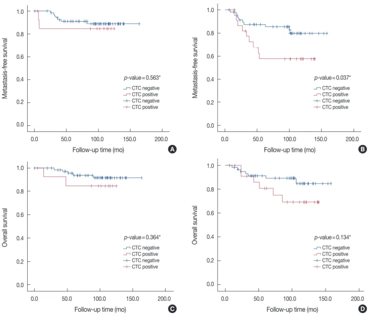
occurred (12.0%). Patients with positive CTC status had sig- nificantly shorter MFS and OS in operable breast cancer (log- rank, p=0.013 and p=0.042, respectively) (Figure 1). Positive CTC was not associated with MFS or OS in patients with either early breast cancer (tumor, node, metastasis [TNM] stage I, II) or advanced breast cancer (TNM stage IIIA) (log-rank, p>0.05) (Figure 2). Positive CTC was not significantly related with MFS or OS in the lymph node-negative patient group, and also was not related with OS in lymph node positive patient group (log-rank, p>0.05) (Figure 3). Positive CTC was asso- ciated with shorter MFS in the lymph node-positive patient group only (log-rank, p=0.037) (Figure 3B).
Figure 2. Different prognostic significances of circulating tumor cells (CTC) according to the American Joint Committee on Cancer tumor stage (early vs. advanced). (A) Metastasis-free survival (MFS) in early breast cancer patients. (B) MFS in patients with advanced breast cancer. (C) Overall survival (OS) in patients with early breast cancer. (D) OS in patients with advanced breast cancer. *log-rank test.
Metastasis-free survival
Follow-up time (mo)
0.0 50.0 100.0 150.0 200.0
1.0
0.8
0.6
0.4
0.2
0.0
p-value=0.144*
CTC negative CTC positive CTC negative CTC positive
A
Metastasis-free survival
Follow-up time (mo)
0.0 20.0 40.0 60.0 80.0 100.0 120.0 140.0 1.0
0.8
0.6
0.4
0.2
0.0
p-value=0.204*
CTC negative CTC positive CTC negative CTC positive
B
Overall survival
Follow-up time (mo)
0.0 50.0 100.0 150.0 200.0
1.0
0.8
0.6
0.4
0.2
0.0
p-value=0.489*
CTC negative CTC positive CTC negative CTC positive
C
Overall survival
Follow-up time (mo)
0.0 20.0 40.0 60.0 80.0 100.0 120.0 140.0 1.0
0.8
0.6
0.4
0.2
0.0
p-value=0.129*
CTC negative CTC positive CTC negative CTC positive
D
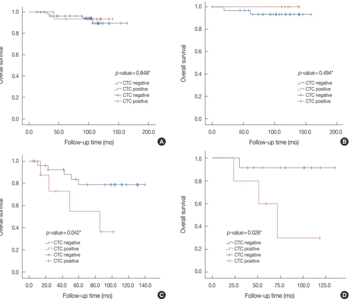
Luminal A and luminal B subtypes did not differ signifi- cantly in MFS (log-rank, p=0.443 and p=0.331, respectively) (Figure 4A, B) and OS (log-rank, p=0.848 and p=0.494, re- spectively) (Figure 5A, B) according to CTC-positive status.
Among patients with triple negative breast cancer, those with CTC-positive status had shorter MFS and OS (log-rank, p=
0.051 and p=0.042, respectively) (Figure 4C, 5C). Patients with CTC-positive status in the HER2 subtype breast cancer group also had shorter MFS and OS (log-rank, p=0.004 and p=0.026, respectively) (Figure 4D, 5D).
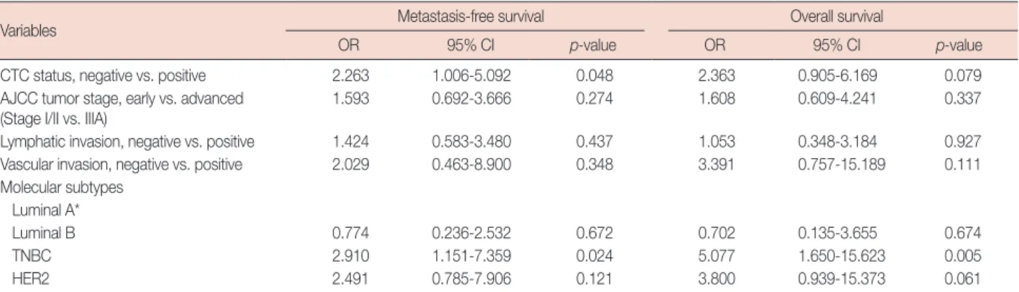
Based on Cox multivariate analysis, CTC status indepen- dently predicted MFS (odds ratio [OR], 2.263; 95% confidence interval [CI], 1.006-5.092; p=0.048) (Table 2) but not OS (OR,
2.363; 95% CI, 0.905-6.169; p=0.079) (Table 2). Among the molecular subtypes of operable breast cancer, only the triple negative subtype independently predicted MFS (OR, 2.910;
95% CI, 1.151-7.359; p=0.024) (Table 2) and OS (OR, 5.077;
95% CI, 1.650-15.623; p=0.005) (Table 2).
DISCUSSION
Using univariate analysis, we found no correlation between CK-20 mRNA-positive CTC and any previously established prognostic factor in operable breast cancer (Table 1). Recent reports present HER2 status as the only primary tumor char- acteristic that reliably predicts the presence of CTCs [9,20].
Figure 3. Different prognostic significances of circulating tumor cells (CTC) according to lymph node status. (A) Metastasis-free survival (MFS) in pa- tients with negative node status. (B) MFS in patients with positive node status. (C) Overall survival (OS) in patients with negative node status. (D) OS in patients with positive node status. *log-rank test.
Metastasis-free survival
Follow-up time (mo)
0.0 50.0 100.0 150.0 200.0
1.0
0.8
0.6
0.4
0.2
0.0
p-value=0.563*
CTC negative CTC positive CTC negative CTC positive
A
Metastasis-free survival
Follow-up time (mo)
0.0 50.0 100.0 150.0 200.0
1.0
0.8
0.6
0.4
0.2
0.0
p-value=0.037*
CTC negative CTC positive CTC negative CTC positive
B
Overall survival
Follow-up time (mo)
0.0 50.0 100.0 150.0 200.0
1.0
0.8
0.6
0.4
0.2
0.0
p-value=0.364*
CTC negative CTC positive CTC negative CTC positive
C
Overall survival
Follow-up time (mo)
0.0 50.0 100.0 150.0 200.0
1.0
0.8
0.6
0.4
0.2
0.0
p-value=0.134*
CTC negative CTC positive CTC negative CTC positive
D
Figure 4. Metastasis-free survival (MFS) curves according to circulating tumor cells (CTC) status by breast cancer subtypes. (A) MFS in the luminal A subtype. (B) MFS in the luminal B subtype. (C) MFS in the triple negative breast cancer subtype. (D) MFS in the HER2 subtype. *log-rank test.
Metastasis-free survival
Follow-up time (mo)
0.0 50.0 100.0 150.0 200.0
1.0
0.8
0.6
0.4
0.2
0.0
p-value=0.443*
CTC negative CTC positive CTC negative CTC positive
A
Metastasis-free survival
Follow-up time (mo)
0.0 50.0 100.0 150.0 200.0
1.0
0.8
0.6
0.4
0.2
0.0
p-value=0.331*
CTC negative CTC positive CTC negative CTC positive
B
Metastasis-free survival
Follow-up time (mo)
0.0 20.0 40.0 60.0 80.0 100.0 120.0 140.0 1.0
0.8
0.6
0.4
0.2
0.0
p-value=0.051*
CTC negative CTC positive CTC negative CTC positive
C
Metastasis-free survival
Follow-up time (mo)
0.0 25.0 50.0 75.0 100.0 125.0 1.0
0.8
0.6
0.4
0.2
0.0
p-value=0.004*
CTC negative CTC positive CTC negative CTC positive
D
Tao et al. [9] used a cell enrichment technique with laser scan- ning cytometry to detect CTC in the peripheral blood. They considered a 2+ staining pattern for HER2/neu as a positive value and did not perform FISH to test the extent of gene am- plification. Lang et al. [20] used the CellSearch® system (Veri- dex, Warren, USA) to detect CTC in 30-mL peripheral blood samples and used FISH to further investigate all cases with 2+
and 3+ staining patterns for HER2 to confirm the presence of gene amplification. However, these results should be interpret- ed with caution due to the small number of cases with HER2 positive status (n=13) in that study. We declined to determine HER2 gene amplification using FISH and excluded patients with HER2 2+ status from our study. This may explain the difference between our results and theirs. Also, the difference
between our result and theirs is well explained by differences in experimental techniques.
We also found that in patients with operable breast cancer, CK-20 mRNA-positive CTC predicted shorter MFS and OS, especially in the triple negative and HER2/neu subtypes. Igna- tiadis et al. [17] reported for the first time that the presence of CK-19 mRNA-positive CTC predicted poor clinical outcomes (relapse and death) in patients with ER-negative but not ER- positive early breast cancer. They explained this as a result that in the ER-negative group micrometastatic cells could be tar- geted only by adjuvant chemotherapy, whereas in the ER-pos- itive subgroup they could be controlled by both adjuvant che- motherapy and hormone treatment. Similarly, we found that our patients with CK-20 mRNA-positive CTC had shorter
MFS and OS (log-rank p<0.05; data not shown) in the ER- negative subgroup only. Ignatiadis et al. [17] also suggested that the presence of CK-19 mRNA-positive CTC was associ- ated with shorter DFS and OS in the triple negative and HER2 positive subgroups but not in the ER-positive/HER2 negative subgroup; this finding is in accordance with our findings [17].
There are some possible explanations for this result. Triple negative and HER2 breast cancer subtypes are well known for their poor prognosis and aggressive behavior [14-16]. Brain metastasis is more often observed from these breast cancer subtypes than other subtypes [21,22]. As such, these subtypes might give CTC in the peripheral blood more opportunities to develop local recurrence with eventual dissemination to distant sites due to failure of systemic therapy and the unknown
intrinsic propensity to distant metastasis. Because HER2 breast cancer and triple negative breast cancer are subgroups of ER- negative breast cancer, these patients did not receive hormone therapy after their surgical procedures. The discontinuity in systemic treatment after surgery may potentially provide time for the CTC to progress and migrate to distant sites. We did not treat our patients with HER2 positive breast cancer with trastuzumab after surgery because the positive trial results were not yet published at that time [23,24]. This may explain in part why patients in the HER2 breast cancer subgroup with CK-20 mRNA-positive CTC had shorter MFS and OS. Further study is required to determine the prognostic significance of CTC in patients with HER2-positive breast cancer who are treated with trastuzumab.
Figure 5. Overall survival (OS) curves according to circulating tumor cells (CTC) status by breast cancer subtype. (A) OS in the luminal A subtype. (B) OS in the luminal B subtype. (C) OS in the triple negative breast cancer subtype. (D) OS in the HER2 subtype. *log-rank test.
Overall survival
Follow-up time (mo)
0.0 50.0 100.0 150.0 200.0
1.0
0.8
0.6
0.4
0.2
0.0
p-value=0.848*
CTC negative CTC positive CTC negative CTC positive
A
Overall survival
Follow-up time (mo)
0.0 50.0 100.0 150.0 200.0
1.0
0.8
0.6
0.4
0.2
0.0
p-value=0.494*
CTC negative CTC positive CTC negative CTC positive
B
Overall survival
Follow-up time (mo)
0.0 20.0 40.0 60.0 80.0 100.0 120.0 140.0 1.0
0.8
0.6
0.4
0.2
0.0
p-value=0.042*
CTC negative CTC positive CTC negative CTC positive
C
Overall survival
Follow-up time (mo)
0.0 25.0 50.0 75.0 100.0 125.0 1.0
0.8
0.6
0.4
0.2
0.0
p-value=0.026*
CTC negative CTC positive CTC negative CTC positive
D
Our results suggest that the prognostic value of preopera- tively detected CTC differs according to the molecular sub- type of breast cancer. Interestingly, however, the biological characteristics of the CTC do not necessarily represent the molecular subtype of the primary tumor. In fact, a change in CTC character with respect to the primary tumor, especially HER2, may serve to monitor responses to targeted therapy and chemotherapy and help clinicians select patients who are most likely to benefit from secondary adjuvant treatment [18, 25,26]. Pestrin et al. [26] reported that 29% (8/28) of HER2- negative primary tumors had HER2-positive CTC and that 42% (5/12) of HER2-positive primary tumors had HER2- negative CTC. They then suggested that a subset of patients with HER2-negative primary tumors develops HER2-positive CTC during disease progression [26]. Unfortunately, we did not characterize the CTC in this study because the necessary RT-PCR reagents were not available.
Methods to detect CTC in the peripheral blood include RT- PCR using mRNA, CellSearch® (Veridex), CTC-chip, AdnaTest BreastCancerTM (AdnaGen, Langenhagen, Germany), CAM method, MAINTRAC® analysis (SYMPO, Bayreuth, Germa- ny), Membrane microfilter assay, Fiberoptic array scanning technology (FAST), Epithelial immunospot assay (EPISPOT), and others [27,28]. Although RT-PCR has high sensitivity, its specificity is generally low and results in higher false positive rates, and molecular profiling assays of CTC could not be con- ducted [18,22,24]. However, we used the CK-20 mRNA with RT-PCR method to detect CTC in the blood because our pre- vious reports revealed that CK-20 was not expressed at all in normal healthy individuals or in patients with benign disease.
This finding showed that CTC as detected by the CK-20 assay originate uniquely in malignant foci [12,19]. Many efforts have been made to enhance the sensitivity and specificity of RT-
PCR for detecting CTC in the peripheral blood [13,18]. Giri- baldi et al. [13] developed a real-time RT-PCR method for de- tecting CK-20 cells with two novel features: 1) a primer over- lapping two adjacent exons to inhibit nonspecific amplifica- tions and 2) a non-end-point first-round amplification to in- crease sensitivity. In addition, the use of multi-marker real-time RT-PCR assays may potentially improve the method even in the case of a single down-regulated gene [18].
This study is limited most significantly by its retrospective design, which reduces the confidence of the study data. In ad- dition, although we intended to assess the prognostic signifi- cance of CTC in the peripheral blood according to breast can- cer molecular subtype, we did not perform FISH to establish HER2 amplification in patients with HER2 2+ status. Instead, we excluded these patients from this study. Third, our study is limited by its small number of patients. Notwithstanding these limitations, to our knowledge, the current study is the first to evaluate the prognostic value of CK-20 mRNA-positive CTC according to molecular subtype based on long-term follow-up of patients with operable breast cancer. Our results may prompt other researchers to seek further correlations between CTC and breast cancer subtype. In the near future, CTC may become a useful parameter for predicting the prognosis of patients with operable breast cancer, particularly those with the triple nega- tive and HER2 subtypes.
CK-20 mRNA-positive CTC may present a clinically rele- vant biological marker of tumor progression, especially in the triple negative and HER2 subtypes of operable breast cancer.
Large scale prospective trials may be justified to assess the val- ue of CTC determinations in planning treatment for and pre- dicting outcome in these cancers.
Table 2. Multivariate analysis for risk factor of metastasis-free survival and overall survival in patients with operable breast cancer
Variables Metastasis-free survival Overall survival
OR 95% CI p-value OR 95% CI p-value
CTC status, negative vs. positive 2.263 1.006-5.092 0.048 2.363 0.905-6.169 0.079
AJCC tumor stage, early vs. advanced
(Stage I/II vs. IIIA) 1.593 0.692-3.666 0.274 1.608 0.609-4.241 0.337
Lymphatic invasion, negative vs. positive 1.424 0.583-3.480 0.437 1.053 0.348-3.184 0.927
Vascular invasion, negative vs. positive 2.029 0.463-8.900 0.348 3.391 0.757-15.189 0.111
Molecular subtypes Luminal A*
Luminal B 0.774 0.236-2.532 0.672 0.702 0.135-3.655 0.674
TNBC 2.910 1.151-7.359 0.024 5.077 1.650-15.623 0.005
HER2 2.491 0.785-7.906 0.121 3.800 0.939-15.373 0.061
OR=odd ratio; CI=confidential interval; CTC=circulating tumor cells; AJCC=American Joint Committee on Cancer; TNBC=triple negative breast cancer; HER2=
human epidermal growth factor receptor 2.
*Reference group.
CONFLICT OF INTEREST
The authors declare that they have no competing interests.
REFERENCES
1. Early Breast Cancer Trialists’ Collaborative Group (EBCTCG). Effects of chemotherapy and hormonal therapy for early breast cancer on re- currence and 15-year survival: an overview of the randomised trials.
Lancet 2005;365:1687-717.
2. Pantel K, Brakenhoff RH, Brandt B. Detection, clinical relevance and specific biological properties of disseminating tumour cells. Nat Rev Cancer 2008;8:329-40.
3. Pantel K, Alix-Panabières C, Riethdorf S. Cancer micrometastases. Nat Rev Clin Oncol 2009;6:339-51.
4. Braun S, Pantel K, Müller P, Janni W, Hepp F, Kentenich CR, et al. Cyto- keratin-positive cells in the bone marrow and survival of patients with stage I, II, or III breast cancer. N Engl J Med 2000;342:525-33.
5. Braun S, Vogl FD, Naume B, Janni W, Osborne MP, Coombes RC, et al.
A pooled analysis of bone marrow micrometastasis in breast cancer. N Engl J Med 2005;353:793-802.
6. Cristofanilli M, Budd GT, Ellis MJ, Stopeck A, Matera J, Miller MC, et al. Circulating tumor cells, disease progression, and survival in meta- static breast cancer. N Engl J Med 2004;351:781-91.
7. Giuliano M, Giordano A, Jackson S, Hess KR, De Giorgi U, Mego M, et al. Circulating tumor cells as prognostic and predictive markers in met- astatic breast cancer patients receiving first-line systemic treatment.
Breast Cancer Res 2011;13:R67.
8. Daskalakis M, Mavroudis D, Sanidas E, Apostolaki S, Askoxylakis I, de Bree E, et al. Assessment of the effect of surgery on the kinetics of circu- lating tumour cells in patients with operable breast cancer based on cy- tokeratin-19 mRNA detection. Eur J Surg Oncol 2011;37:404-10.
9. Tao M, Ma D, Li Y, Zhou C, Zhang Y, Duan W, et al. Clinical significance of circulating tumor cells in breast cancer patients. Breast Cancer Res Treat 2011;129:247-54.
10. Graves H, Czerniecki BJ. Circulating tumor cells in breast cancer patients:
an evolving role in patient prognosis and disease progression. Patholog Res Int 2011;2011:621090.
11. Bostick PJ, Chatterjee S, Chi DD, Huynh KT, Giuliano AE, Cote R, et al.
Limitations of specific reverse-transcriptase polymerase chain reaction markers in the detection of metastases in the lymph nodes and blood of breast cancer patients. J Clin Oncol 1998;16:2632-40.
12. Bae JW, Choi KH, Kim HG, Park SH. The detection of circulating breast cancer cells in peripheral blood by reverse transcriptase-polymerase chain reaction. J Korean Med Sci 2000;15:194-8.
13. Giribaldi G, Procida S, Ulliers D, Mannu F, Volpatto R, Mandili G, et al.
Specific detection of cytokeratin 20-positive cells in blood of colorectal and breast cancer patients by a high sensitivity real-time reverse transcrip-
tase-polymerase chain reaction method. J Mol Diagn 2006;8:105-12.
14. Sorlie T, Tibshirani R, Parker J, Hastie T, Marron JS, Nobel A, et al. Re- peated observation of breast tumor subtypes in independent gene ex- pression data sets. Proc Natl Acad Sci U S A 2003;100:8418-23.
15. Kapp AV, Jeffrey SS, Langerød A, Børresen-Dale AL, Han W, Noh DY, et al. Discovery and validation of breast cancer subtypes. BMC Genom- ics 2006;7:231.
16. Normanno N, De Luca A, Carotenuto P, Lamura L, Oliva I, D’Alessio A.
Prognostic applications of gene expression signatures in breast cancer.
Oncology 2009;77 Suppl 1:2-8.
17. Ignatiadis M, Xenidis N, Perraki M, Apostolaki S, Politaki E, Kafousi M, et al. Different prognostic value of cytokeratin-19 mRNA positive cir- culating tumor cells according to estrogen receptor and HER2 status in early-stage breast cancer. J Clin Oncol 2007;25:5194-202.
18. Riethdorf S, Pantel K. Disseminated tumor cells in bone marrow and circulating tumor cells in blood of breast cancer patients: current state of detection and characterization. Pathobiology 2008;75:140-8.
19. Kim J, Bae JW, Lee JB, Son GS, Koo BH. RT-PCR amplification of CK 20 mRNA in the peripheral blood of breast cancer patients: correlation with established prognostic parameters. Biomed Pharmacother 2005;59 Suppl 2:S380-3.
20. Lang JE, Mosalpuria K, Cristofanilli M, Krishnamurthy S, Reuben J, Singh B, et al. HER2 status predicts the presence of circulating tumor cells in patients with operable breast cancer. Breast Cancer Res Treat 2009;113:501-7.
21. Piccart-Gebhart MJ, Procter M, Leyland-Jones B, Goldhirsch A, Untch M, Smith I, et al. Trastuzumab after adjuvant chemotherapy in HER2- positive breast cancer. N Engl J Med 2005;353:1659-72.
22. Dawood S. Triple-negative breast cancer: epidemiology and manage- ment options. Drugs 2010;70:2247-58.
23. Romond EH, Perez EA, Bryant J, Suman VJ, Geyer CE Jr, Davidson NE, et al. Trastuzumab plus adjuvant chemotherapy for operable HER2- positive breast cancer. N Engl J Med 2005;353:1673-84.
24. Gaedcke J, Traub F, Milde S, Wilkens L, Stan A, Ostertag H, et al. Pre- dominance of the basal type and HER-2/neu type in brain metastasis from breast cancer. Mod Pathol 2007;20:864-70.
25. Ignatiadis M, Rothé F, Chaboteaux C, Durbecq V, Rouas G, Criscitiello C, et al. HER2-positive circulating tumor cells in breast cancer. PLoS One 2011;6:e15624.
26. Pestrin M, Bessi S, Galardi F, Truglia M, Biggeri A, Biagioni C, et al.
Correlation of HER2 status between primary tumors and correspond- ing circulating tumor cells in advanced breast cancer patients. Breast Cancer Res Treat 2009;118:523-30.
27. Kim SI, Jung H. Circulating tumor cells: detection methods and poten- tial clinical application in breast cancer. J Breast Cancer 2010;13:125-31.
28. Riethdorf S, Pantel K. Advancing personalized cancer therapy by detec- tion and characterization of circulating carcinoma cells. Ann N Y Acad Sci 2010;1210:66-77.