ABSTRACT
Purpose: This study aimed to evaluate immediate outcomes and clinical courses of patients with early gastric carcinoma with lymphoid stroma (GCLS) who underwent endoscopic resection.
Materials and Methods: We retrospectively reviewed the medical records of 40 patients (mean age, 56.9 years; 90.0% male) who underwent endoscopic resection and were pathologically diagnosed with GCLS confined to the mucosa or to the submucosa between March 1998 and December 2017.
Results: Forty GCLS lesions in 40 patients were treated using endoscopic resection.
Only 4 (10%) patients received diagnosis of GCLS before endoscopic resection. Fourteen (35.0%) lesions were intramucosal cancers and 26 (65.0%) exhibited submucosal invasion.
En bloc resection (97.5%) was achieved for all lesions except one, with no significant complications. The complete resection rate was 85.0% (34 of 40 lesions). After endoscopic resection, 17 patients were referred for surgery and underwent gastrectomy with lymph node (LN) dissection because of deep submucosal invasion (n=16) and misclassification as undifferentiated cancer (n=1). No LN metastasis was determined in the specimens obtained during surgery. During a mean follow-up period of 49.7 months for 23 patients without surgical treatment, no regional LN enlargements, distant metastases, or gastric cancer-related deaths were found, although 1 metachronous lesion (undifferentiated adenocarcinoma, follow-up duration: 7 months) was observed.
Conclusions: In patients with early GCLS, endoscopic resection is technically feasible and has favorable clinical outcomes. Therefore, endoscopic resection might represent an alternative treatment modality in patients with early GCLS with a low likelihood of LN metastasis.
Keywords: Endoscopy; Stomach; Early gastric cancer; Gastric carcinoma with lymphoid stroma
INTRODUCTION
Gastric carcinoma with lymphoid stroma (GCLS), also known as gastric lymphoepithelioma- like carcinoma, is a histological subtype of gastric cancer that is characterized by
undifferentiated carcinoma mixed with prominent lymphoid infiltration [1]. GCLS has unique clinicopathological features and most GCLS cases are closely associated with Epstein-
Original Article
Received: Sep 18, 2018 Revised: Dec 1, 2018 Accepted: Dec 14, 2018 Correspondence to Jeong Hoon Lee
Department of Gastroenterology, Asan Medical Center, University of Ulsan College of Medicine, 88 Olympic-ro 43-gil, Songpa-gu, Seoul 05505, Korea.
E-mail: [email protected]
Copyright © 2018. Korean Gastric Cancer Association
This is an Open Access article distributed under the terms of the Creative Commons Attribution Non-Commercial License (https://
creativecommons.org/licenses/by-nc/4.0) which permits unrestricted noncommercial use, distribution, and reproduction in any medium, provided the original work is properly cited.
ORCID iDs Hyun Lim
https://orcid.org/0000-0001-6581-6420 Jeong Hoon Lee
https://orcid.org/0000-0002-0778-7585 Hee Kyong Na
https://orcid.org/0000-0001-6764-9099 Ji Yong Ahn
https://orcid.org/0000-0002-0030-3744 Do Hoon Kim
https://orcid.org/0000-0002-4250-4683 Kee Don Choi
https://orcid.org/0000-0002-2517-4109 Ho June Song
https://orcid.org/0000-0002-3195-8794 Hwoon-Yong Jung
https://orcid.org/0000-0003-1281-5859
Hyun Lim 1,2, Jeong Hoon Lee 1, Young Soo Park3, Hee Kyong Na 1, Ji Yong Ahn 1, Do Hoon Kim 1, Kee Don Choi 1, Ho June Song 1, Gin Hyug Lee1, Hwoon-Yong Jung 1
1 Department of Gastroenterology, Asan Medical Center, University of Ulsan College of Medicine, Seoul, Korea
2 Department of Internal Medicine, Hallym University Sacred Heart Hospital, University of Hallym College of Medicine, Anyang, Korea
3Department of Pathology, Asan Medical Center, University of Ulsan College of Medicine, Seoul, Korea
A Single-Center Experience of
Endoscopic Resection for Early Gastric
Cancer with Lymphoid Stroma
Author Contributions
Conceptualization: L.J.H.; Data curation: L.H., P.Y.S., N.H.K., A.J.Y., K.D.H., C.K.D., S.H.J., L.G.H., J.H.; Formal analysis: L.J.H., L.H., P.Y.S.; Supervision: L.J.H.; Writing - original draft: L.H., Writing - review & editing: L.J.H., L.H., P.Y.S., N.H.K., A.J.Y., K.D.H., C.K.D., S.H.J., L.G.H., J.H.
Conflict of Interest
No potential conflict of interest relevant to this article was reported.
Barr virus (EBV) infection [2,3]. It is now widely accepted that GCLS displays a significantly better prognosis than other subtypes of gastric cancer [4,5]. Moreover, recent studies have reported that early GCLS (limited to the mucosa or submucosa) has a lower or equal risk of lymph node (LN) metastasis compared with differentiated early gastric cancer (EGC) [6-8].
Endoscopic resection is largely accepted as a minimally invasive curative treatment for EGC [9]. However, curative endoscopic resection of EGC has been performed only in selected cases depending on the differentiation, size, and depth of invasion of their lesions [10].
Histologically, undifferentiated EGC tends to exhibit more frequent LN metastasis than differentiated EGC and is considered unsuitable for endoscopic resection [10]. GCLS might be classified as undifferentiated cancer owing to its undifferentiated histological features and indistinct histological classification despite its low LN metastasis rate. Recently, several studies have demonstrated the feasibility of endoscopic resection by verifying the rate of LN metastasis and have identified risk factors related to LN metastasis in patients with early GCLS who underwent surgical resection [6-8]. However, to date, only a few reports have investigated clinical outcomes and courses of patients with early GCLS who underwent endoscopic resection.
This study assesses the immediate outcomes and clinical courses of patients with early GCLS who underwent endoscopic resection. In addition, we evaluated the accuracy of pretreatment diagnosis and assessed the histological characteristics in patients with early GCLS.
MATERIALS AND METHODS
Patients
Between March 1998 and December 2017, a total of 756 patients pathologically diagnosed with GCLS underwent surgical or endoscopic resection at our institution. We retrospectively reviewed patients who met the following inclusion criteria: 1) pathologically confirmed GCLS confined to either the mucosa or submucosa, 2) use of endoscopic resection as an initial treatment, 3) availability of complete clinical information for further analysis, including treatment history and outcomes, and 4) availability of gastric cancer tissue specimens for EBV analysis. Forty patients who met the inclusion criteria were enrolled for this study. The study protocol was approved by the Institutional Review Board of our hospital (2017-0312), and informed consent was obtained from all patients.
Endoscopic procedure
The indication for endoscopic resection at our institute was similar to the expanded criteria proposed for endoscopic resection in EGC [10,11]. Patients rejecting surgery or unsuitable for surgery also underwent endoscopic resection despite their lesion not fulfilling the expanded criteria from endoscopic gross and/or endoscopic ultrasonography findings. The endoscopic resection procedures were performed according to the protocol described in our previous study [11].
Before conducting endoscopic resection procedures, we evaluated tumors by
chromoendoscopy using indigo carmine, narrow-band imaging, and/or circumferential mapping biopsies. In addition, endoscopic ultrasonography was performed as needed to assess the submucosal invasion of tumors. Each patient underwent contrast-enhanced computed tomography (CT) before endoscopic resection to evaluate regional LN or distant
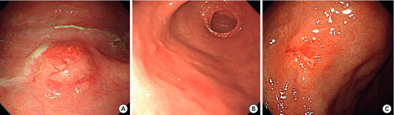
metastases. The longest diameter of the resected tumor specimen was used to define the tumor size. The tumor location was classified according to location as upper, middle, or lower third of the stomach. Furthermore, the Paris classification was used to categorize the macroscopic type of early GCLS as follows: type 0–I (protruded) and type 0–IIa (superficial elevated) for the elevated type; type 0–IIb (flat) for the flat type; and type 0–IIc (superficial depressed) and type 0–III (excavated) for the depressed type (Fig. 1) [12]. The procedure time was defined as the time from circumferential marking of the lesion to the completion of hemostasis following its complete removal.
Histopathological examination and pathological definitions
The resected specimens were fixed in 10% formalin, sectioned serially at 2-mm intervals, and then subjected to histological mapping. GCLS was defined according to the 2010 World Health Organization classification as a lesion of sharply demarcated advancing margins composed of irregular nests or sheets of polygonal tumor cells associated with a prominent lymphoid infiltrate in a non-desmoplastic stroma [13]. We estimated the depth of tumor invasion, lymphovascular and perineural invasion, and tumor involvement of the lateral and vertical margins according to the Japanese Gastric Cancer Association classification [14]. The presence of EBV in cancer cells was assessed via EBV-encoded RNA chromogenic in situ hybridization.
Helicobacter pylori infection was defined as a positive test result when the rapid urease test, urea breath test, or Giemsa stain of pathological specimens displayed a positive result.
A one-piece resection was defined as an en bloc resection, while a complete resection was defined when the microscopic findings of the specimens that achieved en bloc resection exhibited no neoplastic components at the lateral or vertical margins and presented no evidence of lymphovascular or perineural invasion. Incomplete resection was defined when the tumor was resected in multiple fragments, with resected margins positive for cancer, lymphovascular, or perineural invasion. In a case with submucosal invasion, an invasion of
<500 μm was classified as SM1 and an invasion of >500 μm as SM2. Patients with multiple tumors were staged according to the deepest-penetrating tumor.
Follow-up
Endoscopic follow-up examinations were performed routinely at 3, 6, and 12 months and then annually after endoscopic resection to assess the completeness of resection and
A B C
Fig. 1. Macroscopic type lesions of early GCLS. (A) A 10-mm subepithelial tumor-like lesion on the high body (elevated type). (B) A 14-mm hyperemic flat lesion on the antrum (flat type). (C) A 12-mm shallow ulcerative lesion on the angle (depressed type).
GCLS = gastric carcinoma with lymphoid stroma.
to detect local recurrence and metachronous lesions. In addition, abdominal CT scans and chest radiography were performed at 6, 12, and 18 months and then annually after endoscopic resection to assess distant metastasis. During the follow-up period, biopsy samples were obtained from the endoscopic resection ulcer scar or other suspicious mucosal abnormalities. Tumors detected at the resection site during the first or second follow- up endoscopy within 12 months after the resection were defined as residual tumors and considered as an incomplete resection. Local recurrence was defined as recurrent cancer at the resection site after 12 months. Tumors detected at sites other than the primary resection site during follow-up endoscopy were considered metachronous recurrences.
RESULTS
Clinicopathological features and pretreatment diagnosis of early GCLS Table 1 summarizes the clinicopathological features and pretreatment diagnosis of 40 patients with early GCLS who underwent endoscopic resection. The mean age of patients was 56.9 (range, 37–83) years, with 90.0% male predominance. Fourteen (35.0%) patients had intramucosal cancers and 26 (65.0%) presented submucosal invasion. Early GCLS predominantly presented as the depressed macroscopic type (55.0%) and tended to be proximal to tumor locations. The macroscopic type lesion did not present any significant distinction according to the depth of tumor (Table 2). None of the lesions demonstrated lymphovascular or perineural invasion despite the high submucosal tumor invasion.
Among the 40 patients analyzed, EBV and H. pylori infection rates were 90.0% and 79.5%, respectively. The co-infection rate of EBV and H. pylori was 67.5%.
In the pretreatment diagnosis, only 4 (10.0%) patients were suspected or diagnosed with GCLS (Table 1). Among the 40 patients analyzed, the initial endoscopic biopsy of 24 (60.0%) patients revealed differentiated adenocarcinoma or dysplasia. In addition, 8 (20.0%) patients were diagnosed with undifferentiated adenocarcinomas before endoscopic resection and met the expanded criteria in the pretreatment examination [10].
Clinical outcomes of endoscopic resection for early GCLS
Forty GCLS lesions in 40 patients were treated using endoscopic resection. The clinical results of endoscopic resection are shown in Table 3. En bloc resection (97.5%) was achieved in all lesions except one. The mean procedure time was 37.8 (range, 10–157) minutes. Six lesions microscopically revealed tumor involvement of the resection margin: 2 patients with a positive lateral margin, 3 patients with a positive vertical margin, and 1 patient with a positive lateral and vertical margin. The complete resection rate was 85.0% (34 of 40 lesions). Severe complications, such as massive bleeding or perforation, did not develop in our study group.
Clinical courses after endoscopic resection for early GCLS
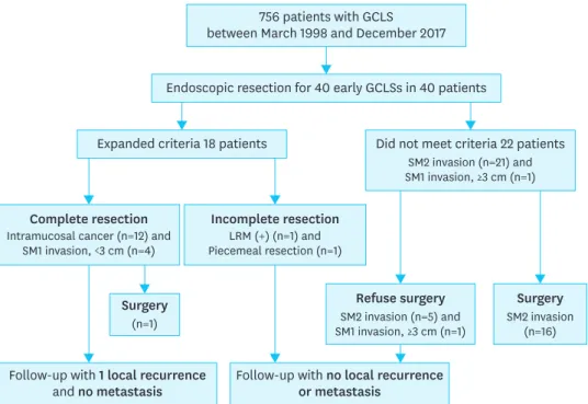
Fig. 2 illustrates the clinical courses of 40 patients with early GCLS who underwent endoscopic resection. Among the 40 patients, 18 met the expanded criteria and 22 did not.
After endoscopic resection, 17 patients were referred for surgery and underwent gastrectomy with LN dissection; 16 patients did not meet the expanded criteria (SM2 invasion), and 1 patient was misclassified as undifferentiated EGC (intramucosal cancer of 3.8 cm in size). All patients who underwent additional surgical resection showed no evidence of LN metastasis on pathological results.
Table 1. Clinicopathological characteristics and pretreatment diagnosis of patients with early GCLS
Factor Value
Age (yr) 56.9 (37–83)
Sex (male) 36 (90.0)
Macroscopic type
Elevated 12 (30.0)
Flat 6 (15.0)
Depressed 22 (55.0)
Location of tumor
Upper third 12 (30.0)
Middle third 20 (50.0)
Lower third 8 (20.0)
Tumor size (cm) 2.2 (0.6–4.8)
Depth of tumor invasion
Mucosal invasion 14 (35.0)
SM1 invasion 5 (12.5)
SM2 invasion 21 (52.5)
Lymphovascular invasion 0 (0)
Perineural invasion 0 (0)
EBV infection 36 (90.0)
H. pylori infection 31 (79.5)
EBV and H. pylori co-infection 27 (67.5)
Pretreatment diagnosis
GCLS 4 (10.0)
Differentiated adenocarcinoma or dysplasia 24 (60.0)
Undifferentiated adenocarcinoma 8 (20.0)
Atypical glands or gastritis 4 (10.0)
Values are presented as median (interquartile range) or number (%).
GCLS = gastric carcinoma with lymphoid stroma; SM1 = penetration <500 μm of the submucosal layer; SM2 = penetration >500 μm of the submucosal layer; EBV = Epstein-Barr virus.
Table 2. Macroscopic type of early GCLS according to the depth of tumor invasion
Macroscopic type Depth of tumor invasion
Mucosal invasion SM1 invasion SM2 invasion
Elevated 3 (21.4) 2 (40.0) 7 (33.3)
Flat 2 (14.3) 1 (20.0) 3 (14.3)
Depressed 9 (64.3) 2 (40.0) 11 (52.4)
Total 14 (100) 5 (100) 21 (100)
Values are presented as number (%).
GCLS = gastric carcinoma with lymphoid stroma; SM1 = penetration <500 μm of the submucosal layer; SM2 = penetration >500 μm of the submucosal layer.
Table 3. Endoscopic and pathologic outcomes of 40 lesions from 40 patients with early GCLS who underwent endoscopic resection
Factor Value
En bloc resection 39 (97.5)
Procedure time (min) 37.8 (10–157)
Positive resection margins
Lateral margin 3 (7.5)
Vertical margin 4 (10.0)
Complete resection 34 (85.0)
Complications
Massive bleeding 0 (0)
Perforation 0 (0)
Values are presented as median (interquartile range) or number (%).
GCLS = gastric carcinoma with lymphoid stroma.
Among the 40 patients, 23 (57.5%) were followed up without additional surgical resection (Fig. 2). The mean follow-up period for the 23 patients was 49.7 (range, 5–120) months.
During the 7-month observation period, 1 metachronous recurrence developed, and endoscopic resection was performed. After the endoscopic resection, the patient was diagnosed with poorly differentiated EGC and underwent additional surgical resection.
The surgical pathological results did not reveal LN metastasis. In the other 22 patients, no evidence of regional LN enlargement or distant metastasis suggesting recurrence, or gastric cancer-related deaths occurred during the follow-up period (mean follow-up duration, 51.6 months; range, 5–120 months).
DISCUSSION
Surgery is the conventional and most specific loco-regional treatment for gastric cancer, but it is associated with significant perioperative complications. Previous studies have reported postoperative complication rates of 17%–30% for open or laparoscopic gastrectomy and major morbidity rates of 5%–10% [15,16]. Moreover, patients who underwent gastrectomy demonstrated a significantly worse quality of life, including impaired social functioning and eating restrictions, even 5 years postoperatively [17]. Endoscopic resection has been widely accepted as a local curative treatment for EGC with a low likelihood of LN metastasis [9,14]. Endoscopic resection achieved similar efficacy and had many advantages compared to surgery for the treatment of EGC [9,14]. However, curative endoscopic resection of EGC has been performed only in selected cases depending on the differentiation, size, and depth of invasion of the lesions [10]. Previously, several studies have demonstrated that early GCLS has a lower risk of LN metastasis than other subtypes of gastric cancer and could be a therapeutic indication for endoscopic resection [6-8]. However, to date, only a few reports have investigated
756 patients with GCLS between March 1998 and December 2017 Endoscopic resection for 40 early GCLSs in 40 patients Expanded criteria 18 patients
Complete resection Intramucosal cancer (n=12) and
SM1 invasion, <3 cm (n=4)
Follow-up with 1 local recurrence
and no metastasis Follow-up with no local recurrence or metastasis
Incomplete resection LRM (+) (n=1) and Piecemeal resection (n=1)
Refuse surgery SM2 invasion (n=5) and SM1 invasion, ≥3 cm (n=1)
Surgery SM2 invasion
(n=16) Surgery
(n=1)
Did not meet criteria 22 patients SM2 invasion (n=21) and SM1 invasion, ≥3 cm (n=1)
Fig. 2. Clinical courses of patients with early GCLS treated with endoscopic submucosal dissection.
GCLS = gastric carcinoma with lymphoid stroma; LRM = lateral resection margin; SM1 = penetration <500 μm of the submucosal layer; SM2 = penetration >500 μm of the submucosal layer.
the clinical outcomes of endoscopic resection in patients with early GCLS [18-20]. This limited series of patients showed favorable clinical outcomes [18-20]. Our study examined the clinical outcomes and courses of 40 patients with early GCLS after endoscopic resection.
In this study, we achieved en bloc resection with endoscopic resection in 97.5% of 40 patients with early GCLS. In addition, no severe complications requiring open surgery, such as perforation or massive bleeding, were reported. The complete resection rate with endoscopic resection was 85.0%, which was similar to the complete resection rate for differentiated EGCs in previous studies [11,14]. Furthermore, this result was higher than that for undifferentiated EGCs that ranged from 60% to >80% in recent studies [21-23]. Advances in endoscopic techniques and devices have increased the immediate clinical outcomes of patients with early GCLS. Our results demonstrated the technical feasibility of endoscopic resection in patients with early GCLS despite its high proximal location and submucosal tumor invasion.
Previous large-scale studies have reported the long-term survival outcomes of endoscopic resection treatment for differentiated EGCs that meet the expanded indications [11,24,25].
Patients with differentiated EGC who underwent endoscopic resection demonstrated similar and excellent long-term survival rates compared with patients who underwent surgical resection, although the endoscopic resection group showed more local or metachronous recurrences [24,25]. In this study, during a mean follow-up period of 49.7 months for 23 patients who did not undergo additional surgical resection, no gastric cancer-related deaths were reported. In addition, none of the patients were diagnosed with regional LN enlargement or distant metastasis during the follow-up period, although 1 patient developed metachronous recurrence (poorly differentiated adenocarcinoma, follow-up duration: 7 months). This result suggested that endoscopic resection for patients with early GCLS who met the expanded criteria has favorable clinical outcomes, and endoscopic resection might represent an alternative treatment method for patients with early GCLS.
Notably, precise pretreatment histological diagnosis and estimation of tumor depth are crucial for formulating a treatment plan for patients with EGC. However, it was challenging to predict the histology and depth of tumor invasion accurately using pretreatment diagnostic modalities in patients with early GCLS [26,27]. In this study, only 4 (10.0%) of 40 patients were diagnosed with GCLS based on the pretreatment histology. This discrepancy in the histological diagnosis was likely caused by the difficulty in rendering a pathological evaluation based on a small sample from the endoscopic biopsy specimen [27].
In addition, unfamiliarity with GCLS owing to its rarity affected the low rate of pretreatment histological diagnosis. With regard to tumor depth, the histological examination of the resected specimens demonstrated a greater depth of tumor invasion than expected based on the initial endoscopic or endoscopic ultrasound (EUS) examination in many cases (data not shown). This corroborates with the findings of a previous study that postulated that an undifferentiated EGC had a significantly higher probability of being understaged by EUS [28].
Further studies investigating the histological features of endoscopic biopsy specimens and EUS findings are warranted.
This study has some limitations. First, the analysis had a retrospective, nonrandomized design. Second, the sample size was small and the follow-up duration after the endoscopic resection in some patients was relatively short. Therefore, there was a possibility of a highly selected endoscopic resection group in this study. Third, LN metastasis was not confirmed in
nonsurgical cases. However, this is a challenging limitation to overcome because of the study design. Fourth, except for 4 cases that were suspected of or diagnosed with GCLS in pretreatment diagnosis, we did not perform endoscopic resection in anticipation of GCLS diagnosis.
In conclusion, despite some limitations to the application of endoscopic resection in early GCLS, including the fact that lesions are often accompanied by a proximal location and most GCLSs invade into the submucosa, making it difficult to estimate the depth of tumor invasion, our results suggest that endoscopic resection might represent an alternative treatment option for early GCLS patients presenting unsuitable conditions for surgery.
REFERENCES
1. Watanabe H, Enjoji M, Imai T. Gastric carcinoma with lymphoid stroma. Its morphologic characteristics and prognostic correlations. Cancer 1976;38:232-243.
PUBMED | CROSSREF
2. Corvalan A, Ding S, Koriyama C, Carrascal E, Carrasquilla G, Backhouse C, et al. Association of a distinctive strain of Epstein-Barr virus with gastric cancer. Int J Cancer 2006;118:1736-1742.
PUBMED | CROSSREF
3. Takada K. Epstein-Barr virus and gastric carcinoma. Mol Pathol 2000;53:255-261.
PUBMED | CROSSREF
4. Lim H, Park YS, Lee JH, Son DH, Ahn JY, Choi KS, et al. Features of gastric carcinoma with lymphoid stroma associated with Epstein-Barr virus. Clin Gastroenterol Hepatol 2015;13:1738-1744.e2.
PUBMED | CROSSREF
5. Song HJ, Srivastava A, Lee J, Kim YS, Kim KM, Ki Kang W, et al. Host inflammatory response predicts survival of patients with Epstein-Barr virus-associated gastric carcinoma. Gastroenterology 2010;139:84- 92.e2.
PUBMED | CROSSREF
6. Lim H, Lee IS, Lee JH, Park YS, Kang HJ, Na HK, et al. Clinical application of early gastric carcinoma with lymphoid stroma based on lymph node metastasis status. Gastric Cancer 2017;20:793-801.
PUBMED | CROSSREF
7. Shin DH, Kim GH, Lee BE, Lee JW, Ha DW, Jeon HK, et al. Clinicopathologic features of early gastric carcinoma with lymphoid stroma and feasibility of endoscopic submucosal dissection. Surg Endosc 2017;31:4156-4164.
PUBMED | CROSSREF
8. Huh CW, Jung DH, Kim H, Kim H, Youn YH, Park H, et al. Clinicopathologic features of gastric carcinoma with lymphoid stroma in early gastric cancer. J Surg Oncol 2016;114:769-772.
PUBMED | CROSSREF
9. Lee JH, Kim JG, Jung HK, Kim JH, Jeong WK, Jeon TJ, et al. Clinical practice guidelines for gastric cancer in Korea: an evidence-based approach. J Gastric Cancer 2014;14:87-104.
PUBMED | CROSSREF
10. Gotoda T, Yanagisawa A, Sasako M, Ono H, Nakanishi Y, Shimoda T, et al. Incidence of lymph node metastasis from early gastric cancer: estimation with a large number of cases at two large centers. Gastric Cancer 2000;3:219-225.
PUBMED | CROSSREF
11. Ahn JY, Jung HY, Choi KD, Choi JY, Kim MY, Lee JH, et al. Endoscopic and oncologic outcomes after endoscopic resection for early gastric cancer: 1370 cases of absolute and extended indications.
Gastrointest Endosc 2011;74:485-493.
PUBMED | CROSSREF
12. The Paris endoscopic classification of superficial neoplastic lesions: esophagus, stomach, and colon:
November 30 to December 1, 2002. Gastrointest Endosc 2003;58 Suppl:S3-S43.
PUBMED | CROSSREF
13. Lauwers G, Carneiro F, Graham D. Gastric carcinoma. In: Bowman FT, Carneiro F, Hruban RH, Theise ND, eds. WHO Classification of Tumours of the Digestive System. 4th ed. Lyon: IARC, 2010.
14. Japanese Gastric Cancer Association. Japanese classification of gastric carcinoma: 3rd English edition.
Gastric Cancer 2011;14:101-112.
PUBMED | CROSSREF
15. Jeong O, Jung MR, Kim GY, Kim HS, Ryu SY, Park YK. Comparison of short-term surgical outcomes between laparoscopic and open total gastrectomy for gastric carcinoma: case-control study using propensity score matching method. J Am Coll Surg 2013;216:184-191.
PUBMED | CROSSREF
16. Eom BW, Kim YW, Lee SE, Ryu KW, Lee JH, Yoon HM, et al. Survival and surgical outcomes after laparoscopy-assisted total gastrectomy for gastric cancer: case-control study. Surg Endosc 2012;26:3273-3281.
PUBMED | CROSSREF
17. Lee SS, Chung HY, Kwon OK, Yu W. Long-term quality of life after distal subtotal and total gastrectomy:
symptom- and behavior-oriented consequences. Ann Surg 2016;263:738-744.
PUBMED | CROSSREF
18. Lee JY, Kim KM, Min BH, Lee JH, Rhee PL, Kim JJ. Epstein-Barr virus-associated lymphoepithelioma- like early gastric carcinomas and endoscopic submucosal dissection: case series. World J Gastroenterol 2014;20:1365-1370.
PUBMED | CROSSREF
19. Moon HS, Kang SH, Seong JK, Jeong HY, Song KS. Lymphoepithelioma-like gastric carcinoma resected by endoscopic submucosal dissection (ESD). Endoscopy 2010;42 Suppl 2:E73-E74.
PUBMED | CROSSREF
20. Gromski MA, Miller CA, Lee SH, Lee TH, Chung IK, Park SH, et al. Gastric lymphoepithelioma-like carcinoma mimicking a subepithelial lesion treated by endoscopic submucosal dissection. Gastrointest Endosc 2012;76:419-421.
PUBMED | CROSSREF
21. Kang HY, Kim SG, Kim JS, Jung HC, Song IS. Clinical outcomes of endoscopic submucosal dissection for undifferentiated early gastric cancer. Surg Endosc 2010;24:509-516.
PUBMED | CROSSREF
22. Ahn JY, Park HJ, Park YS, Lee JH, Choi KS, Jeong KW, et al. Endoscopic resection for undifferentiated-type early gastric cancer: immediate endoscopic outcomes and long-term survivals. Dig Dis Sci 2016;61:1158-1164.
PUBMED | CROSSREF
23. Bang CS, Baik GH, Shin IS, Kim JB, Suk KT, Yoon JH, et al. Endoscopic submucosal dissection for early gastric cancer with undifferentiated-type histology: a meta-analysis. World J Gastroenterol 2015;21:6032-6043.
PUBMED | CROSSREF
24. Pyo JH, Lee H, Min BH, Lee JH, Choi MG, Lee JH, et al. Long-term outcome of endoscopic resection vs.
surgery for early gastric cancer: a non-inferiority-matched cohort study. Am J Gastroenterol 2016;111:240-249.
PUBMED | CROSSREF
25. Choi KS, Jung HY, Choi KD, Lee GH, Song HJ, Kim DH, et al. EMR versus gastrectomy for intramucosal gastric cancer: comparison of long-term outcomes. Gastrointest Endosc 2011;73:942-948.
PUBMED | CROSSREF
26. Mocellin S, Marchet A, Nitti D. EUS for the staging of gastric cancer: a meta-analysis. Gastrointest Endosc 2011;73:1122-1134.
PUBMED | CROSSREF
27. Lim H, Jung HY, Park YS, Na HK, Ahn JY, Choi JY, et al. Discrepancy between endoscopic forceps biopsy and endoscopic resection in gastric epithelial neoplasia. Surg Endosc 2014;28:1256-1262.
PUBMED | CROSSREF
28. Kim JH, Song KS, Youn YH, Lee YC, Cheon JH, Song SY, et al. Clinicopathologic factors influence accurate endosonographic assessment for early gastric cancer. Gastrointest Endosc 2007;66:901-908.
PUBMED | CROSSREF