Copyrights © 2016 by The Korean Gastric Cancer Association www.jgc-online.org This is an open-access article distributed under the terms of the Creative Commons Attribution Non-Commercial License (http://creativecommons.org/
licenses/by-nc/4.0) which permits unrestricted noncommercial use, distribution, and reproduction in any medium, provided the original work is properly cited.
Introduction
Endoscopic submucosal dissection (ESD) is accepted as the primary treatment for early gastric cancer (EGC) lesions that meet the absolute indications for resection and can be considered
as an investigational treatment for lesions that meet the expanded indications and have a negligible risk of lymph node metasta- sis.1,2 After ESD for EGC lesions that meet the final pathological curability criteria for ESD indications, long-term outcomes are favorable and comparable between both criteria for absolute and expanded indications. The 5-year overall survival (OS) rate is 92% to 97% for the patients with absolute indications and 93% to 97% for the patients with expanded indication.3-5 Furthermore, the OS after endoscopic resection (ER) was comparable with that after surgery in patients with EGC lesions that met the curability criteria for absolute6 and expanded indications7 in the final patho- Correspondence to: Il Ju Choi
Center for Gastric Cancer, National Cancer Center, 323 Ilsan-ro, Ilsandong-gu, Goyang 10408, Korea
Tel: +82-31-920-1629, Fax: +82-31-920-0069 E-mail: cij1224@hanmail.net
Received February 13, 2016 Revised March 18, 2016 Accepted March 21, 2016
Discrepancy between Clinical and Final Pathological Evaluation Findings in Early Gastric Cancer Patients Treated
with Endoscopic Submucosal Dissection
Young-Il Kim, Hyoung Sang Kim, Myeong-Cherl Kook, Soo-Jeong Cho, Jong Yeul Lee, Chan Gyoo Kim, Keun Won Ryu, Young-Woo Kim, and Il Ju Choi
Center for Gastric Cancer, National Cancer Center, Goyang, Korea
Purpose: Early gastric cancer cases that are estimated to meet indications for treatment before endoscopic submucosal resection are of- ten revealed to be out-of-indication after the treatment. We investigated the short-term treatment outcomes in patients with early gastric cancer according to the pretreatment clinical endoscopic submucosal resection indications.
Materials and Methods: We retrospectively reviewed the medical records of patients with early gastric cancer that met the pretreatment endoscopic submucosal resection indications, from 2004 to 2011. Curative resection rate and proportion of out-of-indication cases were compared according to the pre-endoscopic submucosal resection indications. Pre-endoscopic submucosal resection factors associated with out-of-indication in the final pathological examination were analyzed.
Results: Of 756 cases, 660 had absolute and 96 had expanded pre-endoscopic submucosal resection indications. The curative resec- tion rate was significantly lower in the patients with expanded indications (64.6%) than in those with absolute indications (81.7%;
P<0.001). The cases with expanded indications (30.2%) were revealed to be out-of-indication more frequently than the cases with absolute indications (13.8%; P<0.001). Age of >65 years, tumor size of >2 cm, tumor location in the upper-third segment of the stomach, and undifferentiated histological type in pre-endoscopic submucosal resection evaluations were significant risk factors for out- of-indication after endoscopic submucosal resection.
Conclusions: Non-curative resection due to out-of-indication occurred in approximately one-third of the early gastric cancer cases that clinically met the expanded indications before endoscopic submucosal resection. The possibility of additional surgery should be empha- sized for patients with early gastric cancers that clinically meet the expanded indications.
Key Words: Endoscopic submucosal dissection; Early gastric cancer; Criteria
logical evaluation. However, these excellent outcomes are based on the post-ESD pathological findings.
ESD indications are determined based on several factors, in- cluding tumor size, histological type, depth of invasion, and the presence of an ulcer.2 However, the current clinical evaluation before ESD has limitations in accurately estimating these fac- tors.8 Owing to the inevitable discrepancies between pretreatment estimation and posttreatment pathological findings, the noncura- tive resection rates for patients who did not meet the pathologi- cal curability criteria for ESD indications have been reported to be between 11% and 21%.3,9-11 An additional surgery is needed for these patients because of the risk of lymph node metastasis.
Recent studies showed that survival was compromised in patients who did not undergo additional surgery after noncurative resec- tion.12,13
In the present study, we investigated discrepancies between pre-ESD clinical indications and post-ESD pathological findings in patients who underwent ESD for EGC lesions that met either the absolute or expanded indications in pre-ESD evaluations.
Materials and Methods
1. Study population
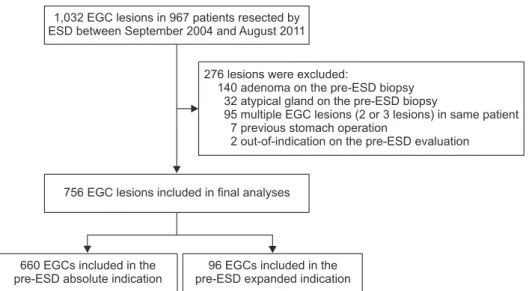
In this retrospective study, we reviewed the medical records of 967 consecutive patients who underwent ESD for 1,032 EGC lesions between September 2004 and August 2011 at the National Cancer Center, Korea. Of these EGC lesions, 276 were excluded because of the following: 1) were not adenocarcinomas as deter- mined after a diagnostic endoscopic biopsy (adenoma lesions and atypical gland), 2) were diagnosed at the remnant stomach after subtotal gastrectomy, 3) were out-of-indication per the diagnostic evaluations, or 4) were multiple in same patients.
The baseline demographic characteristics; pre-ESD diagnostic findings, including endoscopy and pathological results; and final pathological evaluation results after ESD were obtained from the prospectively collected database. This study was approved by the Institutional Review Board (IRB) of the National Cancer Center, Korea (NCC2015-0059). Informed consent was waived for all the patients because the IRB assessed this study as low-risk.
2. Definition of pre-endoscopic submucosal dissection (ESD) indications and pathological curability criteria for post-ESD indications Based on pre-ESD evaluations and final pathological evalua-
tion results after ESD, ESD indications were divided into the ab- solute and expanded indications according to the Japanese Gastric Cancer Treatment Guidelines.2 The pre-ESD depth of tumor invasion was clinically determined by using conventional white light endoscopy (WLE), abdominal computed tomography (CT), and/or endoscopic ultrasonography (EUS).
Pre-ESD absolute indication was defined as a differentiated tumor measuring ≤2 cm that has no ulcer and is confined to the mucosal layer.2 The pre-ESD expanded indications included the following types of tumors confined to the mucosal layer: 1) a differentiated tumor measuring >2 cm without an ulcer, 2) a differentiated tumor measuring ≤3 cm with an ulcer, and 3) an undifferentiated tumor measuring ≤2 cm without an ulcer.2 The presence of ulcer was defined when a definite visible ulcer was detected on the tumor upon endoscopic examination.
The final pathological curability criteria for absolute indications (post-ESD absolute criteria) and expanded indications (post- ESD expanded criteria) were defined as follows: The post-ESD absolute criteria were the same as the pre-ESD absolute indica- tion criteria for the lesions without lymphovascular invasion. The post-ESD expanded criteria included the 3 aforementioned pre- ESD expanded indication criteria plus the criterion of a differen- tiated histological type with minute submucosal invasion (<500 μm) and a tumor size of ≤3 cm. All lesions that meet the post- ESD expanded criteria should have no lymphovascular invasion.2 Tumors out-of-indication were defined as tumors that did not meet the expanded criteria in the final pathological evaluations.
3. Pathological evaluation
The ESD procedures, described in a previous study,14 were performed by 4 experienced gastroenterologists. All the endos- copists were certified specialty board members of the Korean Society of Gastrointestinal Endoscopy and have performed more than 5,000 diagnostic endoscopic procedures. Resected ESD specimens were fixed in 10% formalin and then embedded in a paraffin block, which was serially sliced at 2-mm intervals. The paraffin slices were then stained with hematoxylin and eosin, and a single specialized pathologist (MC Kook) performed the patho- logical evaluation. The World Health Organization classification of gastric cancer was used for determination of tumor histological subtypes.15 Subsequently, well-differentiated and moderately dif- ferentiated tubular adenocarcinomas, as well as papillary adeno- carcinoma, were included in the differentiated histological type.
By contrast, poorly differentiated tubular adenocarcinoma, signet
ring cell carcinoma, and mucinous adenocarcinoma were included in the undifferentiated histological type.2 Tumor histological types were determined according to the major component that consti- tuted >50% of the tumor in cases of mixed histological types.2,15
4. Definitions of endoscopic submucosal dissection outcomes
En bloc resection was defined as removal of the tumor in one piece without fragmentation. Complete resection was defined as removal of the tumor using en bloc resection, with negative hori- zontal and vertical tumor resection margins.16 Curative resection was achieved when tumors were completely resected and final pathological evaluation results met the curability criteria for abso- lute or expanded indications of ER.2
5. Statistical analyses
Data were compared between the pre-ESD absolute and ex- panded indication groups by using the chi-square or Fisher exact test for categorical variables and the Student t-test or Mann- Whitney U-test for continuous variables. Univariate and multi- variate logistic regression analyses were performed to investigate pre-ESD risk factors associated with out-of-indication that were identified upon final pathological evaluation after ESD. The covariates for the multivariate logistic regression analysis were pre-ESD variables that showed a statistical significance in the univariate analyses. P-values <0.05 were considered statistically significant. All data were analyzed by using Stata 12.1 (Stata, Col- lege Station, TX, USA).
Results
1. Baseline characteristics
Of the 967 EGC lesions, 756 were included in the final analy- ses (Fig. 1). These tumors were further classified into the pre- ESD absolute and expanded indication groups on the basis of diagnostic evaluations. EGC lesions met the pre-ESD absolute indications in 660 cases (87.3%) and the pre-ESD expanded in- dications in 96 cases (12.7%).
The median age of all the included patients was 64 years, and the proportion of male patients was 78.0%. Compared with the patients in the pre-ESD absolute indication group, those in the pre-ESD expanded indication group were significantly older (median age, 63 vs. 67 years; P<0.001) and had a larger mean tumor size (1.24 vs. 2.24 cm; P<0.001; Table 1). No significant differences in sex, comorbid disease, tumor type, and tumor lo- cation were observed between the two groups.
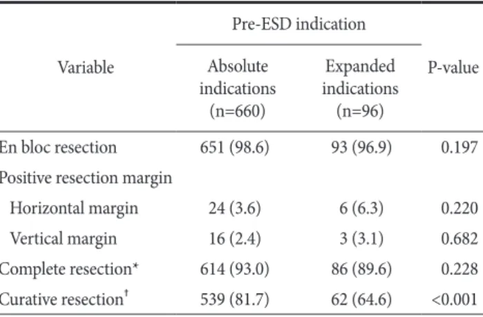
2. Short-term outcomes of endoscopic submucosal dissection (ESD) according to pre-ESD indication The en bloc resection rate in all the included lesions was 98.4%, and the rates did not different between the pre-ESD ab- solute and expanded indication groups (98.6% vs. 96.9%, respec- tively; P=0.197). The final pathological evaluation results of the resected specimens indicated no significant difference (P=0.228) in the complete resection rate between the pre-ESD absolute indication group (93.0%) and the pre-ESD expanded indication group (89.6%). However, the curative resection rate was signifi- cantly lower in the lesions with the pre-ESD expanded indica- tions than in those with the pre-ESD absolute indications (64.6%
1,032 EGC lesions in 967 patients resected by ESD between September 2004 and August 2011
276 lesions were excluded:
140 adenoma on the pre-ESD biopsy 32 atypical gland on the pre-ESD biopsy
95 multiple EGC lesions (2 or 3 lesions) in same patient 7 previous stomach operation
2 out-of-indication on the pre-ESD evaluation
660 EGCs included in the pre-ESD absolute indication
96 EGCs included in the pre-ESD expanded indication 756 EGC lesions included in final analyses
Fig. 1. Flowchart of the study. EGC = early gastric cancer; ESD = endoscop- ic submucosal dissection.
vs. 81.7%, respectively; P<0.001; Table 2). 3. Discrepancies between pre-endoscopic submucosal dissection (ESD) and post-ESD criteria
The discrepancies between the pre-ESD indications and post- ESD criteria are shown in Table 3. Overall, the final pathological evaluation results indicated that 15.9% of tumors met the out-of- indication criteria. Of the tumors in the pre-ESD absolute indi- cation group, only 61.7% were correctly estimated according to the post-ESD pathological evaluation results, and the remaining 38.3% of the lesions were upgraded to either expanded indication (24.5%) or out-of-indication (13.8%). Of the tumors in the pre- ESD expanded indication group, only 52.1% were correctly esti- mated, and 30.2% met the out-of-indication criteria in the post- ESD pathological evaluation. Tumors with a pre-ESD expanded indication had a significantly higher rate of post-ESD out-of- Table 1. Baseline demographic characteristics and endoscopic and
histological findings before ESD for EGC lesions Variable
Pre-ESD indication
P-value Absolute
indications (n=660)
Expanded indications (n=96)
Age (yr) 63 (29~83) 67 (36~87) <0.001
Sex 0.612
Male 517 (78.3) 73 (76.0)
Female 143 (21.7) 23 (24.0)
Comorbid disease 280 (42.4) 44 (45.8) 0.528
Hypertension 212 (32.1) 36 (37.5) 0.294
Diabetes mellitus 79 (12.0) 11 (11.5) 0.885 Cardiovascular disease 25 (3.8) 5 (5.2) 0.505 Chronic liver disease 32 (4.9) 4 (4.2) 0.769 Chronic lung disease 16 (2.4) 3 (3.1) 0.682
Tumor type 0.988
Elevated 89 (13.5) 13 (13.5)
Flat or depressed 571 (86.5) 83 (86.5)
Tumor size (cm) 1.24±0.43 2.24±0.88 <0.001
Tumor location 0.111
Lower one-third 508 (77.0) 76 (79.2) Middle one-third 94 (14.2) 17 (17.7)
Upper one-third 58 (8.8) 3 (3.1)
Values are presented as median (interquartile range), number (%), or mean±standard deviation. ESD = endoscopic submucosal dissection;
EGC = early gastric cancer.
Table 2. Short-term outcomes according to pre-ESD indications Variable
Pre-ESD indication
P-value Absolute
indications (n=660)
Expanded indications (n=96)
En bloc resection 651 (98.6) 93 (96.9) 0.197
Positive resection margin
Horizontal margin 24 (3.6) 6 (6.3) 0.220
Vertical margin 16 (2.4) 3 (3.1) 0.682
Complete resection* 614 (93.0) 86 (89.6) 0.228 Curative resection† 539 (81.7) 62 (64.6) <0.001 Values are presented as number (%). ESD = endoscopic submucosal dissection. *Defined when en bloc resection with a negative resection margin was achieved. †Defined when tumors were completely resected and final pathological results were met with absolute or expanded criteria for endoscopic resection.
Table 3. Discrepancy between the pre-ESD indications and the post-ESD criteria
Variable Total Post-ESD criteria
Absolute criteria Expanded criteria Out-of-indication Pre-ESD indication
Absolute indication 660 407 (61.7) 162 (24.5) 91 (13.8)
Expanded indication 96 17 (17.7) 50 (52.1) 29 (30.2)
I: M, differentiated, size >2 cm, UL (−) 62 12 (19.4) 33 (53.2) 17 (27.4)
II: M, differentiated, size ≤3 cm, UL (+) 19 2 (10.5) 13 (68.4) 4 (21.1)
III: M, undifferentiated, size ≤2 cm, UL (−) 15 3 (20.0) 4 (26.7) 8 (53.3)
Total 756 424 (56.1) 212 (28.0) 120 (15.9)
Values are presented as number only or number (%). ESD = endoscopic submucosal dissection; M = mucosa; UL = ulcer.
indication than those with a pre-ESD absolute indication (13.8%
vs. 30.2%, respectively; P<0.001). Among the three subgroups of pre-ESD expanded indications, the undifferentiated histological type subgroup had the highest rate of post-ESD out-of-indica- tion (53.3%).
After ESD, the presence of lymphovascular and deep submu- cosal tumor invasions (≥500 μm) were the main causes of the out-of-indication in both pre-ESD indication groups. Details of the post-ESD criteria, determined via pathological evaluations, are compared with the pre-ESD indications in Table 4.
4. Pre-endoscopic submucosal dissection (ESD) characteristics associated with the post-ESD out- of-indication
According to univariate analyses of the final pathological evaluation results, significant risk factors associated with out- of-indication include age of >65 years, tumor size of >2.0 cm, tumor in the upper-third segment of the stomach, and an undifferentiated histological type in the pre-ESD evaluations.
Multivariate analysis confirmed that age of >65 years (adjusted odd ratio [aOR], 1.84; P=0.004), tumor size of >2.0 cm (aOR, 2.51; P=0.003), tumor in the upper-third segment of the stomach (aOR, 4.68; P<0.001), and an undifferentiated tumor (aOR, 6.47;
P=0.001) were independent pre-ESD risk factors associated with out-of-indication (Table 5).
Discussion
EGC lesions that are estimated to meet the criteria for ESD indications are often found to be out-of-indication in post-ESD pathological evaluations. This situation results in the need for ad- ditional curative-intent surgery. In the present study, 15.9% of all patients who had met the pre-ESD indications for treatment in the diagnostic evaluations were identified as out-of-indication according to the post-ESD pathology results. Out-of-indication rates determined via final pathological evaluations were 30.2% in tumors that met the pre-ESD expanded indications and 13.8% in those that met pre-ESD absolute indications (P<0.001). The out- of-indication rate after ESD was highest (53.3%) in the tumors that met the pre-ESD expanded indications of undifferentiated tumors measuring ≤2 cm and without an ulcer.
The ESD indication for EGC lesions were based on the neg- ligible risks of lymph node metastasis that were derived from lymph node risk analysis in a large number of cases of surgically resected specimens.17 Tumor characteristics, including size, his- tological type, depth of invasion, and presence of ulceration are Table 4. Details of the post-ESD criteria used in the final pathological examination according to pre-ESD indications
Variable Pre-ESD indications
P-value Absolute indications (n=660) Expanded indications (n=96)
Post-ESD criteria <0.001*
Absolute criteria 407 (61.7) 17 (17.7)
Expanded criteria 162 (24.6) 50 (52.1)
I: M, differentiated, size >2 cm, UL (−) 108 (16.4) 31 (32.3)
II: M, differentiated, size ≤3 cm, UL (+) 4 (0.6) 12 (12.5)
III: M, undifferentiated, size ≤2 cm, UL (−) 43 (6.5) 3 (3.1)
IV: SM1, differentiated, size ≤3 cm 7 (1.1) 4 (4.2)
Out-of-indication 91 (13.8) 29 (30.2)
LVI (+) 43 (6.5) 9 (9.4)
LVI (−) with other conditions
SM2 or more 41 (6.2) 13 (13.5)
SM1, size ≥3 cm 4 (0.6) 2 (2.1)
SM1, undifferentiated 1 (0.2) 0 (0)
Undifferentiated, size ≥2 cm 2 (0.3) 5 (5.2)
Values are presented as number (%). ESD = endoscopic submucosal dissection; M = mucosa; UL = ulcer; SM = submucosa; LVI = lymphovascular invasion. *P-value for distribution of post-ESD criteria between the pre-ESD absolute and expanded indications.
major factors to be considered in the estimation of lymph node metastasis risks.18 Clinical evaluations for determining the afore- mentioned factors before ESD include conventional WLE for tumor size estimation, ulcer findings, biopsy for tumor histology, and imaging studies (abdominal CT and/or EUS) for predicting depth of tumor invasion. However, current diagnostic modalities have limitations in accurate assessment, such as the underestima- tion of tumor size via conventional WLE19 and the limited ef- ficacy of CT and EUS in predicting depth of tumor invasion.11,20,21 Hence, discrepancies between clinical indications of ESD and the final criteria for curative resection seems inevitable.
Previous studies that compared outcomes of ESD according to the post-ESD criteria reported that the en bloc resection rate did
not differ between both post-ESD criteria, but complete resection rates were significantly lower in the expanded criteria than in the absolute criteria groups.3,9,10 In the present study, the en bloc and complete resection rates did not differ between both pre-ESD indication groups according to the pre-ESD evaluation results.
As the endoscopists decided to perform ESD only for EGC le- sions that met the ER indications in the pre-ESD evaluations, the complete resection rate did not differ between the expanded- and absolute indication groups in the present study. However, the curative resection rate was significantly lower in the pre-ESD expanded indication group than in the pre-ESD absolute indica- tion group (64.6% vs. 81.7%, respectively). In addition, a guideline states that ESD for EGC lesions that meet the expanded indica- Table 5. Risk factors associated with out-of-indication after ESD among variables of pre-ESD stage
Variable Total Univariate analysis* Multivariate analysis*
cOR 95% CI P-value aOR 95% CI P-value
Age (yr)
≤65 439 1.00 1.00
>65 317 1.87 1.27~2.78 0.002 1.84 1.22~2.77 0.004
Sex
Male 590 1.00
Female 166 1.10 0.69~1.75 0.692
Comorbidity
No 432 1.00
Yes 324 1.02 0.69~1.52 0.909
Tumor type
Elevated 102 1.00
Flat or depressed 654 1.48 0.78~2.80 0.225
Tumor size (cm)
≤2.0 692 1.00 1.00
>2.0 64 2.07 1.14~3.74 0.016 2.51 1.36~4.62 0.003
Tumor location
Lower one-third 584 1.00 1.00
Middle one-third 94 1.34 0.77~2.32 0.296 1.28 0.72~2.26 0.403
Upper one-third 61 3.93 2.22~6.94 <0.001 4.68 2.59~8.43 <0.001
Tumor histological type
Differentiated 741 1.00 1.00
Undifferentiated 15 6.42 2.28~18.05 <0.001 6.47 2.21~18.94 0.001
Ulceration
Absence 737 1.00
Presence 19 1.43 0.47~4.38 0.534
ESD = endoscopic submucosal dissection; cOR = crude odd ratio; CI = confidence interval; aOR = adjusted odd ratio. *Logistic regression analysis.
tion is defined as an investigational treatment.2 Therefore, caution should be exercised when choosing ESD as the primary treatment for patients with EGC lesions that meet the expanded indications in clinical evaluations, even though performing ESD for these le- sions seems technically feasible.
In our study, the highest rate of post-ESD out-of-indication was observed for tumors classified under the pre-ESD expanded indication group of the undifferentiated histologic type subgroup (53.3%). This result is similar to those in previous studies that reported low curative resection rates (55%~65%) in patients who underwent ER for undifferentiated lesions.22-24 Many possible causes of noncurative resection after ESD for undifferentiated lesions have been suggested. First, tumor sizes tend to be larger than those estimated before ESD. For example, undifferentiated lesions have been reported to have significantly larger size dis- crepancies between pre-ESD and post-ESD evaluations than the differentiated lesions.20,25 Second, the submucosal tumor invasion could play a role in noncurative resection. In patients who under- go ESD for undifferentiated lesions, final pathological evaluations revealed submucosal invasion rates of 20% to 28%.13,23,25 In ad- dition, previous studies reported significantly lower accuracies of pre-ESD prediction of depth of tumor invasion in lesions of the undifferentiated histologic type than in those of the differentiated histologic type.11 Magnifying endoscopy with narrow band imag- ing might be useful for accurate demarcation of undifferentiated EGC lesions.26 However, endoscopists should be careful in decid- ing whether to perform an ESD in patients with undifferentiated EGC.
In the present study, both lymphovascular and submuco- sal tumor invasions were the most common causes of out-of- indication. Of the components that comprised ER indications, diagnostic evaluations before ESD have limited roles in predict- ing the depth of tumor invasion and lymphovascular invasion.
In predicting the depth of tumor invasion, the reported accuracy rates of pre-ESD evaluations are low, ranging from 63% to 74%
as determined via conventional endoscopy11,27 and from 67% to 72% as determined via EUS.11,21,27,28
In addition, the presence of lymphovascular invasion is the single, most important risk fac- tor of lymph node metastasis in EGC patients. However, this risk factor cannot be evaluated or predicted with pre-ESD evaluation;
it can only be determined by performing a pathological evaluation of the resected specimen after ESD. These limitations of pre- ESD evaluations are the main reasons for the discrepancies be- tween pre-ESD indication and post-ESD criteria. Further studies
to investigate more-accurate methods for predicting the depth of tumor invasion and presence of lymphovascular invasion, if pos- sible, are needed.
Previous studies reported that large tumor size, tumor loca- tion in the upper-third segment of the stomach, submucosal tumor invasion, and ulcer findings are the factors associated with noncurative resection after ESD.29-31 Similarly, the multivariate analysis in our study revealed that a tumor size of >2 cm, tu- mor location in the upper-third segment of the stomach, and an undifferentiated histologic type in the pre-ESD evaluations were independent risk factors. These results suggest that before ESD is performed, patients who have the aforementioned pre-ESD risk factors should be fully informed of the possibility of out-of- indication in the final pathological evaluation results and the pos- sible need for additional surgical treatment.
An additional surgery is needed in patients with EGC le- sions that are judged as out-of-indication after ESD.2 Patients who underwent noncurative resection after ESD who did not receive an additional surgical treatment had significantly lower OS than those who underwent an additional surgery.12,13 Despite this knowledge, a considerable number (44%~68%) of patients who undergo noncurative resection after ESD do not undergo ad- ditional surgery.4,10,12,13 Thus, endoscopists should carefully select candidates for ESD for EGC lesions based on precise pre-ESD diagnostic evaluations and explain to the patient that the expand- ed indications of ESD is for an investigational treatment.
The strength of the present study is that it investigated the discrepancies between pre-ESD indication and post-ESD criteria in a large number of EGC cases. In addition, pre-ESD risk fac- tors of out-of-indication after ESD were analyzed. These results may be helpful for endoscopists to more precisely decide on ESD indications for EGC. The limitations of this study include the potential for selection bias caused by its retrospective design and single-center setting. Furthermore, not all patients underwent additional pre-ESD diagnostic evaluations by using narrow band imaging or EUS to estimate tumor size and depth of invasion.
Another limitation is that the results of the pre-ESD evaluation to identify lesion characteristics might have slightly differed between endoscopists.
In conclusion, noncurative resection because of out-of-indica- tion occurred in as many as one third of the cases of EGC lesions that clinically met the pre-ESD expanded indications. There- fore, the possibility of additional surgery should be emphasized to patients with these types of EGC lesions before they undergo
ESD treatment, and minimally invasive surgery rather than ESD should be considered as the primary treatment for undifferenti- ated EGCs.
Acknowledgments
This work was supported by a grant (No. NCC-1310280) from the National Cancer Center, Republic of Korea.
Conflicts of Interest
No potential conflict of interest relevant to this article was re- ported.
References
1. Lee JH, Kim JG, Jung HK, Kim JH, Jeong WK, Jeon TJ, et al.
Clinical practice guidelines for gastric cancer in Korea: an evidence-based approach. J Gastric Cancer 2014;14:87-104.
2. Japanese Gastric Cancer Association. Japanese gastric cancer treatment guidelines 2010 (ver. 3). Gastric Cancer 2011;14:113- 123.
3. Ahn JY, Jung HY, Choi KD, Choi JY, Kim MY, Lee JH, et al. En- doscopic and oncologic outcomes after endoscopic resection for early gastric cancer: 1370 cases of absolute and extended indications. Gastrointest Endosc 2011;74:485-493.
4. Isomoto H, Shikuwa S, Yamaguchi N, Fukuda E, Ikeda K, Nishiyama H, et al. Endoscopic submucosal dissection for early gastric cancer: a large-scale feasibility study. Gut 2009;58:331- 336.
5. Gotoda T, Iwasaki M, Kusano C, Seewald S, Oda I. Endoscopic resection of early gastric cancer treated by guideline and ex- panded National Cancer Centre criteria. Br J Surg 2010;97:868- 871.
6. Choi IJ, Lee JH, Kim YI, Kim CG, Cho SJ, Lee JY, et al. Long- term outcome comparison of endoscopic resection and sur- gery in early gastric cancer meeting the absolute indication for endoscopic resection. Gastrointest Endosc 2015;81:333-341.
7. Kim YI, Kim YW, Choi IJ, Kim CG, Lee JY, Cho SJ, et al. Long- term survival after endoscopic resection versus surgery in early gastric cancers. Endoscopy 2015;47:293-301.
8. Min YW, Lee JH. Endoscopic resection for early gastric cancer beyond absolute indication with emphasis on controversial is- sues. J Gastric Cancer 2014;14:7-14.
9. Park CH, Shin S, Park JC, Shin SK, Lee SK, Lee YC, et al. Long- term outcome of early gastric cancer after endoscopic submu- cosal dissection: expanded indication is comparable to absolute indication. Dig Liver Dis 2013;45:651-656.
10. Choi J, Kim SG, Im JP, Kim JS, Jung HC. Long-term clinical outcomes of endoscopic resection for early gastric cancer. Surg Endosc 2015;29:1223-1230.
11. Choi J, Kim SG, Im JP, Kim JS, Jung HC, Song IS. Comparison of endoscopic ultrasonography and conventional endoscopy for prediction of depth of tumor invasion in early gastric can- cer. Endoscopy 2010;42:705-713.
12. Kusano C, Iwasaki M, Kaltenbach T, Conlin A, Oda I, Gotoda T. Should elderly patients undergo additional surgery after non-curative endoscopic resection for early gastric cancer?
Long-term comparative outcomes. Am J Gastroenterol 2011;106:1064-1069.
13. Ohnita K, Isomoto H, Shikuwa S, Yajima H, Minami H, Mat- sushima K, et al. Early and long-term outcomes of endoscopic submucosal dissection for early gastric cancer in a large patient series. Exp Ther Med 2014;7:594-598.
14. Lee JY, Choi IJ, Cho SJ, Kim CG, Kook MC, Lee JH, et al. Rou- tine follow-up biopsies after complete endoscopic resection for early gastric cancer may be unnecessary. J Gastric Cancer 2012;12:88-98.
15. Hamilton SR, Lauri L. Pathology and genetics of tumours of the digestive system. World Health Organization Classification of Tumours. Lyon: IARC, 2000.
16. Lian J, Chen S, Zhang Y, Qiu F. A meta-analysis of endoscopic submucosal dissection and EMR for early gastric cancer. Gas- trointest Endosc 2012;76:763-770.
17. Gotoda T, Yanagisawa A, Sasako M, Ono H, Nakanishi Y, Shi- moda T, et al. Incidence of lymph node metastasis from early gastric cancer: estimation with a large number of cases at two large centers. Gastric Cancer 2000;3:219-225.
18. Kwee RM, Kwee TC. Predicting lymph node status in early gastric cancer. Gastric Cancer 2008;11:134-148.
19. Shim CN, Song MK, Kang DR, Chung HS, Park JC, Lee H, et al. Size discrepancy between endoscopic size and pathologic size is not negligible in endoscopic resection for early gastric cancer. Surg Endosc 2014;28:2199-2207.
20. Choi JI, Joo I, Lee JM. State-of-the-art preoperative staging of gastric cancer by MDCT and magnetic resonance imaging.
World J Gastroenterol 2014;20:4546-4557.
21. Kim JH, Song KS, Youn YH, Lee YC, Cheon JH, Song SY, et al.
Clinicopathologic factors influence accurate endosonographic assessment for early gastric cancer. Gastrointest Endosc 2007;66:901-908.
22. Oka S, Tanaka S, Higashiyama M, Numata N, Sanomura Y, Yo- shida S, et al. Clinical validity of the expanded criteria for en- doscopic resection of undifferentiated-type early gastric cancer based on long-term outcomes. Surg Endosc 2014;28:639-647.
23. Abe S, Oda I, Suzuki H, Nonaka S, Yoshinaga S, Odagaki T, et al. Short- and long-term outcomes of endoscopic submucosal dissection for undifferentiated early gastric cancer. Endoscopy 2013;45:703-707.
24. Kim JH, Kim YH, Jung da H, Jeon HH, Lee YC, Lee H, et al. Follow-up outcomes of endoscopic resection for early gastric cancer with undifferentiated histology. Surg Endosc 2014;28:2627-2633.
25. Kang HY, Kim SG, Kim JS, Jung HC, Song IS. Clinical out- comes of endoscopic submucosal dissection for undifferenti- ated early gastric cancer. Surg Endosc 2010;24:509-516.
26. Horiuchi Y, Fujisaki J, Yamamoto N, Shimizu T, Miyamoto Y, Tomida H, et al. Accuracy of diagnostic demarcation of undifferentiated-type early gastric cancers for magnifying en- doscopy with narrow-band imaging: endoscopic submucosal dissection cases. Gastric Cancer 2015. doi: 10. 1007/ s10120-
015-0568-y [In print].
27. Yanai H, Noguchi T, Mizumachi S, Tokiyama H, Nakamura H, Tada M, et al. A blind comparison of the effectiveness of endo- scopic ultrasonography and endoscopy in staging early gastric cancer. Gut 1999;44:361-365.
28. Lee JY, Choi IJ, Kim CG, Cho SJ, Kook MC, Ryu KW, et al.
Therapeutic decision-making using endoscopic ultrasonogra- phy in endoscopic treatment of early gastric cancer. Gut Liver 2016;10:42-50.
29. Ohnita K, Isomoto H, Yamaguchi N, Fukuda E, Nakamura T, Nishiyama H, et al. Factors related to the curability of early gastric cancer with endoscopic submucosal dissection. Surg Endosc 2009;23:2713-2719.
30. Hirasawa K, Kokawa A, Oka H, Yahara S, Sasaki T, Nozawa A, et al. Risk assessment chart for curability of early gastric cancer with endoscopic submucosal dissection. Gastrointest Endosc 2011;74:1268-1275.
31. Toyokawa T, Inaba T, Omote S, Okamoto A, Miyasaka R, Watanabe K, et al. Risk factors for non-curative resection of early gastric neoplasms with endoscopic submucosal dissec- tion: Analysis of 1,123 lesions. Exp Ther Med 2015;9:1209- 1214.