재발한 접형동염에 의한 시신경염 1예
메리놀병원 이비인후과
권민상·김정근·전 준·조중환
A Case of Optic Neuritis Caused by Recurrent Sphenoiditis
Min Sang Kwon, MD, Jeong Geun Kim, MD, Joon Jeon, MD and Joong Hwan Cho, MD Department of Otorhinolaryngology-Head and Neck Surgery, Maryknoll General Hospital, Busan, Korea -
--
- ABSTRACT ----
Optic neuritis is an inflammatory optic neuropathy, of which the multiple sclerosis is the most common cause.
Other causes include infection, tumor, granuloma, vascular disease and so on. A 61 year old male patient was admitted to our hospital with headache for 4 days and decreased visual acuity of left eye for 2 days. He took an operation for sinusitis 5 years ago at other hospital. His maximal corrected visual acuity was 0.1 at admis- sion. A full of soft tissue shadow in bilateral sphenoidal sinuses was identified on paranasal computer to-mo- graphic scan. We operated an endoscopic sinus surgery. His visual acuity was getting better, so the visual field analysis test and visual evoked potential test were improved. We report a successful treatment of optic neuritis from sphenoidal sinusitis with a review of literature. (J Clinical Otolaryngol 2005;16:311-315)
KEY WORDS:Optic neuritis·Sphenoid sinus·Recurrent.
서 론
시신경염은 염증, 감염, 탈수질과정(demyelination) 등 이 시신경을 침범하여 생기는 질환으로 다발성 경화증이 가장 흔한 원인이며 그 외 감염, 종양, 육아종, 혈관성 질 환 등이 원인이 될 수 있다.1) 시신경염은 해부학적 위치 에 따라 구후신경염(retrobulbar neuritis), 유두염(pa- pillitis), 시신경망막염(neuroretinitis) 등으로 분류되며 탈수질과 변성이 일어나 시력손실이 생기는 질환이다.2)
저자들은 진균성 부비동염이나 부비동내의 점액낭종
이 아닌 재발된 세균성 부비동염에 의해 시신경염이 유 발되었던 환자 1예를 내시경 부비동 수술 및 항생제, 스 테로이드 등을 사용하여 성공적으로 치료하였기에 문헌 고찰과 함께 보고하는 바이다.
증 례
61세 남자 환자가 4일 전부터의 두통, 2일 전부터 좌 안의 시력저하로 안과를 경유하여 내원하였다. 과거력상 환자는 5년 전 타병원에서 양측 부비동염으로 수술 받 은 병력이 있었고 그 후 간헐적으로 비폐색과 후비루가 있었으나 특별한 치료는 받지 않았다.
전비경 검사와 비내시경 검사상 양측에 구상돌기와 중비갑개가 부분적으로 제거되어 있었고 양측 중비도 및 접사함요에서 농성 분비물이 관찰되었다. 안과 검사상 교정시력 검사에서 우안 0.8, 좌안 0.1이었고 안저소견 논문접수일:2005년 09월 03일
심사완료일:2005년 10월 13일
교신저자:전 준, 600-730 부산광역시 중구 대청동 4가 12번지 메리놀병원 이비인후과
전화:(051) 461-2205・전송:(051) 461-0297 E-mail:[email protected]
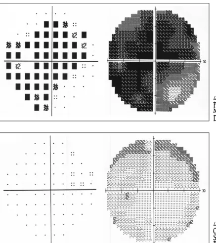
은 양측 모두 정상이었다. 시야검사상 좌안의 전반적 시 야협착을 보였고(Figs. 1 and 2), 시유발전위검사(visual evoked potential)상 좌안에서 p100 value가 관찰되지 않아(Fig. 3) 시력소실의 소견을 뒷받침 하였다. 부비동 전산화단층촬영상 양측 상악동내 점막의 비후 소견과 양측 사골동, 접형동내에 연조직 음영으로 가득 찬 소견 을 보였다(Fig. 4).
환자는 입원하여 항생제와 스테로이드를 투여하였으 나 시력 호전 없어 입원 4일째 전신마취하에 수술을 시 행하였다. 내시경을 이용하여 양측 구상돌기 절제술 및 사골동절제술을 시행한 후 접형동의 전벽을 제거하였으 며 농성 분비물이 관찰되어 이를 제거하였다(Fig. 5). 상 악동 자연개구부 확장술을 시행하고 상악동내 농성 분 비물을 제거한 후 수술을 마쳤다. 접형동내 분미물의 균 배양 검사상 gram-negative bacillus가 배양되었다.
술 후 항생제와 스테로이드를 투여하고 2일째 비강내 패킹을 제거하였다. 술 후 4일째 측정한 교정시력 검사 에서 좌안이 0.4로 호전되었고, 시야 검사 및 시유발전 위검사 소견도 호전되는 양상을 보였다. 술 후 5개월이 지난 현재 재발된 소견 및 시력저하는 관찰되지 않고 추적관찰 중이다.
고 찰
시신경염이란 시신경 수초의 염증이나 탈수초화로 인 한 시신경의 질환으로, 전형적인 시신경염의 경우 20대 에서 50대 사이에 호발하며, 여자에게 많고, 급성 단안 성의 시력감소를 주증상으로 하며, 많은 경우 다발성 경 화증과 동반되어 나타나거나 그 병력을 가지고 있다고 알려져 있다.3)
Fig. 1. Visual field analysis test (preo- perative state). The visual field was much limited due to optic nerve compression.
Fig. 2. Visual field analysis test (post- operative 2 weeks state). The limi- tation of the visual field was re- solved.
시신경염은 해부학적 위치 및 검안경의 검사소견에 따 라 구후신경염, 유두염, 시신경 망막염 등으로 분류되며, 이 중 구후신경염이 가장 흔한 형태이고 다른 형태의 시 신경염과는 달리 시신경 유두가 정상 소견을 보인다.1)
시신경염은 약 50%에서는 원인을 모르나 다발성 경 화증이 가장 흔하고 부비동염에 의한 경우는 드문 것으 로 알려져 있다.2)4) 그 외 사골동이나 접형동의 점액 낭 종, 농종 등이 시신경을 압박하는 경우,5) 세균 감염 후 발 Fig. 3. The change of the visual
evoked potential. A:Visual evok- ed potential of left eye shows no visible p100 value which means abnormal p100 value (Preopera- tive state). B:It shows that p100 value is normalized (Postoperative 2 weeks state).
A AA A
BB BB
Fig. 4. Axial view of PNS CT scan.
A:The image shows inhomogenous soft tissue density in both sphenoid sinuses (Preoperative state). B:
Both sphenoidotomy was done
(Postoperative 2 weeks state). AAAA BB BB
생한 면역 염증 부산물이 정맥이나 림프관을 통해 시신 경으로 유입되는 경우,6) 바이러스성 발진 같은 바이러 스 감염 후에 나타나는 parainfection2)5) 등이 그 원인 으로 알려져 있다. 이 중 부비동내의 염증이 직접 시신경 으로 전파되는 경우가 가장 흔한 경로로 알려져 있다.7) 본 증례에서는 재발된 접형동염이 해부학적으로 가까운 위치에 있는 시신경관에 파급되어 골염을 유발시키고 이 로 인해 시신경내로 염증이 전파되었을 것으로 생각된다.
시신경염의 주된 증상은 갑작스런 시력소실이고 그 경중은 다양하게 나타난다. 또한 안구주위에 동통이 특 징적으로 나타나며 중심암점, 색각이상, 구심성동공반사 이상 등이 나타난다. 시신경염에 동반되는 시력 및 시야 장애는 매우 빠른 속도로 진행되어 발병 후 대개 2일에 서 7일 사이에 가장 심한 시력 및 시야 장애를 보이게 되는데, 이때의 시력은 경한 시력 저하에서부터 0.01 내 지 안전수동, 심한 경우 광각상실까지 이른다고 보고되 고 있다.8)
진단은 정상적인 초자방(vitreous chamber)의 소견 을 보이면서 대개 1주 이내의 지속되지 않은 갑작스런 시력감소, 구심성 동공장애, 안구운동시의 동통 등의 소 견이 있을 때 가능하며, 시유발전위검사(visual evoked potential:VEP), 뇌 자기공명 영상, 루푸스나 매독 진
단을 위한 혈청학적 검사, 요추 천자 등이 진단에 도움 이 될 수 있다.2)4) 시신경염과 감별해야될 질환으로는 뇌 혈관질환, 뇌종양, 기타 전신질환과 연관되어 나타난 시 신경질환 등이 있다.9) 그 외 급성 부비동염에 의해 시력 소실 및 안구돌출, 안검부종, 결막부종 등의 연부조직 침습증상을 보이는 안와봉와직염, 골막하농양, 안와농양 및 안와첨증후군과 감별해야 한다. 본 환자의 경우 안구 돌출, 안검부종, 결막부종 및 외안근 마비 등의 증상은 보이지 않아 위의 질환들과 감별할 수 있다.
부비동염에 속발한 시신경염의 치료로는 항생제, 스테 로이드, 부비동 수술 등을 단독 혹은 적절히 병합하여 사 용되어져 왔으며, 이 중 수술적 처치가 가장 중요한 부 분을 차지한다. 수술적 처치로는 단순한 배액술6) 혹은 접형-사골동 절제술5) 등이 시행되고 있다. 본 환자의 경 우 내시경을 이용하여 접형-사골동 절제술을 시행한 후 항생제, 스테로이드 등을 사용하여 치료하였다.
시신경염의 예후는 유발 원인에 따라 다르지만, 전반 적으로 좋은 것으로 알려져 있다. 탈수질과정에 의한 시 신경염은 75%에서 시력이 회복가능하며, 이환기간 중 광감응이 없을 경우에도 회복될 수 있다.10) 감염에 의 한 시신경염은 원인을 치료할 때 대부분 회복된다.10) 최종 시력의 예후 인자로는 초진 시의 시력이 가장 중 요한 인자이나 초기 시력 저하가 매우 심한 경우라도 대부분의 환자들은 술 후 만족할 만한 시력 회복을 보 인다고 한다.8) 또한 시유발전위검사로 환자의 시력상태 및 예후를 알 수 있는데,11) 시력이 감소되어 있어도 latency와 곡선상이 정상에 가까우면 회복될 가능성이 높다.
저자들은 재발된 부비동염에 의해 시신경염이 유발되 었던 환자 1예를 내시경 부비동 수술 및 항생제, 스테로 이드 등을 사용하여 성공적으로 치료하였기에 문헌고찰 과 함께 보고하는 바이다.
중심 단어:시신경염・접형동・재발.
REFERENCES
1) Kim CH, Cho MJ, Kim HJ, Ahn JH. A case of optic neuri- tis secondary to fungal sphenoid sinusitis. Korean J Oto- laryngol 2004;47:594-7.
2) Kanski JJ. Clinical Ophthalmology. 4th ed. Butterworth- Heinemann: Reed Educational and Professional Publish- Fig. 5. Endoscopic view showing mucopurulent dis-
charge (arrow) in left sphenoid sinus.
ing;1999. p.590-3.
3) Ebers GC. Optic neuritis and multiple sclerosis. Arch Neurol 1985;42:702-4.
4) Beck RW. Inflammatory optic neuropathies and neurore- tinitis. In: Janoff M, Duker JS. editors. Ophthalmology. 2nd ed. London: Mosby;1999. p.1161-4.
5) Rothstein J, Maisel RH, Berlinger NT, Wirtschafter JD.
Relationship of optic neuritis to disease of the paranasal sinuses. Laryngoscope 1984;94:1501-8.
6) Awerbuch G, Labadie EL, Van Dalen JT. Reversible optic neuritis secondary to paranasal sinusitis. Eur Neurol 1989;
29:189-93.
7) Kountakis SE, Maillard AA, Steinberg CM. Optic neuritis secondary to sphenoethmoiditis: Surgical treatment. Am J
Otolaryngol 1995;16:422-7.
8) Beck RW, Cleary PA, Backlund JC. The Optic Neuritis Study Group: The course of visual recovery after optic neuritis. Ophthalmology 1994;101:1771-8.
9) Lightman S, Mcdonald WI, Bird AC, Francis DA, Hoskins A, Butchelor JR, et al. Retinal venous sheating in optic neuritis: Its significance for the pathogenesis of multiple sclerosis. Brain 1987;110: 405-14.
10) Jack JK. Neuro-ophthalmology. In: Clinical ophthalmology:
A systemic Approach. 5th ed. Butterworth Heinemann;2003.
p.601-3.
11) Cho SH, Cho KK, Lee KS. A case of sphenoid sinus mu- cocele with visual field defect. Korean J Neurosurg 1992;
21:1338-42.