pISSN 2288-0585⋅eISSN 2288-6850
Rapid Diagnosis of Mycobacterium abscessus Bacteremia Using Matrix-Assisted Laser Desorption Ionization
Time-of-Flight Mass Spectrometry
Eun Jeong Won*, Yong Jun Choi*, Soo Hyun Kim, Jong Hee Shin
Department of Laboratory Medicine, Chonnam National University Medical School, Gwangju, Korea
Mycobacterium abscessus was isolated from cultures of seven blood samples from a 64-year-old diabetic female who was admitted due to steroid-unrespon- sive adrenal insufficiency. The isolates were difficult to identify using the conventional commercial sys- tems, VITEK 2 (bioMérieux, France) or MicroScan (Siemens Healthcare Diagnostics, USA), but were ra- pidly identified as M. abscessus by a matrix-assisted laser desorption ionization time-of-flight mass spec- trometry (MALDI-TOF MS)-based Bruker Biotyper system (Bruker Daltonics, USA). Identification of M.
abscessus was confirmed by a reverse hybridization- based assay (Genotype Mycobacterium CM/AS 12,
Hain Lifescience) and direct sequencing of a heat- shock protein gene. After removal of her central ve- nous catheter, the patient was successfully treated with a combination therapy comprising clarithromycin, amikacin, cefoxitin, and imipenem. Our findings dem- onstrate that MALDI-TOF MS can facilitate rapid and accurate identification of M. abscessus from blood cultures, which enables prompt administration of ap- propriate therapy following catheter removal. (Ann Clin Microbiol 2016;19:77-81)
Key Words: Bacteremia, MALDI-TOF, Mycobacterium abscessus
77
Received 18 July, 2016, Revised 27 August, 2016, Accepted 28 August, 2016
Correspondence: Jong Hee Shin, Department of Laboratory Medicine, Chonnam National University Medical School, 42 Jebong-ro, Dong-gu, Gwangju 61469, Korea. (Tel) 82-62-220-5342, (Fax) 82-62-224-2518, (E-mail) [email protected]
*These authors contributed equally to this work.
ⓒ The Korean Society of Clinical Microbiology.
This is an Open Access article distributed under the terms of the Creative Commons Attribution Non-Commercial License (http://creativecommons.org/licenses/by-nc/4.0) which permits unrestricted non-commercial use, distribution, and reproduction in any medium, provided the original work is properly cited.
INTRODUCTION
Mycobacterium abscessus comprises a group of rapidly grow- ing (within 7 days on solid media), multidrug-resistant, non- tuberculous mycobacteria (NTM) species that are ubiquitous in soil and water [1]. M. abscessus causes pulmonary and skin and soft-tissue infections [2,3]. Although M. abscessus bacteremia is rare in Korea, the number of M. abscessus bacteremia cases re- ported worldwide has increased [3-5]. Correct identification of M. abscessus has clinical relevance, especially because there are closely related species, M. massiliense and M. bolletii, which showed different susceptibility to clarithromycin, and their pathogenic potentials [6]. However, diagnosis of M. abscessus bacteremia can be challenging, because identification of this rapidly growing NTM using conventional identification systems is problematic [5]. Recently, the introduction of matrix-assisted
laser desorption ionization time-of-flight mass spectrometry (MALDI-TOF MS) provides a rapid identification for clinical isolates of NTM. The identification ability of MALDI-TOF MS for NTM has been assessed and improved by several studies in various setting of the composition of database, or sample pre- treatment [7,8]. Here, we report a rare case of M. abscessus bac- teremia in which blood isolates were identified by MALDI-TOF MS. The findings demonstrate that MALDI-TOF MS can facili- tate diagnosis of M. abscessus bacteremia.
CASE REPORT
A 64-year-old female suffering from adrenal insufficiency was transferred to our hospital because of non-responsiveness to steroid replacement therapy. She had a history of hypertension, diabetes mellitus, and hypoglycemic encephalopathy. On admis-
Fig. 2. (A) Macroscopic and microscopic appearance of the isolate. Growth on sheep blood agar as non-hemolytic, white pinpoint colonies after 72 h of incubation at 35oC in an aerobic environment. (B) The isolates yielded positive results of acid-fast staining (×1,000).
Fig. 1. Hospital course of patient with Mycobacterium abscessus bacteremia.
Solid lines with diamonds indicate body temperature, and dotted lines with circles indicate the period of antimicrobial agent usage. Empty boxes denote negative cultures, and gray-shaded boxes denote positive cultures. Abbreviations: CVC, periph- eral central venous catheter; B, peri- pheral blood culture; BC, catheter blood culture; T, catheter tip culture.
Numbers in boxed indicate number of cultures performed on that day.
sion, she was febrile and, 3 months prior, had undergone in- sertion of a peripheral central venous catheter (CVC), which re- mained in situ. A chest X-ray showed no evidence of active lung lesions. Seven cultures blood of samples obtained on ad- mission days 1 (one of two blood sets), 2 (two of two blood sets), and 6 (four of four blood sets) yielded a gram-positive ba- cillus (Fig. 1). Antibiotic therapy with cefazolin was started on the day of admission; on day 6, this therapy was changed to er-
tapenem and teicoplanin, and, on day 10, it was changed to a combination of clarithromycin, amikacin, cefoxitin, and imipenem.
The CVC was removed on day 6, and a culture of the catheter tip yielded no growth. The patient suffered from intermittent fe- ver (temperature, 37.4-39.3oC) from the day of admission, but this gradually subsided and blood cultures became negative on day 10. The patient’s condition improved and she was dis- charged from the hospital on day 20.
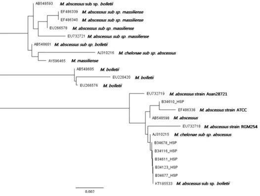
Fig. 3. Phylogenic trees of hsp65 se- quences of the six isolates (B34610_
HSP, B34678_HSP, B34116_HSP, B34611_HSP, B34123_HSP, B34677_
HSP) obtained from the patient in this study.
The isolates grew on sheep blood agar as non-hemolytic, white pinpoint colonies after 72 h of incubation at 35oC in an aerobic environment (Fig. 2). The bacteria produced catalase.
The isolates were identified as Kocuria rosea (94% probability) using VITEK 2 with a GP card (bioMérieux, Marcy-l’Étoile, France) and as Micrococcus and related species using MicroScan (Siemens Healthcare Diagnostics, West Sacramento, CA, USA) systems. Using MALDI Biotyper (software version 3.1, refer- ence database version 4.0.0.1, Bruker Daltonics, Billerica, MA, USA) with direct colony extraction method by which the mi- crobes are directly applied as a thin film on a spot of the target slide (a reusable 96 wells stainless steel target slide, Bruker Daltonics), allowed to dry at room temperature, followed by the addition of 1 μL of HCCA (α-cyano-4-hydroxycinnamic acid) matrix onto each sample spot dried at room temperature. The isolates were identified as follows: Mycobacterium abscessus (score: 1.731), Tissierella praeacuta (1.333), Rhodococcus ruber (1.325), and other species (scores lower than 1.3). When the iso- lates were retested with protein extraction method using etha- nol/formic acid, they were identified as Mycobacterium ab- scessus (score: 2.055). The isolate was found to be acid-fast.
The identification as M. abscessus was confirmed by reverse hy- bridization-based assay (Genotype Mycobacterium CM/AS 12, Hain Lifescience), and direct DNA sequencing of the hsp65 re- gion [9]. A BLAST search of the sequence revealed 100% se-
quence homology with a Mycobacterium abscessus strain (HM454214.1), Mycobacterium abscessus subsp. abscessus strain (KT185518.1), and Mycobacterium abscessus subsp. bol- letii strain (KT185533.1), whereas the similarity with Nocardia farcinica strain HN11062 (KF432771.1) was 99.77% (440/441 bp) (Fig. 3). Antimicrobial susceptibility testing at the Korean Institute of Tuberculosis [10] indicated that the isolate exhibited inducible resistance to clarithromycin (MIC, 2 μg/mL and >64 μg/mL); resistance to ciprofloxacin (>16 μg/mL), doxycy- cline (>32 μg/mL), and moxifloxacin (8 μg/mL); and inter- mediate resistance to amikacin (>32 μg/mL), cefoxitin (>32 μg/mL), and imipenem (8 μg/mL).
DISCUSSION
To date, about 20 species of rapidly growing NTM that are capable of causing bloodstream infections have been identified [5]. However, reports of cases of bloodstream infections by rap- idly growing NTM have been limited [4,5] which may reflect, in part, the difficulty in identifying this species from blood cultures. M. abscessus isolates recovered from blood cultures could be regarded as skin contaminants or misidentified as Corynebacterium spp., because they are Gram-positive, non- motile, and catalase-positive bacilli. In the present study, the isolates were misidentified by VITEK 2 with a GP card and
Microscan, but they were correctly identified as M. abscessus by MALDI-TOF MS. The identification of M. abscessus was supported by a positive AFB stain result. Our case highlighted that MALDI-TOF MS can be considered as a rapid diagnostic tool of bacteremia caused by rapid growing NTM including M.
abscessus.
Recent studies have shown that M. abscessus and other myco- bacterial species can be identified by MALDI-TOF MS [7,8,11,12]. In study which compares MALDI-TOF MS and re- verse hybridization-based assay for the identification of NTM, the concordance analysis between the two methods showed agreement in 96.9% [7]. However, these studies utilized colo- nies grown on Lowenstein medium or Middlebrook 7H11 agar, which are used for mycobacterial culture, because most isolates were from respiratory specimens. The present study showed that M. abscessus blood isolates grown on blood agar plates could be easily identified by MALDI-TOF MS. Compared with >24 h for sequencing, the short turnaround time of the MALDI-TOF MS method (15 min) enables rapid diagnosis of M. abscessus bacteremia.
Our patient exhibited multiple risk factors for M. abscessus infection, including immunosuppression, diabetes mellitus, and the long-term presence of a CVC [3,13]. The isolates were re- sistant to various antibiotics, including inducible resistance to clarithromycin, but CVC removal and combination therapy yielded a successful clinical outcome. Although the CVC tip culture yielded no growth, we postulate a catheter-related blood- stream infection based on the following: first, this isolate was present continuously in seven blood cultures from the day of ad- mission until that of CVC removal. Second, the fever subsided after CVC removal, which was prior to initiation of treatment of M. abscessus. Third, no typical radiological findings of M.
abscessus pulmonary infection were evident, and sputum cul- tures and AFB stains were negative. In conclusion, this case shows that M. abscessus bacteremia case can be rapidly diag- nosed by MALDI-TOF MS, albeit the automated bacterial iden- tification systems fail to identify them.
ACKNOWLEDGMENTS
We are grateful to Bong-Ju Kang, M.T. at the Laboratory of Clinical Microbiology of Chonnam National University Hospital
for technical assistance. This work was partially supported by the Fund of Annals of Clinical Microbiology.
REFERENCES
1. Wagner D and Young LS. Nontuberculous mycobacterial in- fections: a clinical review. Infection 2004;32:257-70.
2. Brown-Elliott BA and Wallace RJ Jr. Clinical and taxonomic status of pathogenic nonpigmented or late-pigmenting rapidly growing mycobacteria. Clin Microbiol Rev 2002;15:716-46.
3. Lee MR, Sheng WH, Hung CC, Yu CJ, Lee LN, Hsueh PR.
Mycobacterium abscessus complex infections in humans. Emerg Infect Dis 2015;21:1638-46.
4. Lee MR, Ko JC, Liang SK, Lee SW, Yen DH, Hsueh PR.
Bacteraemia caused by Mycobacterium abscessus subsp. abscessus and M. abscessus subsp. bolletii: clinical features and suscep- tibilities of the isolates. Int J Antimicrob Agents 2014;43:438-41.
5. El Helou G, Viola GM, Hachem R, Han XY, Raad II. Rapidly growing mycobacterial bloodstream infections. Lancet Infect Dis 2013;13:166-74.
6. Kim HY, Kook Y, Yun YJ, Park CG, Lee NY, Shim TS, et al.
Proportions of Mycobacterium massiliense and Mycobacterium bolletii strains among Korean Mycobacterium chelonae-Mycobac- terium abscessus group isolates. J Clin Microbiol 2008;46:3384-90.
7. Mediavilla-Gradolph MC, De Toro-Peinado I, Bermúdez-Ruiz MP, García-Martínez Mde L, Ortega-Torres M, Montiel Quezel-Guerraz N, et al. Use of MALDI-TOF MS for identification of nontuber- culous Mycobacterium species isolated from clinical specimens.
Biomed Res Int 2015;2015:854078.
8. Mareković I, Bošnjak Z, Jakopović M, Boras Z, Janković M, Popović-Grle S. Evaluation of matrix-assisted laser desorption/
ionization time-of-flight mass spectrometry in identification of nontuberculous mycobacteria. Chemotherapy 2016;61:167-70.
9. Telenti A, Marchesi F, Balz M, Bally F, Böttger EC, Bodmer T.
Rapid identification of mycobacteria to the species level by polymerase chain reaction and restriction enzyme analysis. J Clin Microbiol 1993;31:175-8.
10. CLSI. Susceptibility testing of mycobacteria, nocardiae and other aerobic actinomycetes: approved standard-second edition. CLSI document M24-A2. Wayne, PA: Clinical and Laboratory Standards Institute; 2011.
11. Teng SH, Chen CM, Lee MR, Lee TF, Chien KY, Teng LJ, et al.
Matrix-assisted laser desorption ionization-time of flight mass spectrometry can accurately differentiate between Mycobacterium masilliense (M. abscessus subspecies bolletti) and M. abscessus (sensu stricto). J Clin Microbiol 2013;51:3113-6.
12. Panagea T, Pincus DH, Grogono D, Jones M, Bryant J, Parkhill J, et al. Mycobacterium abscessus complex identification with matrix-assisted laser desorption ionization-time of flight mass spectrometry. J Clin Microbiol 2015;53:2355-8.
13. De Groote MA. Disease resulting from contaminated equipment and invasive procedures. In: Pedley S, Bartram J, Rees G, Dufour A, Cotruvo J, eds. Pathogenic Mycobacteria in Water: A Guide to Public Health Consequences, Monitoring and Management. Padstow, Cornwall; TJ International, 2004:131-42.
=국문초록=
말디토프 질량분석법으로 신속 진단한
Mycobacterium abscessus
에 의한 혈류 감염 1예전남대학교 의과대학 진단검사의학교실 원은정*, 최용준*, 김수현, 신종희
Mycobacterium abscessus는 신속 발육하는 비결핵성 마이코박테리움에 속하며, 주로 폐감염과 창상 감염 등을 유발한다.
저자들은 국내에서 말디토프 질량분석법에 의해 최초로 진단된 M. abscessus 균혈증을 보고하고자 한다. 본 증례에서는 스테로이드 치료에 반응하지 않는 부신기능저하증으로 입원한 64세 여자, 당뇨 환자에서 7회에 걸친 혈액 배양 결과에 서 M. abscessus가 분리되었다. 분리된 균주는 전통적인 동정 시스템인 VITEK 2 (bioMérieux, France)나 MicroScan (Siemens Healthcare Diagnostics, USA)으로는 잘못 동정되었으나, 말디토프 질량분석법에 의해 M. abscessus로 정확하게 동정되었으며, 이는 heat shock protein 유전자 염기서열 분석으로 확인되었다. 환자는 중심정맥관 제거와 함께 clari- thromycin, amikacin, cefoxitin, imipenem 병합요법으로 치료되었다. 본 증례는 혈액배양에서 분리되는 M. abscessus의 동 정이 말디토프 질량분석법을 통해 신속하고 정확하게 이뤄질 수 있으며, 이러한 동정이 중심정맥관 제거와 함께 시기 적절한 치료로 이어져 임상적으로 유용할 수 있음을 시사하였다. [Ann Clin Microbiol 2016;19:77-81]
교신저자 : 신종희, 61469, 광주시 동구 제봉로 42 전남대학교 의과대학 진단검사의학교실 Tel: 062-220-5342, Fax: 062-224-2518 E-mail: [email protected]