J Korean Soc Radiol 2017;76(1):48-53 https://doi.org/10.3348/jksr.2017.76.1.48
INTRODUCTION
Lumbosacral radiculopathy is a common medical and socio- economic problem with a lifetime prevalence estimated to be around 40–60% (1-3). Intervertebral disc herniation and degen- erative lumbar spinal stenosis are the two most common causes of lumbosacral radiculopathy (4). Degenerative lumbar spinal
stenosis is a common disease in the elderly population, which is rapidly increasing (5). However, in most cases, the initial clini- cal symptoms and imaging findings are nonspecific, discordant or insufficient for diagnosis (6-9). In addition, some patients complain about multi-level radiculopathy.
Currently, epidural steroid injection (ESI) is used for the treat- ment of both acute and chronic lower back pain. Its use relieves
Comparison of the Therapeutic Effect between a Transforaminal along with a Caudal Epidural Injection, as Well as Two-Level Transforaminal Epidural Injections in a Radiculopathy Patient
요천추병증에 대한 미골부 경막 외 스테로이드 주사의 기존 추간공 경막 외 스테로이드 주사와의 치료 효과 비교
Junghan Hwang, MD
1, Cheol Mog Hwang, MD
1*, Young Jun Cho, MD
1, Keum Won Kim, MD
1, Young Joong Kim, MD
1, Jae Young Seo, MD
1, Seong Joo Lim, MD
1, Byeong Seong Kang, MD
21Department of Diagnostic Radiology, Konyang University Hospital, Deajeon, Korea
2Department of Radiology, University of Ulsan College of Medicine, Ulsan University Hospital, Ulsan, Korea
Purpose: The aim of this study was to evaluate the therapeutic effect of a transfo- raminal epidural steroid injection (TFESI) along with a caudal epidural steroid injec- tion (ESI), compared to two-level TFESIs in a multi-level radiculopathy patient.
Materials and Methods: A total of 895 lumbar ESIs were performed in 492 pa- tients with multi-level radiculopathy from January 2012 to January 2015. Before in- jections were performed, the initial Numeric Rating Scale (NRS) score was assessed in all patients, categorized into no pain (excellent), mild (good, NRS: 1–3), moderate (fair, NRS: 4–6), and severe pain (poor, NRS: 7–10). Therapeutic effects were exam- ined for two groups: one-level TFESI along with caudal and ESI two-level TFESIs. Pa- tient outcomes were assessed by NRS in a serial follow-up at one, three, and six months.
Results: One TFESI along with caudal ESI was performed in 274 patients and two TFESIs for 218. For the former group with one TFESI along with caudal ESI, excellent results were shown: 219 (79.9%) patients after one month, 200 (72.9%) after three, and 193 (70.4%) after six months. In the patient group with two TFESIs (n = 218) the outcomes were also very good: 152 (69.7%) after one month, 131 (60.0%) after three months, and 123 (56.4%) patients after six months. The therapeutic effect of one TFESI along with caudal ESI was better than two TFESIs in for one, threes, and six months (p < 0.01).
Conclusion: Transforaminal epidural steroid with caudal epidural injection is a more effective tool for lumbosacral radiculopathy than two level transforaminal in- jections in multi-level radiculopathy patients.
Index terms Spine Low Back Pain Epidural Injection
Received May 3, 2016 Revised June 15, 2016 Accepted October 13, 2016
*Corresponding author: Cheol Mog Hwang, MD Department of Diagnostic Radiology, Konyang University Hospital, 158 Gwanjeodong-ro, Seo-gu, Daejeon 35365, Korea.
Tel. 82-42-600-9762 Fax. 82-42-600-9193 E-mail: [email protected]
This is an Open Access article distributed under the terms of the Creative Commons Attribution Non-Commercial License (http://creativecommons.org/licenses/by-nc/3.0) which permits unrestricted non-commercial use, distri- bution, and reproduction in any medium, provided the original work is properly cited.
symptoms attributed to lumbosacral radiculopathy. In multi-level radiculopathy patients, therapeutic effect of one level epidural steroid injection has been limited. Thus, we focused on addi- tional epidural injections for treating multi-level herniated disc patients. The objectives of our study were comparing the thera- peutic effect and complication rates between two level injec- tions and a one level injection along with caudal injection.
MATERIALS AND METHODS
Patients
Lumbar ESIs were performed from January 2012 to January 2015. Among the patients, 895 consecutive lumbar ESIs in 492 were reviewed. We received approval from Institutional Review Board. Total 895 lumbar ESIs (285 men, 207 women; mean age, 63.9 years) were used for analysis. There is no significant differ- ence in the two groups’ age range. The initial Numeric Rating Scale (NRS) score was assessed in every patient (Table 1), divid- ing them into no, mild (NRS: 1–3), moderate (NRS: 4–6), and severe pain (NRS: 7–10) categories. We classified the patients into two groups based on the NRS anddefined marked improve- ment with no pain as a satisfactory result for therapeutic effect.
Retrospectively, the therapeutic effect difference in the groups was evaluated: one level transforaminal epidural steroid injec- tion (TFESI) along with caudal epidural injection and two level TFESIs for lumbosacral radiculopathy. The inclusion criteria were presence of low back or leg pain, clear radiculopathy, such as evidenced by computed tomography (CT) or MRI, which was identified by radiologic reports, and the presence of follow- up medical records after caudal ESI. Exclusion criteria were un-
clear descriptions of symptoms, the absence of evidence of ra- diculopathy on cross-sectional images or of follow-up data.
Injections for two groups were performed for patients with multi-level radiculopathy and contralateral mild radiculopathy or chronic back pain. There was no significant statistical differ- ence in two patient groups’ symptoms, who were injected the same steroid dose at each time. Therapeutic effects were ana- lyzed at one, three, and six months after injection in the two com- parison groups and assessed by using the NRS.
Technique
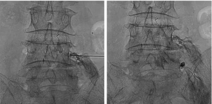
All therapeutic TFESI were conducted under fluoroscopic guidance by a musculoskeletal radiologist experienced in per- forming spinal interventions. All the injections were performed as outpatient procedures, after obtaining informed consent. With a patient in susceptible position, the tube was rotated obliquely to ensure the facet joint injection pars interarticularis and a clear- ly visiblesite for it. Next, the skin and subcutaneous tissues over- lying the desired pedicle and nerve root were anesthetized with lidocaine. The needle was then advanced under intermittent flu- oroscopy into the neural foramen. If it is not possible to inject in a safe triangle, we had a procedure employing the postero- lateral approach. Serial lateral views were obtained to ensure that the antero-posterior position of the needle tip was appropri- ate. The needle position was checked through fluoroscopy us- ing injections of contrast materials’ small amounts. Postero-an- terior and lateral spot radiographs were obtained to document contrast material distribution in epidural space. The steroid was slowly and gently injected (Fig. 1).
Caudal injection was performed in same manner as above, palpating the sacral cornua and localizing the entry point of the needle on A–P view. Sacral cornua were used as landmarks to identify the sacral hiatus, as the sacral hiatus is bounded bilater- ally by them. Then, the needle position was confirmed on the oblique, lateral view at the level S2–3. Nonionic contrast materi- als were injected to the sacral epidural space; then, a mixture of steroid and normal saline was slowly injected (Fig. 2).
Clinical Assessment
Before the injection was performed, the radiologist recorded the duration of symptoms, level, and cause of nerve root im- pingement on the CT or MR images and the affected dermato- Table 1. Baseline Characteristic of Enrolled Patients
Patients Two Level Injection One level + Caudal Gender
Male 135 150
Female 83 124
Age, yr
≤ 60 88 124
> 60 130 150
Primary NRS 1–3 4–6 7–10
18 40 160
24 50 200 NRS = Numeric Rating Scale
mal distribution on the medical charts. Patient outcomes were assessed at one, three, and six month serial follow-up using a NRS that ranged from 0 to 10, subjectively expressed by the pa- tients for the degree of improvement: excellent, good, fair, and poor. An excellent degree represented marked improvement, much improved (> 3-point NRS decreases), good was improve- ment but with a lesser effect (1–2 point NRS decreases). A fair degree indicated no interval change since the first injection and a poor one meant worsening of pain during the study.
The patients were not informed about the injection method to ensure a blinded protocol for the study. We defined satisfac- tory results as patients with a reduction of pain as reflected in
NRS and no residual pain. After epidural steroid injection, the above-described one, three, and six month serial follow-ups were conducted at a clinical department, as proposed in the literature and based on the duration of therapeutically effective cortico- steroids. Follow-up after ESI was scheduled for one month. At follow-up, the outcome was measured on a four-point patient satisfaction scale and recorded on the medical chart. If there was no pain, observations without repeated ESI were considered. If we decided this, the next follow-up was scheduled for three and sixe months later. We advised patients to return to our hospital immediately, even before the routine follow up date, if symp- toms recurred.
Statistical Analysis
The Student t-test was used to evaluate age differences be- tween two groups. Statistical analysis was also performed using Fisher’s exact test and multiple logistic regression analysis to evaluate differences between the two level TFESIs and one level TFESI subsamples along with caudal epidural injection.
The Fisher’s exact test was used to determine differences in sex, therapeutic effects, and the complication rate between the two patient groups. Multiple logistic regression analysis was also performed to assess therapeutic effects. Analyses were performed for both short- and mid-term therapeutic effects. Analyses too place by using the software program SPSS, version 10.0 (SPSS Inc., Chicago, IL, USA). A p value of less than 0.05 was consid- ered to indicate statistical significance.
RESULTS
274 (55.6%) of 492 patients were treated by one TFESI along with caudal epidural injections; 218 (44.4%) were subjected to two level TFESIs. 219 (79.9%) and they achieved a satisfactory result after one TFESI along with caudal epidural injections (group 1) after one month, and 152 (69.7%) of the 218 patients after two level TFESIs (group 2). After a three month follow-up, 200 (72.9%) of the 274 patients obtained a good outcome after one TFESI along with caudal epidural injections and 131 (60.0%) of the 218 patients achieved a satisfactory result after two level TFESIs. After the six month follow-up, 193 (70.4%) of the 274 subjects as well, after one TFESI along with caudal epidural in- jections, and the findings were likewise for 123 (56.4%) of the Fig. 1. Radiographs of 69-year-old man with radiating pain to right
buttock and lower leg in the S1 dermatome. MR image (not shown) revealed right L4/5 and L5/S1 disc herniations with compression of right L5/S1 nerve root. Initial NRS score is 10 point. The patient re- ceived one level TFESI, he experienced less pain for approximately 1 months (NRS: 6). PA spot radiograph shows contrast material has spread to the epidural space.
NRS = Numeric Rating Scale, PA = posteroanterior, TFESI = transfo- raminal epidural steroid injection
Fig. 2. Radiographs of 65-year-old man with severe back pain and ra- diating pain to right buttock. The patient received one level TFESI along with caudal epidural injection. Initial NRS score is 10 point. The patient received one level TFESI and ESI, he experienced no residual pain for approximately 1 months (NRS: 0). After on PA spot radiographs during TFESI, the spinal needle is located in the sacral canal, and the contrast agent is spreading into the epidural space of the lumbar spine.
ESI = epidural steroid injection NRS = Numeric Rating Scale, PA = posteroanterior, TFESI = transforaminal epidural steroid injection
218 patients after two level TFESIs. Statistical differences were significant for results of therapeutic effects’ difference between the groups (p < 0.05). Univariate analysis showed that the one TFESI along with caudal epidural injections group demonstrated a superior effect than did the two level injection only subsample at short-term follow-up (p < 0.01) (Table 2).
DISCUSSION
Nonsurgical treatment for spinal stenosis varies, but it in- cludes bed rest, nonsteroidal anti-inflammatory drugs, analge- sics, oral administration of corticosteroids, physical therapy, and ESI. According to a cohort study by Simotas et al. (11) of non- surgically treated patients with lumbar spinal stenosis, three years after treatment, nine of the 49 patients had undergone surgical interventions. Twelve of the 40 patients not operated also reported no or only mild pain. The authors concluded that aggressive non-operative treatment for spinal stenosis remains a reasonable option (11).
The indication and efficacy of ESI still remains controversial.
This technique is currently used as an intermediate treatment for back pain of various causes and duration. It is not consid- ered curative, but a number of patients have reported long and short-term pain relief (12-14). Many reports have been pub- lished about the use of TFESI in patients with chronic lower back pain (15); however, to the best of our knowledge, few re- ports are available about the outcome predictors of TFESI treat- ment, especially in terms of the injection level numbers. The therapeutic effect of conventional TFESI is limited to multi-lev- el herniated disc patients, because it is a target-specific treat-
ment for leveled originated radiculopathy. Caudal injections are performed for multi-level disc patients and–previously–two level injections did not achieve a sufficient therapeutic effect.
Caudal injection is designed for maximizing the therapeutic effect and to cover broad disc pain. Lee et al. (16) reported that approximately 85% of patients showed improvements after an initial caudal ESI and some 55% displayed excellent ameliorai- ton after a series of caudal ESI. According to a study by Delport et al. (17), of the 140 participants who underwent TFESI or cau- dal injection, 32% reported more than three months of pain re- lief, 39% less than that, while 29% stated that there was no elief . Manchikanti et al. (18) stated that significant pain relief (≥ 50%) was demonstrated in 55–65% of the patients with spinal steno- sis after the use of caudal injection, While Botwin et al. (19) had published that 65% of patients after six weeks, 62% after six months, and 54% after 12 months had achieved a successful outcome, reporting an at least over-50% reduction in visual an- alog pain scores after caudal injection, but our results appear more optimistic. Fukusaki et al. (20) reported that ESI had no beneficial effect on the pseudoclaudication associated with spinal canal stenosis. On the other hand, Kim et al. (21) could demon- strate that fluoroscopy-guided intra-articular facet joint injec- tion exhibited excellent immediate effectiveness and good pro- longed pain relief (> two months) in patients with chronic low back pain. According to our study, one TFESI along with caudal epidural injection was more effective than two level TFESIs. This finding supports the idea that the former can more directly to reduce inflammation and relieve pain.
Our study also has several limitations. First, it was not a pro- spective study and follow-ups was not long-term designed, al- though it is difficult to organize a regular schedule for TFESI. We think that it is better for patients to receive TFESI when symp- toms appear or worsen. We still believe that the midterm fol- low-up is sufficient to evaluate the therapeutic effects. Second, initial patient groups had severe back pain. Up to 80% patient’s initial NRS scores were 10 points. Thus, the injection effect ap- pears to be overestimated. We trust that a prospective analysis is the next logical step to further analyze such findings. Further- more, there was no significant difference in therapeutic effect by symptom duration at a short-term follow-up.
In conclusion, the transforaminal epidural steroid with cau- dal epidural injection is a more effective tool for multi-level ra- Table 2. Comparison of Therapeutic Effect between the 2 Groups
after 1, 3, and 6 Months Follow Up
Excellent (%) Good (%) Fair (%) Poor (%) 1 month
1 TFESI + caudal 79.9 10.1 7.0 3.0
2 TFESI 69.7 15.3 9.3 6.7
3 months
1 TFESI + caudal 72.9 17.1 8.5 1.5
2 TFESI 60.0 16.4 15.3 8.3
6 months
1 TFESI + caudal 70.4 17.1 8.5 1.5
2 TFESI 56.4 15.4 18.0 10.2
p-value: 0.01.
TFESI = transforaminal epidural steroid injection
diculopathy and contralateral mild radiculopathy or chronic back pain than two level TFESIs.
REFERENCES
1. Vad VB, Bhat AL, Lutz GE, Cammisa F. Transforaminal epi- dural steroid injections in lumbosacral radiculopathy: a pro- spective randomized study. Spine (Phila Pa 1976) 2002;27:
11-16
2. Silbergleit R, Mehta BA, Sanders WP, Talati SJ. Imaging- guided injection techniques with fluoroscopy and CT for spinal pain management. Radiographics 2001;21:927-939;
discussion 940-942
3. Frymoyer JW, Wiesel SW. The adult and pediatric spine.
Philadelphia: Lippincott Williams & Wilkins, 2004:929-944 4. Cyteval C, Fescquet N, Thomas E, Decoux E, Blotman F,
Taourel P. Predictive factors of efficacy of periradicular cor- ticosteroid injections for lumbar radiculopathy. AJNR Am J Neuroradiol 2006;27:978-982
5. Robecchi A, Capra R. [Hydrocortisone (compound F); first clinical experiments in the field of rheumatology]. Minerva Med 1952;43:1259-1263
6. Goebert HW Jr, Jallo SJ, Gardner WJ, Wasmuth CE. Painful radiculopathy treated with epidural injections of procaine and hydrocortisone acetate: results in 113 patients. Anesth Analg 1961;40:130-134
7. Pfirrmann CW, Oberholzer PA, Zanetti M, Boos N, Trudell DJ, Resnick D, et al. Selective nerve root blocks for the treatment of sciatica: evaluation of injection site and effec- tiveness--a study with patients and cadavers. Radiology 2001;221:704-711
8. Rydevik B, Garfin S. Spinal nerve root compression. In Szabo RM. Nerve compression syndromes: diagnosis and treatment. New York: Slack Medical, 1989:247-261 9. Wong DA, Errico T, Saal J, Sims W, Watters W. Clinical
guideline on low back pain. Rosemont, IL: American Acad- emy of Orthopedic Surgeons, 1996:147-161
10. Botwin KP, Gruber RD, Bouchlas CG, Torres-Ramos FM, Sanelli JT, Freeman ED, et al. Fluoroscopically guided lumbar transformational epidural steroid injections in degenerative
lumbar stenosis: an outcome study. Am J Phys Med Rehabil 2002;81:898-905
11. Simotas AC, Dorey FJ, Hansraj KK, Cammisa F Jr. Nonoper- ative treatment for lumbar spinal stenosis. Clinical and out- come results and a 3-year survivorship analysis. Spine (Phila Pa 1976) 2000;25:197-203; discussions 203-204
12. Jamison RN, VadeBoncouer T, Ferrante FM. Low back pain patients unresponsive to an epidural steroid injection: iden- tifying predictive factors. Clin J Pain 1991;7:311-317 13. Manchikanti L. Transforaminal lumbar epidural steroid in-
jections. Pain Physician 2000;3:374-398
14. Lew HL, Coelho P, Chou LH. Preganglionic approach to transforaminal epidural steroid injections. Am J Phys Med Rehabil 2004;83:378
15. Benzon HT. Epidural steroid injections for low back pain and lumbosacral radiculopathy. Pain 1986;24:277-295 16. Lee JW, Kim SH, Choi JY, Yeom JS, Kim KJ, Chung SK, et al.
Transforaminal epidural steroid injection for lumbosacral radiculopathy: preganglionic versus conventional approach.
Korean J Radiol 2006;7:139-144
17. Delport EG, Cucuzzella AR, Marley JK, Pruitt CM, Fisher JR.
Treatment of lumbar spinal stenosis with epidural steroid injections: a retrospective outcome study. Arch Phys Med Rehabil 2004;85:479-484
18. Manchikanti L, Cash KA, McManus CD, Pampati V, Abdi S.
Preliminary results of a randomized, equivalence trial of fluoroscopic caudal epidural injections in managing chronic low back pain: part 4--spinal stenosis. Pain Physician 2008;
11:833-848
19. Botwin K, Brown LA, Fishman M, Rao S. Fluoroscopically guided caudal epidural steroid injections in degenerative lumbar spine stenosis. Pain Physician 2007;10:547-558 20. Fukusaki M, Kobayashi I, Hara T, Sumikawa K. Symptoms of
spinal stenosis do not improve after epidural steroid injec- tion. Clin J Pain 1998;14:148-151
21. Kim S, Lee JW, Chai JW, Lee GY, You JY, Kang HS, et al.
Fluoroscopy-guided intra-articular facet joint steroid in- jection for the management of low back pain: therapeutic effectiveness and arthrographic pattern. J Korean Soc Ra- diol 2015;73:172-180
요천추병증에 대한 미골부 경막 외 스테로이드 주사의 기존 추간공 경막 외 스테로이드 주사와의 치료 효과 비교
황정한
1· 황철목
1* · 조영준
1· 김금원
1· 김영중
1· 서재영
1· 임성주
1· 강병성
2목적: 여러 층 신경근증 증상을 가진 환자군에서 추간공 경막 및 미골부 경막 외 스테로이드 주사를 함께 시행했을 경우 와 두 층 추간공 경막 외 스테로이드 주사를 시행하였을 때의 치료 효과의 차이를 알아보고자 한다.
대상과 방법: 2012년 1월에서 2015년 1월 사이 492명의 여러 층 신경근증 증상이 있는 환자를 대상으로 하였다. 주사 전, 모든 환자에서 초기 통증평가척도(Numeric Rating Scale; 이하 NRS)를 측정하여 경미한(NRS: 1~3), 중등도의(NRS:
4~6), 그리고 심각한(NRS: 7~10) 정도로 나누었다. 환자들은 두 레벨의 경막 외 스테로이드 주사, 그리고 한 레벨의 경 막 외 스테로이드 주사와 함께 미골부 경막 외 스테로이드 주사를 병행한 두 가지의 환자군으로 나누어 치료 효과를 비교 하였다. 각각의 환자군은 시술 후 1, 3, 6개월에 통증평가척도를 통해 치료 반응을 평가하였다. 평가는 환자의 시술 후 주 관적인 통증 해소 정도에 따라 ‘훌륭한, 좋은, 보통의, 악화된’의 4단계로 나누어 평가하였다.
결과: 274명에서 추간공 경막 외 스테로이드 주사와 더불어 미골부 스테로이드 주사를 받았고, 218명에서 두 층 추간공 경막 외 스테로이드 주사를 시행받았다. 추간공 경막 외 스테로이드 주사와 더불어 미골부 스테로이드 주사를 시행받은 274명의 환자 중, 시술 후 1개월 후에 219(79.9%)명이, 3개월 후에 200(72.9%)명이, 그리고 6개월 후에 193(70.4%)명 이 ‘훌륭한’ 정도의 치료 반응을 보였다. 두 층 추간공 경막 외 스테로이드 주사를 시행받은 218명의 환자 중, 시술 후 1개 월 후에 152(69.7%)명이, 3개월 후에 131(60.0%)명이, 그리고 6개월 후에 123(56.4%)명이 ‘훌륭한’ 정도의 치료 반응 을 보였다. 추간공 경막 외 스테로이드 주사와 더불어 미골부 스테로이드 주사를 병행한 경우가 더 나은 치료 효과를 보이 는 것이 확인되었다.
결론: 여러 층 신경근증 증상을 가진 환자군에서 추간공 경막 및 미골부 경막 외 스테로이드 주사를 함께 시행하였을 때 의 치료 효과가 두 층 추간공 경막 외 스테로이드 주사보다 더 효과적이다.
1건양대학교병원 영상의학과, 2울산대학교 의과대학 울산대학교병원 영상의학과