J Korean Soc Radiol 2018;78(3):193-199 https://doi.org/10.3348/jksr.2018.78.3.193
INTRODUCTION
Recently, percutaneous ablation has become a commonly used method in the treatment of renal cysts, and our preferred method for this procedure is multiple session sclerotherapy us- ing ethanol. In this case, in order to decide upon a treatment plan, it is very important for both the patient and the physician to know whether the average length of stay in hospital or the number of ethanol injections needed can be predicted, but there has not yet been any research on criteria for making these predictions prior to treatment. Thus, the present study aimed to
examine findings from pre-treatment imaging that could pre- dict the number of ethanol injections.
MaTeRIals aND MeThODs
Patient Population
This retrospective study was approved by our Institutional Review Board, and the need for written informed consent was waived (CBNUH 2016-05-006). The study protocol conforms to the ethical guidelines of the 1975 declaration of Helsinki as reflected in a prior approval by the Institutional Human Re-
Predictors of the Frequency of Ethanol Injections for Renal Cyst Ablation: A Preliminary Study
신장 낭종 경화술에서 에탄올 주입의 횟수에 영향을 미치는 효과적인 예측인자에 대한 고찰: 초기 보고
Yong Hun Kim, MD
1, Bum Sang Cho, MD
1,2*
1Department of Radiology, Chungbuk National University Hostpital, Cheongju, Korea
2Department of Radiology, College of Medicine, Chungbuk National University, Cheongju, Korea
Purpose: To assess the factors facilitating the prediction of the frequency of etha- nol injections in successful renal cyst ablation.
Materials and Methods: We retrospectively reviewed the computed tomography and ultrasonography scans of 37 renal cysts. Two radiologists evaluated the charac- teristics of the presenting renal cysts, including size, calcification, septation, and lobulated configuration. Patients were divided into an “above-four” group and “be- low-three” group, according to the number of ethanol injections.
Results: Among the 37 renal cysts, six belonged to the “above-four” group and 31 to the “below-three” group. The mean volume of “above-four” group was 409.48 cc and that of the other group was 301.64 cc. Seven renal cysts included three belong- ing to the “above-four” group, which showed calcification. Twelve renal cysts pre- sented a multilocular appearance with thin septa, and included a cyst classified un- der the “above-four” group. Three renal cysts had lobulated configuration, with one of them belonging to the “above-four” group.
Conclusion: No significant correlation was found among the different factors in the prediction of the frequency of renal cyst ablation. However, calcification was the most useful parameter for prediction of the number of renal cyst ablations needed.
Index terms Kidney Diseases Cysts
Ablation Techniques Sclerotherapy Ethanol
Received September 5, 2017 Revised October 10, 2017 Accepted October 22, 2017
*Corresponding author: Bum Sang Cho, MD Department of Radiology, Chungbuk National University Hostpital, 776 1sunhwan-ro, Seowon-gu, Cheongju 28644, Korea.
Tel. 82-43-262-6487 Fax. 82-43-269-6479 E-mail: [email protected]
This is an Open Access article distributed under the terms of the Creative Commons Attribution Non-Commercial License (http://creativecommons.org/licenses/by-nc/4.0) which permits unrestricted non-commercial use, distri- bution, and reproduction in any medium, provided the original work is properly cited.
search Committee.
This study initially included a total of 39 patients (16 males and 23 females) with 40 renal cysts, who underwent percutane- ous renal cyst ablation at our hospital between December 2007 and May 2016. The mean age of the patients was 62 years (range:
29–81 years).
The included patients were diagnosed with renal cysts either at our hospital or another hospital using abdominal computed tomography (CT) or abdominal ultrasonography (USG). Three patients without pre-procedure CT or USG images were ultimate- ly excluded from the study, and the remaining 36 patients (37 renal cysts) were included in the analysis. The mean time be- tween imaging and treatment was 24 days (range: 1–130 days).
Twenty-two cysts were asymptomatic, or were discovered in- cidentally during examinations for other diseases, while the presence of fifteen cysts were accompanied by symptoms of flank pain, flank discomfort, abdominal pain, or hematuria.
For patients with CT images, we only included those patients with benign cysts corresponding to Bosniak classification I or II. For the two patients without CT images, prior to including them in the study, we confirmed that there was no solid portion or thick wall of 1 mm or greater in size on USG examination.
Renal cyst cytology was performed on the fluid obtained dur- ing renal cyst ablation, and malignant cells were not observed in any of the patients. The majority of patients underwent treat- ment due to symptoms such as abdominal or flank pain. As- ymptomatic patients were treated when the cyst measured at
least 5 cm in diameter, and/or when the patients themselves wanted treatment.
Imaging Analysis
Two genitourinary radiologists confirmed the radiological findings retrospectively, with a conclusion reached by consen- sus when there were conflicting opinions. CT findings were considered when CT images were available, while USG findings were used in the analysis of the two patients who did not have CT images. A 64-channel multiple detector CT system (Bril- liance 64; Philips Medical Systems, Best, Netherlands) was used for CT imaging with the following parameters: detector config- uration, 64 × 0.625 mm; section thickness, 3.0 mm; reconstruc- tion interval, 2 mm; table speed, 46.8 mm per rotation; effective amperage setting, 150 mA; rotation time, 0.75 s; tube voltage, 120 kVp; and matrix, 512 × 512.
A workstation (Marosis M-view; Infinitt, Seoul, Korea) and picture archiving communication system were used to examine the radiological findings.
Radiological findings were analyzed and the presence or ab- sence of internal septa, calcification, and lobulated contours was noted (Figs. 1–3). The volume of each renal cyst was calculated using the ellipsoid formula (width × height × length × 0.523) following measurement of the length in three axes based on the available images.
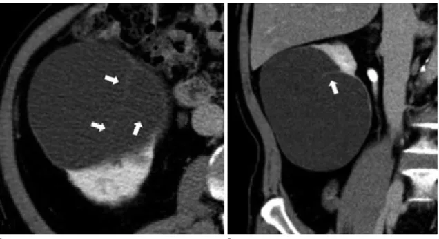
Fig. 1. A 55-year-old woman with a Bosniak classification II lesion in the right kidney and she was performed four times ethanol injections.
A, B. The renal cyst in the right kidney shows calcification (white arrows) and septa (black arrows) in non-contrast-enhanced (A) and contrast- enhanced (B) axial computed tomography scans. In addition, it has a lobulated margin (white arrowheads) with at least one lobule.
C. Like the axial axis view, the coronal axis view shows a lobulated margin (arrowheads) with at least one lobule; since the lobulated margin is visible in two planes, this cyst was classified as a lobulated renal cyst with calcification and thin septa.
A B C
Procedure
All patients were hospitalized for the procedure. After mark- ing the position of the cyst with the patient in the prone posi- tion under USG guidance, 2% lidocaine was used to induce lo- cal anesthesia at the puncture site. A 21-gauge puncture needle was used to puncture the cyst, and then a pigtail catheter (8.5Fr;
Cook Medical, Bloomington, IN, USA) was placed inside the cyst. In all patients, a contrast agent was injected to check that there was no communication with the pelvicalyceal collecting system, extravasation, or leakage of the contrast agent into the retroperitoneum. At this point, the fluid in the cyst was aspirat-
ed as much as possible and, after noting the volume of the aspi- rated fluid, it was subjected to cytological and biochemical ex- aminations. Following aspiration, sclerotherapy was performed using 99% ethanol; the amount of ethanol injected was one- third the total volume of the aspirated fluid, but no more than 100 cc. Following the injection of the ethanol into the renal cyst, to achieve an even application, the patient was placed in the supine, prone, and both decubitus positions for five minutes each, and then the ethanol was removed. The drainage volume after 24 hours was inspected via the catheter, and after using USG to check for any remaining fluid in the cyst, aspiration was
Fig. 2. A 57-year-old man with a Bosniak classification I lesion in the left kidney and he was performed two times ethanol injections.
A. The contrast-enhanced computed tomography axial scan shows a lobulated margin (arrowheads).
B, C. The coronal scan also shows a renal cyst with a lobulated margin (arrowheads) renal cyst; however, this is not accompanied by internal sep- ta or calcification. Hence, this case was classified as a lobulated renal cyst in the left kidney without internal septa or calcification.
D. Similarly, a sonogram performed during sclerotherapy also demonstrated a lobulated contour (arrowheads).
A
C
B
D
performed with a 10 cc syringe. When the total drainage vol- ume after 24 hours was over 5 cc, ethanol was injected to a vol- ume of one-third the total drainage volume, but no more than 100 cc, and further follow-up examinations were performed af- ter another 24 hours. When the total drainage volume was 5 cc or less, the procedure was considered a complete success and the pigtail catheter was removed (1).
Statistical Analysis
We examined the correlation between the number of ethanol injections, and the following four criteria: volume, septa (or not), calcification (or not), and margin (lobulated or smooth).
The mean total number of ethanol injections was obtained, and the subjects were divided into an “above” group and a “below”
group based on the rounded mean; the volume, septa (or not),
calcification (or not), and margin (lobulated or smooth) were then compared between the two groups.
An independent sample t-test was used to compare the volume between the two groups. Due to the small sizes of the two groups, Fisher’s exact test was used to compare the values of each vari- able between the groups.
SPSS ver. 18.0 (SPSS Inc., Chicago, IL, USA) was used for all statistics. p-values < 0.05 were considered to be statistically sig- nificant.
ResUlTs
A total of 37 renal cysts were included in the study, and renal cyst ablation was performed 2.5 times on average (range: 1–7 times). And all patients were no significant problem except mi- Table 1. Image Features of Renal Cysts and the Comparison between the Two Groups
Group Mean Volume (cc) Calcification Septa Lobulated
Positive Negative Positive Negative Positive Negative
Above-four 409.48 3 3 1 5 1 5
Below-three 301.64 4 27 11 20 2 29
Total 319.12 7 30 12 25 3 34
p-value 0.408 0.068 0.350 0.421
Numbers are mean volume and number of patients. p-values are for the difference between results for the “above-four” group and the “below-three”
group. The subjects were divided according to the number of rounds of renal cyst ablation, with the “above-four” group including those who underwent at least four ethanol injections, and the “below-three” group including those who underwent three or fewer ethanol injections.
Fig. 3. A 71-year-old man with a Bosniak classification II lesion in the right kidney and he was performed two times ethanol injections.
A, B. The axial and coronal contrast-enhanced CT scans show a renal cyst with internal linear density (white arrows). This was classified as a smooth margined renal cyst with thin septa and no calcification.
A B
nor pain at procedure site. The mean number of procedures was rounded to three, and the subjects were divided into two groups based on this criterion. There were six cases in the
“above-four” group and 31 cases in the “below-three” group.
The mean volume of the renal cysts was 319.12 cc, with a mean volume of 409.48 cc in the “above-four” group and 301.64 cc in the “below-three” group. The difference between the two groups was not statistically significant (p = 0.408) (Table 1).
A total of seven cases showed calcification, with three such cases in the “above-four” group and four cases in the “below- three” group. This was not a statistically significant difference (p
= 0.068) (Table 1).
A total of 12 cases showed septa, of which only one case was in the “above-four” group and 11 cases were in the “below- three” group. With a p-value of only 0.350, this was also not a statistically significant difference (Table 1).
Finally, there were three cases in total with lobulated margins, of which only one case was in the “above-four” group and the other two cases were in the “below-three” group. With a p-value of 0.421, this was not a statistically significant difference (Table 1).
DIsCUssION
Renal cysts are the most common disease in the kidneys, and are known to occur in approximately 50% of the population aged 50 years and older. Treatment is indicated in patients with symptoms such as pain, hematuria, hypertension, or pelvicaly- ceal dilatation due to pressure from the cyst, as well as in some patients in who the diameter of the cyst is at least 5 cm (2-6). Re- nal cyst sclerotherapy is a minimally invasive method for the treatment of renal cysts. It is being used increasingly often due to its high performance relative to cost and good success rate (3).
Drugs used for renal cyst sclerotherapy include ethanol, mi- nocycline hydrochloride, povidone iodine, acetic acid, ethanol- amine oleate and holmium-166 chitosan complex. Of these, eth- anol is the most widely used because it is considered to be safer and more effective than the other drugs (7-18). The present study used 99% ethanol.
When a sclerosing agent such as ethanol is injected into a renal cyst, one of the best-known mechanisms of cyst ablation is ne- crosis of the fluid-secreting epithelium via coagulation (19, 20).
In addition, the space for the cyst to grow into is believed to be
reduced by adhesion due to secondary inflammation; although the effectiveness of this mechanism is not clear, it seems possi- ble given the effects of pleurodesis in the lungs (21, 22).
The methods of percutaneous renal cyst sclerotherapy in- clude single-session sclerotherapy and multiple session sclero- therapy. Chung et al. (5) reported that multiple sessions of per- cutaneous sclerotherapy had a superior effect as compared to the effect of a single session, and the present authors also imple- ment multiple session sclerotherapy with positive outcomes. In interviews conducted between the patient and physician prior to treatment, we are often asked about the expected length of stay in the hospital. However, there has not yet been enough re- search conducted to predict the average number of ethanol in- jections and the average number of rounds of renal cyst sclero- therapy that could be required.
In the present study, 2.5 ethanol injections on average were completed until the total drainage volume reached a level of less than 5 cc. And average length of hospital stay was 6.8 days. Using the rounded average of three injections as the criterion value, the authors divided the patients into two groups, and compared a series of variables between these groups. Specifically, the com- parative analysis showed p-values of 0.408, 0.068, 0.350, and 0.421 for renal cyst size, calcification, septa, and lobulated mar- gin, respectively, meaning that there were no statistically signifi- cant results for any of these variables. However, although the p- value of 0.068 for calcification was higher than the significance level of 0.05, it was lower than 0.1. Given that this was a prelim- inary study involving only small groups of patients, this is con- sidered a relatively meaningful result that will require addition- al research.
The reason that calcification showed a more significant result than the other factors is not clear. Calcification is more likely to develop in older cysts than acute cysts, and can be caused by a particular patient history, such as the onset of previous inflam- mation in the cyst. Taking this into account, research will be re- quired with respect to the association between calcification and time since onset or history of inflammation (23, 24).
Moreover, although it is not clear, when considering the mechanism of pleurodesis discussed above, calcification could have caused an increase in the duration of treatment by inter- fering with cyst adhesion.
This was a single-center preliminary study, in advance of a
multicenter study, to identify predictive factors for the number of ethanol injections administered in percutaneous renal cyst sclerotherapy. Hence, the study’s greatest limitation was the small number of cases included. Another limitation of this study is that the cyst features were examined by USG for two cases, which could have resulted in differences between these cases and the cases examined by CT.
The average number of ethanol injections for successful renal cyst sclerotherapy was 2.5 (average length of hospital stay was 6.8 days), and this number was not greatly affected by features including renal cyst volume, intracystic septa, or a lobulated mar- gin. However, wall and septal calcification showed a relatively significant p-value of 0.068, suggesting the need for future re- search in a multicenter study with a larger patient population.
In conclusion, there were no significantly correlated factors for predicting the number of renal cyst ablations. However, in our study, calcification was determined to be the most useful factor for predicting the number of renal cyst ablations that may be performed. Additional research is needed to clarify this correlation.
ReFeReNCes
1. Korean Society of Interventional Radiology. Interventional radiology. 2nd ed. Seoul: Ilchokak 2014:681-689
2. Mohsen T, Gomha MA. Treatment of symptomatic simple renal cysts by percutaneous aspiration and ethanol sclero- therapy. BJU Int 2005;96:1369-1372
3. Okeke AA, Mitchelmore AE, Keeley FX, Timoney AG. A com- parison of aspiration and sclerotherapy with laparoscopic de-roofing in the management of symptomatic simple re- nal cysts. BJU Int 2003;92:610-613
4. el-Diasty TA, Shokeir AA, Tawfeek HA, Mahmoud NA, Nabeeh A, Ghoneim MA. Ethanol sclerotherapy for symptomatic simple renal cysts. J Endourol 1995;9:273-276
5. Chung BH, Kim JH, Hong CH, Yang SC, Lee MS. Comparison of single and multiple sessions of percutaneous sclerother- apy for simple renal cyst. BJU Int 2000;85:626-627 6. Touloupidis S, Fatles G, Rombis V, Papathanasiou A, Balaxis E.
Percutaneous drainage of simple cysts of the kidney: a new method. Urol Int 2004;73:169-172
7. Ali TA, Abdelaal MA, Enite A, Badran YA. Ultrasound-guided
percutaneous sclerotherapy of simple renal cysts with n-bu- tyl cyanoacrylate and iodized oil mixture as an outpatient procedure. Urol Ann 2016;8:51-55
8. Kim SH, Kim SH, Cho JY. Cyst ablation using a mixture of N- butyl cyanoacrylate and iodized oil in patients with autoso- mal dominant polycystic kidney disease: the long-term re- sults. Korean J Radiol 2009;10:377-383
9. Seo TS, Oh JH, Yoon Y, Lim JW, Park SJ, Chang SG, et al. Ace- tic acid as a sclerosing agent for renal cysts: comparison with ethanol in follow-up results. Cardiovasc Intervent Radiol 2000;23:177-181
10. Uemasu J, Fujihara M, Munemura C, Nakamura E, Kawasaki H. Cyst sclerotherapy with minocycline hydrochloride in pa- tients with autosomal dominant polycystic kidney disease.
Nephrol Dial Transplant 1996;11:843-846
11. Peyromaure M, Debré B, Flam TA. Sclerotherapy of a giant renal cyst with povidone-iodine. J Urol 2002;168:2525 12. Kim JH, Lee JT, Kim EK, Won JY, Kim MJ, Lee JD, et al. Percu-
taneous sclerotherapy of renal cysts with a beta-emitting radionuclide, holmium-166-chitosan complex. Korean J Ra- diol 2004;5:128-133
13. Bean WJ. Renal cysts: treatment with alcohol. Radiology 1981;138:329-331
14. Bozkurt FB, Boyvat F, Tekin I, Aytekin C, Coskun M, Ozkardes H. Percutaneous sclerotherapy of a giant benign renal cyst with alcohol. Eur J Radiol 2001;40:64-67
15. Lee YR, Lee KB. Ablation of symptomatic cysts using absolute ethanol in 11 patients with autosomal-dominant polycystic kidney disease. Korean J Radiol 2003;4:239-242
16. El-Kader OA, Mohyelden K, Metwally AH, Sherif MH, Elnash- er A, Abdelhameed H, et al. Ethanolamine oleate vs. abso- lute ethanol as sclerosing agents for treating symptomatic simple renal cysts. Arab J Urol 2014;12:294-298
17. Demir E, Alan C, Kilciler M, Bedir S. Comparison of ethanol and sodium tetradecyl sulfate in the sclerotherapy of renal cyst. J Endourol 2007;21:903-905
18. Egilmez H, Gok V, Oztoprak I, Atalar M, Cetin A, Arslan M, et al. Comparison of CT-guided sclerotherapy with using 95%
ethanol and 20% hypertonic saline for managing simple renal cyst. Korean J Radiol 2007;8:512-519
19. Omerovic´ S, Zerem E. Alcohol sclerotherapy in the treat- ment of symptomatic simple renal cysts. Bosn J Basic Med
Sci 2008;8:337-340
20. Lee SE, Cho JH. The effect of two-injection ethanol sclero- therapy with 5-minute duration of exposure time in sim- ple renal cysts. J Korean Soc Radiol 2017;77:113-120 21. Rodriguez-Panadero F, Segado A, Martin Juan J, Ayerbe R,
Torres Garcia I, Castillo J. Failure of talc pleurodesis is asso- ciated with increased pleural fibrinolysis. Am J Respir Crit Care Med 1995;151(3 Pt 1):785-790
22. Genofre EH, Marchi E, Vargas FS. Inflammation and clinical
repercussions of pleurodesis induced by intrapleural talc ad- ministration. Clinics (Sao Paulo) 2007;62:627-634 23. Levine E, Grantham JJ. Calcified renal stones and cyst calci-
fications in autosomal dominant polycystic kidney disease:
clinical and CT study in 84 patients. AJR Am J Roentgenol 1992;159:77-81
24. Weyman PJ, McClennan BL, Lee JK, Stanley RJ. CT of calcified renal masses. AJR Am J Roentgenol 1982;138:1095-1099
신장 낭종 경화술에서 에탄올 주입의 횟수에 영향을 미치는 효과적인 예측인자에 대한 고찰: 초기 보고
김용훈
1· 조범상
1,2*
목적: 신장 낭종 경화술에서 어떤 인자가 횟수를 예측할 수 있는 효과적인 인자인지를 고찰하고자 하였다.
대상과 방법: 전산화단층촬영 혹은 초음파를 시행한 37개의 신장 낭종을 후향적으로 검토하였다. 두 명의 영상의학과 의 사가 신장 낭종의 크기, 석회화, 중격 그리고 소엽모양의 외형을 평가하였다. 환자는 에탄올을 주입한 횟수에 따라서 ‘4번 이상’ 그리고 ‘3번 이하’의 군으로 나누었다.
결과: 37개의 신장 낭종 중 6개가 ‘4번 이상’군에 속하였으며, 31개가 ‘3번 이하’군에 속하였다. ‘4번 이상’군은 평균 크 기가 409.48 cc였으며, 다른 군은 301.64 cc였다. 7개의 신장 낭종에서 석회화가 있었으며, 이 중 3개의 신장 낭종이 ‘4 번 이상’군에 속하였다. 중격이 있는 신장 낭종은 12개가 있었으며, 이 중 1개만이 ‘4번 이상’군에 속하였다. 소엽모양을 보 이는 신장 낭종은 3개가 있었으며, 이 중 1개만이 ‘4번 이상’군에 속하였다.
결론: 신장 낭종 경화술의 횟수에 영향을 주는 중요한 인자는 확인되지 않았다. 그러나, 석회화는 신장 난종 경화술의 시 술 횟수를 예측할 수 있는 인자가 될 수 있을 것으로 생각된다.
1충북대학교병원 영상의학과, 2충북대학교 의과대학 영상의학교실