Computed Tomography of the Left Atrium and Left Atrial Appendage: A Pictorial Essay on the Anatomy, Normal
Variants, and Pathology
좌심방과 좌심방이의 전산화단층촬영 소견:
해부학, 정상변이 및 질환에 관한 임상화보
Minji Song, MD
1, Sung Jin Kim, PhD
1,2, Hyun Jung Koo, PhD
3, Moon Young Kim, MD
4, Jin Young Yoo, MD
1*
1Department of Radiology, Chungbuk National University Hospital, Cheongju, Korea
2Department of Radiology, Chungbuk National University Hospital, Chungbuk National University College of Medicine, Cheongju, Korea
3Department of Radiology and Research Institute of Radiology, Cardiac Imaging Center, Asan Medical Center, University of Ulsan College of Medicine, Seoul, Korea
4Department of Radiology, SMG-SNU Boramae Medical Center, Seoul, Korea
Current advances in CT techniques allow thorough evaluation of the beating heart. The strengths of cardiac CT relative to echocardiography and magnetic resonance imaging are its high availability in most institutions, rapid production of high-quality images, and outstanding delineation of the anatomy. For many normal variants and pathologic conditions, such as thrombi, masses, and congenital abnormalities of the left atrium, CT findings are sufficient to make a presumptive diagnosis. Assessments of the left atrium and left atrial appendage are par- ticularly important for the management of atrial fibrillation, as various catheter-based proce- dures are aimed at the mechanical and electrical isolation of these structures. CT offers informa- tion crucial to a successful catheter-based procedure or surgery. Therefore, a comprehensive review of the geometry (shape, size, and relative position), along with various CT imaging fea- tures of pathologic states, should be provided in radiology reports to be of clinical value.
Index terms Left Atrium; Atrial Appendage; Cardiac Catheterization; Atrial Remodeling;
Heart Neoplasms; Atrial Fibrillation; Computed Tomography, X-Ray
Received December 24, 2019 Revised February 5, 2020 Accepted March 19, 2020
*Corresponding author Jin Young Yoo, MD Department of Radiology, Chungbuk National University Hospital, 776 1sunhwan-ro, Seowon-gu, Cheongju 28644, Korea.
Tel 82-43-269-6365 Fax 82-43-269-6479 E-mail [email protected] This is an Open Access article distributed under the terms of the Creative Commons Attribu- tion Non-Commercial License (https://creativecommons.org/
licenses/by-nc/4.0) which permits unrestricted non-commercial use, distribution, and reproduc- tion in any medium, provided the original work is properly cited.
ORCID iDs Minji Song https://
orcid.org/0000-0002-6546-7918 Sung Jin Kim
https://
orcid.org/0000-0001-9474-7786 Hyun Jung Koo
https://
orcid.org/0000-0001-5640-3835 Moon Young Kim
https://
orcid.org/0000-0003-3025-0409 Jin Young Yoo
https://
orcid.org/0000-0003-0007-1960
서론
최근 들어 좌심방(left atrium)의 해부학적 및 기능적 평가에 대한 관심이 증대되었다. 이 르네 상스를 일으킨 주요 원인은 두 가지로 요약해 볼 수 있다. 먼저, 과거에는 끊임없이 박동하는 심장 에 대해 고해상도의 이미지를 얻는 것이 어려웠는데 심전도 동조화(electrocardiogram; 이하 ECG-gating) 기술을 결합한 진보된 심장 CT (cardiac CT) 검사 기법들이 개발되면서 심장 안팎에 대해 시공간적으로 높은 해상도의 이미지를 얻을 수 있게 되었기 때문이다(1). 둘째, 심방세동과 좌심방 및 좌심방이(left atrial appendage) 사이의 연관성을 밝힌 연구들이 축적되고 다양한 카테 터 기반의 시술들(냉각도자 혹은 전극도자를 이용한 카테터 절제술, 좌심방이 폐색술 등)이 발전 함에 따라 심방세동(atrial fibrillation)의 치료 및 관리법이 개선되어 임상적인 이득을 가져다주 고 있기 때문이다. 특히 CT는 심장 관련 시술 전 목표하는 부위에 대한 정확한 해부학적 평가를 가 능하게 하고 시술 후 합병증 평가에도 우수한 영상기법임(imaging modality)이 입증되었다(2).
따라서 영상의학과 의사는 좌심방의 다양한 CT 소견들을 발견하고 적절한 진단을 내려 임상의 에게 환자 관리에 중요한 정보를 제공할 수 있어야 한다. 본 임상화보에서는 1) 다른 영상기법들과 비교한 심장 CT의 가치, 2) 좌심방 본체 및 좌심방이의 정상 해부학 그리고 3) 좌심방에 생길 수 있 는 병변들과 정상 변이(normal variants)들의 특징적인 CT 소견 및 해석상의 함정에 대해 다루고 자 한다.
정상 해부학
좌심방은 직육면체 형상이며 심장의 후상방에 위치한다. 바깥에서 보면 좌심방의 전방에는 대 동맥 뿌리(aortic root)와 심막횡동(transverse pericardial sinus)이 있으며 후방에는 기관 분기 점, 식도 및 하행 흉부 대동맥이 있다. 격벽은 오른쪽 심방과 공유한다. 관상정맥동(coronary si- nus)은 좌심방 후하벽의 심장바깥막(epicardium)을 따라 뻗어 나가는 반면 좌회선동맥(left cir- cumflex artery)과 대심장정맥(great cardiac vein)은 좌심방과 좌심실 사이의 방실구(atrioven- tricular groove)를 따라 주행한다(3).
좌심방은 본체와 그로부터 돌출된 주머니 형상의 부속 구조물인 좌심방이로 구성된다. 본체는 폐정맥과 연속되는 폐정맥 부분(pulmonary venous part), 심방중격(interatrial septum) 그리고 좌심방의 출구를 둘러싸고 있는 전정부(vestibule) 등 세 부분으로 구성되어 있는데 이들은 연속 된 구조물로 명확한 해부학적 경계가 없다. 좌심방 몸체의 내벽은 매끄럽지만 두께가 불규칙하다.
이전 연구에서 포르말린으로 고정된 심장 표본의 두께를 측정해보았을 때 상부벽의 부피가 가장 컸으며 가장 얇은 곳은 전벽으로 특히 대동맥 바로 뒷부분이 현저하게 얇아 천공의 위험이 있는 것으로 밝혀졌다(3, 4).
좌심방이는 해부학적 및 생리학적으로 본체와 구별된다(5). 전체적으로 손가락 모양을 하며 입
구(orifice), 몸체 및 닫힌 끝으로 구성된다. 좌심방이의 모양, 크기(부피, 길이, 너비 및 구멍 크기)
그리고 공간 관계에는 상당한 변이가 존재하는데 삼차원 다평면재구성(multiplanar reconstruc-
tion) 및 볼륨 렌더링(volume rendering) 기술과 결합된 심장 CT는 이러한 복잡한 구조에 대해 뛰어난 해부학적 묘사를 제공해준다. 대부분 좌심방이는 좌심방의 전벽과 측벽 사이에 위치하며 그 끝이 전상방을 향해있어 좌전하행동맥(left anterior descending coronary artery), 우심실 유 출로(right ventricular outflow tract) 및 폐동맥(pulmonary trunk)과 근접하게 된다. 끝부분이 후외측을 향해 있는 경우도 흔히 관찰된다(6).
입구는 좌심방이에서 가장 두꺼운 부분으로 경계가 분명하고 타원형인 경우가 많다. 좌측 폐정 맥의 전하부에 위치하며 그와의 사이에 좌측 능선(left lateral ridge)을 두고 있다. 입구부의 모양 과 크기는 기구를 이용한 좌심방이 폐색술을 시행할 때 폐색기(occlusion device)의 크기를 결정 하므로 중요하다. 일반적으로 경식도 심초음파(transesophageal echocardiography)를 이용해 개구부의 최대 직경을 측정한다. 하지만 최근 연구에 따르면 심장 CT가 2D 초음파 혹은 혈관조영 술보다 정확한 측정값을 제공하며(7) 이를 통한 시술 결과도 우수하다(8). 또한 CT는 폐색 기구의 삽입으로 손상될 수 있는 이웃 구조물에 대한 영상을 함께 제공해주는 이점이 있다. 폐색술 전후 로 좌측 상폐정맥(left superior pulmonary vein), 좌회선동맥, 대심장정맥 그리고 좌측 횡격막 신경(phrenic nerve) 등과 같은 인접 부위에 대한 평가를 반드시 시행해야 한다(9).
몸통 부위는 형태학적으로 닭 날개(chicken wing), 바람자루(windsock), 선인장(cactus) 및 콜 리플라워(cauliflower) 등 네 가지 유형으로 분류된다. 좌심방이의 형태는 뇌혈관 질환의 위험 평 가에 직접적인 영향을 미치며 심방세동 환자에서 경구 항응고제 사용 시 고려되는 요소이기도 하 다. 네 가지 유형 중 닭 날개가 가장 흔하고(9) 안전하며, 그 외 세 가지 유형들(non-chicken wing types)은 낮은 CHA2DS2-VASc 점수[심방세동 환자들에서 허혈성 뇌졸중의 위험도를 평가하는 점수 체계로 울혈성 심부전·좌심실 기능부전, 고혈압, 75세 이상, 당뇨병, 뇌졸중·일과성허혈발작·
혈전증, 심혈관질환, 65~74세, 및 여성에 해당하는 경우 점수가 부여되며 위험도에 따라 항응고 치료 여부와 사용되는 약제가 달라진다(10)]의 환자들에서 혈전색전증의 위험을 유의하게 증가시 키는 것으로 보고되었다(5). 하지만 CT를 이용한 기존의 형태학적 분류법은 관찰자 간(interob- server) 및 관찰자 내부(intraobserver) 변이 폭이 크므로 해석에 주의해야 한다(4, 11).
심방세동과 연관된 좌심방과 좌심방이의 병태생리학적 변화
좌심방 재형성(left atrial remodeling)은 다양한 “외부 스트레스 요인”에 대한 반응으로 생기는
좌심방의 구조, 기능 및 전기 신호의 연속적인 변화 과정으로 정의된다. 스트레스 요인에 대한 노
출 기간은 재형성의 가역성에 영향을 미치는 중요한 조절 변수이다. 하지만 아직까지 구체적으로
어떠한 요소들이 이러한 병태생리학적 변화를 구성하고 이들을 어떻게 모니터하며 어느 시점부
터 치료적 개입을 해야 하는지 합의가 되어있지 않은 실정이다(12). CT는 구조적 변화를 잘 보여
주지만 심근섬유증(myocardial fibrosis)과 같은 조직학적 변화나 다양한 부정맥에서 발생하는
전도장애(conduction disorder)를 설명하지 못한다. 따라서, 다양한 영상기법과 전기해부학적 지
도화(mapping) 기술을 결합하여 좌심방 재형성의 구조적, 기능적 및 전기생리학적 특징들에 대
한 포괄적인 분석을 해야 한다.
좌심방확장(left atrial chamber enlargement)은 구조적 재형성의 특징적 소견으로 만성적인 압력 혹은 체액 과부하로 심벽에 가해지는 장력이 증가하여 좌심방에 형태학적 변화가 생긴 것이 다(13). 좌심방확장은 다수의 심혈관 질환의 바이오 마커(biomarker)로 확립되었으며 심방세동 과 좌심실 이완기 기능장애(left ventricular diastolic dysfunction)의 결정적인 예후인자이다. 이 처럼 중요한 소견이지만 아직까지 좌심방확장에 대해 합의된 정량적 정의는 없다. 좌심방의 크기 는 단일 시점에 한 번 측정하는 것보다 추적 관찰을 통해 시간에 따른 변화 추이를 관찰하는 것이 중요하다(14). 과거 연구들에서는 심초음파 혹은 심장 MR로 측정한 좌심방 부피가 기준선 대비 15% 이상 증가했을 때 유의한 크기 변화가 있다(12)고 정의하였다. 또한 영상기법의 문제도 있는 데, 정상 동율동(normal sinus rhythm)에서 CT는 MR 만큼 정확하게 좌심방 부피를 측정할 수 있 으며 경흉부 심초음파(transthoracic echocardiography)보다 월등한 능력을 보인다(15). 반면 지 속성 심방세동의 경우에는 CT가 부정확한 결과값을 내기도 하는데 이는 고해상도 320-slice CT와 고정 위상 분석 기술(fixed-phase analysis technique)을 통해 보완이 가능하다(12).
좌심방벽 두께(left atrial wall thickness)는 심방 재형성의 잠재적인 바이오 마커로서 심장의 전기생리학적 특성에 영향을 미치는 것으로 추정된다(16). 심장 CT를 기반으로 수행된 과거 연구 에서 정상에 비해 발작성(paroxysmal) 심방세동 환자의 좌심방 전벽 두께가 유의하게 증가되어 있었고, 발작성에서 지속성(persistent) 심방세동으로 진행함에 따라 전벽 두께가 유의하게 감소 했다. 심장 CT는 좌심방 벽에 대한 정량적 평가를 위한 최적의 영상기법일 뿐 아니라 좌심방의 부 하 조건(loading condition)에 대한 정보를 제공하고 주변 구조물과의 관계를 파악하는 데에 도 움을 준다. 이러한 정보들은 모두 고주파 전극도자 절제술을 시행할 때 벽 두께에 최적화된 에너 지 값을 설정하기 위해 필수적이다. 성공적인 절제술은 심근전층 병변(transmural lesion)을 형성 하는 것을 목표로 하며, 천공을 일으키지 않을 정도로만 에너지를 가하는 것이 핵심이다. 그러나 CT는 인접한 구조물(예를 들어, 심외막과 심내막, 심방벽과 대동맥벽 혹은 식도) 간에 하운스필드 수치(Hounsfield number, Hounsfield unit; 이하 HU)가 유사할 때 이들을 구분해내지 못하는 단점이 있다. 따라서 미래에는 이런 제한점을 극복하고 좌심방 벽 두께를 보다 섬세하며 재현 가 능한 방법으로 평가할 수 있는 새로운 CT 기술의 개발이 요청된다(17).
좌심방이 재형성(left atrial appendage remodeling)은 좌심방이의 확장, 빗살근(pectinate
muscle) 부피와 수의 감소 및 내막의 섬유탄력증(endocardial fibroelastosis)으로 구성되는 변화
를 가리킨다(18). 빗살근 부피와 수의 변화는 좌심방이 수축 기능 부전을 초래하고 내막의 결합 조
직 및 탄력 섬유 양이 증가하면서 전도 장애가 생긴다(19). 이는 주로 심방세동 환자에서 수반되는
변화로 발작성 심방세동 환자의 좌심방이 크기가 정상의 경우보다 유의하게 크며 심방세동 환자
에서 좌심방이의 부피와 경부 면적(단경 × 장경)의 증가는 혈전 형성을 촉진시키며 심장성 뇌경색
(cardioembolic infarction)의 빈도 증가와 강하게 연관되어 있다(5). 또한 좌심방이 부피의 뇌졸
중 예측력은 CHADS2 혹은 CHA2DS2-VASc 점수와 유사하다(20, 21).
좌심방과 좌심방이의 질환들
혈전(Thrombus)과 혈류 정체(Blood Stasis)
좌심방이 혈전(left atrial appendage thrombus)은 심방세동 환자에서 생기는 혈전의 90%를 차지하며 비판막성 심방세동의 경우에는 그 비율이 더욱 증가하여 91~100%를 차지한다(5). 뿐만 아니라 심부전 혹은 판막 질환과 같이 심방의 충만압(filling pressure)을 증가시키는 병적 상태에 서 발생하는 혈전도 대부분 좌심방이에서 기원한다. 혈전 형성의 선행 요인(predisposing factor) 에는 1) 좌심방이의 구조적 변화, 2) 좌심방이의 수축력 감소 그리고 3) 심방압 증가 등이 있다(22).
따라서 위에서 나열한 선행 요인들의 직접 혹은 간접적인 징후들이 보인다면 좌심방이 혈전의 가 능성을 염두에 두어야 한다. 현재 임상 현장에서는 좌심방이 혈전의 평가에 경식도 초음파가 지배 적으로 사용되지만 CT의 진단능은 경식도 초음파와 동등하다. 혈전은 CT에서 낮은 감쇠(attenu- ation)의 충만결손으로 나타나며(23) 다중검출기 CT는 혈전 식별에 100% 민감도를 보이는 것으 로 보고된다. 최신 가이드라인은 임상 상황에 맞게 두 가지 영상기법 중 하나를 선택하여 좌심방 이 혈전을 평가하도록 권고하고 있다(24).
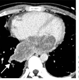
좌심방이 혈전의 감별진단에는 혈류 정체와 빗살근에 의한 난류(turbulent flow)가 포함되며(9) 두 가지 모두 CT에서 충만결손으로 보인다. 보고된 바에 의하면 단일 시기(monophasic) 영상의 양성예측도(positive predictive value)는 41~91%로 변이도가 크기 때문에 신뢰하기 어렵다. 반면 조영제 투여 60초 혹은 90초 후의 지연기(delayed phase) 영상을 추가적으로 획득할 경우 양성예 측도가 92%로 증가할 뿐 아니라 일관되게 높은 값으로 보고되어 신뢰할 수 있다. 혈류 정체로 인 한 충만결손은 지연기 영상에서 조영제에 의해 가려져 없어지지만(Fig. 1) 혈전에 의한 충만결손 은 지연기에서도 여전히 낮은 밀도의 결손으로 보인다(Fig. 2) (5). 더 나아가 3D 재구성 기법을 적
A B
Fig. 1. LAA blood stasis in a 57-year-old man with a history of atrial fibrillation.
A. CT angiography in the arterial phase shows a suspicious, irregular filling defect (arrow) at the LAA near the apex.
B. The defect is obscured on the delayed-phase image. It is probably a pseudolesion caused by blood stasis.
LAA = left atrial appendage
용한 CT 영상으로 좌심방이의 내부 유속(flow velocity)과 구혈률(ejection fraction)을 계산하여 혈전을 유발할 수 있는 기능적 상태를 평가할 수 있다(22, 24, 25).
좌심방 내 혈전(intra-atrial thrombus)은 좌심방이 혈전에 비해 드물며 양측 심방 어디에서나 생길 수 있지만 좌측에서 더 빈번하게 생기는 것으로 보고된다. 심방 내 혈전의 선행 요인들에는 혈전성향증(hypercoagulability), 부정맥, 국소 벽운동 이상(regional wall motion abnormality) 을 동반한 수축기 기능장애 및 인공기구 삽입 과거력이 해당된다. 조영증강 CT에서 심방 내부에 저감쇠의 충만 결손으로 보이는데 신생물로 오인될 수 있어 해석에 주의를 요한다. 운동성이 없고 다발성으로 나타나는 경우 종양보다는 혈전을 시사한다. 특이할 점은 만성 혈전에서 드물게 내부 에 석회화가 생기거나 혈관신생(neovascularization)으로 인해 주변부가 조영증강되어 점액종 (myxoma)처럼 보일 수 있다는 것이다. 따라서 다양한 선행 요인의 유무, 넓은 부착 부위, 모양 그 리고 위치 등을 포괄적으로 고려해서 감별진단해야 한다(26, 27).
종괴(Cardiac Mass)
현재의 가이드라인에 따르면 심장 종괴의 평가 시 권장되는 주된 촬영 기법은 심장 초음파와 MR이다. CT는 일반적으로 MR에 대한 금기 사항이 있거나 다른 영상 검사 결과가 모호한 환자에 적응된다. 하지만 보다 중요한 것은 CT 영상이 전체적인 해부학적 지형을 묘사해 줌으로써 전이성 병변이나 수술 전 평가에 이득을 준다는 것이다. CT에서 심장 종괴가 발견되었을 때, 임상 정보와 크기, 수, 위치(심장의 방 안에 위치하는지, 심낭막 혹은 심장 밖으로 침범하고 있는지), 모양, 경계 및 조직의 특성(석회화, 지방, 혈관)과 같은 영상 소견들을 종합적으로 평가하여 감별진단할 수 있 다. 심장의 종괴는 크게 신생물과 비신생물로 나뉘고 신생물은 양성 혹은 악성으로 분류된다(26).
A B
Fig. 2. LAA thrombus in a 57-year-old woman with a history of atrial fibrillation and right middle cerebral ar- tery infarction.
A. CT angiography in the arterial phase reveals a 5.4 × 3.0 cm-sized irregular filling defect (arrow) at the LAA.
B. The defect (arrow) persists on the delayed-phase image, which is diagnostic of a thrombus.
LAA = left atrial appendage
점액종(myxoma)은 원발성 심장 종양 중 가장 흔하다(25~50%). 대부분의 점액종은 매끄럽거 나(smooth) 분엽성(lobulated)의 타원형 종괴가 얇은 경부를 통해 심장 내벽에 부착되어 있는 소 견으로 보고된다. 흔한 부착 부위는 심방 내벽 혹은 중격이며 주로 단일 병변으로 존재한다. 조영 증강 전 영상에서 혈액과 유사한 하운스필드 수치를(Fig. 3A) 보이며 간혹 내부에 석회화를 포함 하기도 한다. 조영증강 영상에서는 심방 내 충만 결손(Fig. 3B)으로 보이고 괴사, 낭성 및 출혈 등 다양한 변성으로 인해 불균질한 조영증강을 나타낸다. 감별진단에는 혈전과 판막 증식증(valvu- lar vegetation)이 있다. 점액종은 방실판막을 통해 심실로 탈출할 수 있으며 영화자기공명영상 (cine MRI)나 심초음파를 통해서 점액종이 심장과 혈액의 움직임에 따라 운동하는 모습을 관찰할 수 있다(26, 28).
A B
A B
Fig. 3. Myxoma in a 54-year-old woman.
A. The precontrast image shows an indistinct, round, hypoattenuated (mean CT attenuation value: 23.1 HU) lesion (arrow) at the LA.
B. The lesion is better delineated on the arterial-phase image, appearing as a 2.7 × 2.7 cm-sized smooth, round intracavitary filling defect (arrow) with poor enhancement (mean CT attenuation value: 53.1 HU) and suspicious attachment to the septal wall of the LA (not clearly shown on this image).
HU = Hounsfield unit, LA = left atrium
Fig. 4. Fibroelastoma in an 81-year-old woman with multifocal embolic infarction.
A. CT angiography shows a multilobulated, hypoattenuated lesion at the left atrium (arrow), measuring 1.2 × 1.6 cm.
B. There is a narrow stalk (arrow) attaching the lesion to the mitral valve.
유두상 섬육탄력종(papillary fibroelastoma)은 세 번째로 흔한 원발성 양성 심장 신생물로 병 리적으로 무혈관성의 결합조직이 종려잎 모양으로 밀집되어 있는 것이 특징이다. 약 90% 이상의 경우 판막 표면에서 기원하고 좌측이 우측보다 발생 빈도가 약간 높은 것으로 알려져 있다. 심장 초음파 검사에서 분엽성의 작고(직경 15 mm 미만) 유동성을 보이는 단일 종괴가 짧은 줄기를 통 해 판막이나 심내막에 붙어있는 소견으로 나타난다(Fig. 4). 하지만 작은 병변의 크기와 부착 부위 인 판막의 지속적인 움직임 때문에 CT나 MR에서 발견되는 경우는 매우 드물다(26, 29).
심방중격지방종성비후(lipomatous atrial septal hypertrophy)는 난원와(fossa ovalis)를 제외 한 심방중격의 근세포(myocyte)에 비정상적으로 지방세포(adipocyte)가 침윤하여 생기는 비피 막형(nonencapsulated) 양성 종양이다. 조영증강 CT에서 매끈한 덤벨(dumbbell) 모양의 조영증 강되지 않는 저밀도(-50 HU 미만) 종괴로 나타난다. 중격을 넘어서 존재하지 않으며 보통 꼬리 쪽 보다 머리 쪽이 두껍다(Fig. 5). 풍부한 심외막하 지방(subepicardial fat)이 흔히 동반되고 나이와 체중과 상관관계가 있을 것으로 추정된다(30). 대표적인 감별 질환으로 지방종(lipoma)이 있는데, 지방종은 피막을 갖고 있는 원형의 균일한 종괴로 심근섬유를 침윤하지 않는다. 그 외 지방육종 (liposarcoma)과 과거 심근 경색으로 인한 지방 화생(fatty metaplasia)도 감별 질환에 포함된다.
악성 종양의 심장 전이(metastatic tumors)는 심장에서 가장 흔하게 발견되는 신생물로(26) 전 이 경로는 림프성, 혈행성, 직접 침범 및 경정맥(transvenous) 경로 등으로 나눌 수 있다. 특히 좌 심방에 생기는 이차성 심장 종양은 대부분 인접 기관에서 악성 종양이 직접 침범해 오거나 폐정맥 을 타고 와서 생긴다. 직접 침범의 경우 주로 거대한 기관지원성 암종(bronchogenic carcinoma) 이 원인이 되는 경우가 많고, 이 경우 T4 병기에 해당한다. CT 혹은 MR 영상에서 종양에 의한 폐 정맥의 폐쇄가 확인되면 좌심방으로의 전이를 의심해야 한다. 이 외에 식도암, 종격동 림프종, 및 유방암도 주변 구조물을 파괴하며 좌심방으로 직접 전이할 수 있다(31). 경정맥 전이는 원발성 혹
Fig. 5. Lipomatous hypertrophy of the interatrial septum in a 63-year-old woman.
A. The precontrast image shows a smooth, dumbbell-shaped fat density lesion (long arrows) confined to the septum, accompanied by abundant subepicardial fat deposits (short arrows).
B. On postcontrast image, the fatty lesion (long arrows) is slightly hypodense relative to the surrounding soft tissues.
A B
은 이차성 폐암의 종양혈전(tumor thrombus)이 폐정맥을 통해 좌심방 강내에 파종되는 것으로 영상에서 폐정맥에서부터 좌심방으로 이어지는 형태의 종양을 관찰할 수 있다(Fig. 6) (31). 전이 성 신생물에 특이적인 CT 영상 소견은 없다. 하지만 높은 병기의 암 환자에서 조영증강되는 심장 종괴가 발견되고 심낭삼출을 동반하며 직접 침범 혹은 경정맥 전이 소견이 있는 경우 전이성 심장 종양을 강하게 의심할 수 있다(32).
횡문근육종(rhabdomyosarcoma)은 횡문근에서 기원하는 악성 신생물로 원발성 악성 심장 종 양 중 가장 흔하다(20%). 횡문근에 생기기 때문에 심근에서 찾아볼 수 있지만 심장판막, 심외막의 지방(epicardial fat), 심장막 공간(pericardial space) 혹은 심장 외 구조물들을 침범하기도 한다.
발생 빈도는 심장의 4가지 방(chamber) 모두 비슷한 수준이며 과반수에서 병변이 여러 곳에서 발 견된다. 심장 CT에서 횡문근육종은 주로 크고 불규칙한 경계와 낮은 감쇠를 나타내는 심방 내 종
Fig. 7. Spindle cell rhabdomyosarcoma with myxoid stroma in a 40-year-old man.A. There is a large (8 × 4.7 cm), broad-based mass with irregular margin in the LA (short arrows). The mass infiltrates the anterior wall of the LA, appearing as diffuse wall thickening with suspicious nodular irregulari- ties (long arrow).
B. The tumor is attached to the LA roof and further invades the endocardium, which is diffusely thickened with eccentric mass-forming components (arrows).
LA = left atrium, LV = left ventricle
A B
LV
LA LA
Fig. 6. Metastatic osteosarcoma in a 28-year-old man.
Contrast-enhanced axial CT reveals a bulky, heteroge- neously enhancing cardiac mass extending into the LA via the right inferior pulmonary vein (arrow).
LA = left atrium LA
괴로 보인다(Fig. 7). 괴사와 출혈 변화도 흔히 동반되는 소견이다. 초기에는 심근이 단순히 두꺼워 진 것처럼 보일 수 있으나 크기가 커지거나 공격적인 성장을 보이는 경우 주변으로 침윤하는 모습 을 관찰할 수 있다(26).
선천성 기형
삼심방증(cor triatriatum sinister)은 매우 희귀한 질환으로 심방이 두꺼운 섬유근막(fibro- muscular membrane)으로 이분되는 선천 기형을 뜻한다(Fig. 8). 소아 환자의 경우 80%에서 폐 정맥 환류이상(anomalous pulmonary venous return), 심방중격결손 및 활로 4징(tetralogy of Fallot) 등과 같은 선천성 기형을 동반한다. 섬유근막은 온전한 벽에서부터 다공성의 막까지 넓은 스펙트럼으로 존재하여 이분할된 심방 내 구획들 간의 개통성에 영향을 미치고, 이는 증상의 중증 도를 결정한다. 삼심방증은 승모판막 협착증(mitral stenosis), 판상부환(supravalvular mitral ring) 혹은 폐정맥 협착증(pulmonary venous stenosis)과 같이 폐정맥계와 좌측 심장 사이에 혈 류 장애를 일으키는 질환들과 유사한 혈역학적 변화를 유발한다(33, 34). 따라서 환자들은 운동 시 호흡곤란이나 폐부종의 징후들 혹은 부정맥으로 병원을 찾게 된다. 무증상인 경우에도 시간이 지 나면 결국 섬유근막의 구멍에 석회화와 섬유성 변화가 일어나기 때문에 증상이 발현된다. 삼차원 볼륨 렌더링 기술을 결합한 심장 CT는 섬유근막의 해부학적 구조 및 수술 전 평가를 위한 고품질 이미지를 제공해주며 심방 내 혈전, 점액종 혹은 삼심방증에 동반되는 기타 선천 기형도 함께 진 단해낼 수 있다는 장점이 있다. 하지만 CT는 혈역학 혹은 생리적 정보를 제공해주지 못하는 제한 점이 있고(35) 특히 소아 환자에서 방사선 피폭선량 관리에 신경 써야 한다.
난원공 개존증(patent foramen ovale; 이하 PFO)은 생후 일차심방중격(septum primum)과 이차심방중격(septum secundum)의 불완전한 융합으로 초래되는 기형이다. 현재까지는 경식도 초음파가 진단의 황금기준(gold standard)이나 다중검출기 CT도 우수한 진단능을 보인다. CT에 서 심방중격에 위치한 개방된 통로를 통해 조영제가 한 쪽 심방에서 다른 쪽으로 빠르게 흐르는
Fig. 8. Cor triatriatum sinister in a 21-year-old woman.A, B. CT angiography reveals markedly enlarged LA with a thin fibromuscular membrane (arrows) bisecting the cavity. The proximal chamber is contiguous to the pulmonary venous confluence, and bulging distal chamber is connected to an enlarged LAA. The RA is also enlarged.
C. A small fenestration (13 mm, arrowhead) is detected at the lower portion of the membrane, allowing decompression of the pulmonary veins.
LA = left atrium, LAA = left atrial appendage, LV = left ventricle, RA = right atrium
A B C
LV RA LV
LAA
Fenestration Distal LA
Distal LA Proximal LA
Proximal LA
(jet flow) 것이 관찰된다면 진단할 수 있다(Fig. 9). 추가적으로 좌우단락(left-to-right shunt)의 난 원공개존의 경우 일차심방중격의 유리피판(free flap)이 짧거나 심방중격동맥류가 흔히 동반되므 로 이러한 소견을 찾아 진단에 도움을 받을 수 있다. 난원공개존의 유병률은 전체 인구의 약 25~30%로 추정될 정도로 흔한 기형에 해당하고 일반적으로는 치료하지 않는다. 젊은 환자에서 나타나는 원인불명 뇌경색, 전조증상이 있는 편두통 그리고 잠수병(decompression sickness) 등 과 강한 연관성이 있는 것으로 생각되며 이러한 경우엔 경피적 난원공 폐쇄술(percutaneous PFO closure)을 시행해볼 수 있으나 아직까지 해당 시술의 적응증에 관해 합의된 내용은 없다 (36). 한편으로 열려있는 난원공이 오히려 카테터가 지나는 통로가 되어주어 카테터 기반 시술 시 활용 가치가 있다는 평가도 있으며 특히 이런 경우 CT를 통해 난원공개존의 진단과 시술 전 해부 학적 평가를 모두 할 수 있는 장점이 있다. 또한 CT는 비침습적이고 빠르다는 장점이 있으나 한 번 의 호흡 정지 동안 구득해야 하기 때문에 우좌단락을 유발하기 위해 발살바 조작을 할 수 없다는 제 한점이 있다(3, 37).
심방중격주머니(atrial septal pouch) 일차 심방중격과 이차 심방중격의 불완전한 융합에 의해 형성된 주머니형 구조이다. 융합 부위에 따라 입구가 좌측, 우측 혹은 양측 심방 모두로 열릴 수 있 다. 중격주머니는 형성 과정상 난원공개존과 공존할 수 없으며 두 가지는 양측 심방 간 연결 통로 유무로 간단하게 구별할 수 있다. 조영증강 심전도 동조화 다중검출기 CT (contrast enhanced ECG-gated multi detector CT)는 생리식염수 주입과 발살바 조작을 하면서 시행한 경식도초음파 와 비교해보았을 때 좌측 심방중격주머니를 식별해내는 능력은 유사하였으나 난원공개존과 구분 하는 데에는 열등한 결과를 보였다(38). 좌측에 생긴 심방중격주머니는 혈전을 형성하는 잠재적 병 소로 알려져 있을 뿐 아니라, 주머니 끝(apex) 부위의 반흔 조직은 정상 전기전도에 간섭을 일으키 는 것으로 생각되어 현재 허혈성 뇌졸중, 심방세동 및 편두통과의 연관성을 두고 연구들이 진행되 고 있다. 반면 우측에 생긴 심방중격주머니는 임상적으로 문제를 일으키지는 않는 것으로 보고되 고 있다(39).
심방중격동맥류(atrial septal aneurysm)는 심방중격이 양측 심방 중 일측 혹은 양측 모두로 중 격 평면에서 10 mm 이상 돌출된 비정상적인 상태를 말한다(Fig. 10) (36).
Fig. 9. Patent foramen ovale incidentally detect- ed in a 66-year-old woman.
The CT angiography multiplanar reformatted im- age shows a slit-like channel at the interatrial septum with a visible jet-flow (arrow) of the con- trast agent from the LA to the RA.
LA = left atrium, RA = right atrium LA
RA
심방중격결손증(atrial septal defect)은 성인에서 가장 흔하게 발견되는 선천성 심장 기형 중 하 나로 배아발생 단계에서 조직 형성에 실패하여 심방중격에 양측 심방을 연결하는 구멍이 남은 것 이다. 빈도순으로 이차공(secundum), 일차공(primum), 정맥동(sinus venosus) 및 관상정맥동 (coronary sinus) 심장중격결손 등 네 가지 아형이 있다(Fig. 11) (37). 각 유형의 심방중격결손증은 다른 심장 또는 심장 외 기형을 흔히 동반하는 것으로 보고된다. 무증상일 때는 치료적 개입을 하지 않지만 크기가 크거나 혈역학적 이상이 발생할 경우 수술적 교정을 해주어야 한다(27, 37, 40).
정상 변이와 해석상의 함정들
좌심방이 부속물(accessory left atrial appendage)과 게실(diverticulum)은 좌심방에서 튀어 나온 작은 구조물들로 정상 변이에 해당한다. 일상적인 판독 시에도 자주 볼 수 있을 만큼 흔한데, 각각의 유병률은 6.5%에서 28% 그리고 16.7%에서 38.2%로 보고된다(41). 먼저 좌심방이 부속물 은 내벽의 빗살근에 의해 불규칙한 윤곽으로 나타나며 좁고 명확한 개구부를 갖고 있다(Fig. 12).
주로 좌심방의 전벽이나 측벽에서 관찰되며 좌심방이와 별개의 구조물로 존재한다. 반면, 게실은 주머니형으로 매끈한 윤곽으로 보이고 개구부가 넓으며 좌심방의 전상부벽 혹은 측벽에서 발견 된다(Fig. 13). 이 두 가지 변이들은 한 환자에서 함께 발견되기도 하고 한 가지 변이가 여러 개 존 재하는 경우도 있다. 아직까지는 관련 연구가 빈약하고 그나마 있는 연구들 간에도 모순적인 결과 를 보이기 때문에 두 가지 정상 변이가 심방세동, 혈전색전증 혹은 심부전 등의 위험을 증가시키 거나 해당 질환들의 불량한 결과와 연관이 있는지 알 수 없다(41). 그러나 이러한 변이들이 관찰될 경우 판독문에 포함해주어야 하는데 특히 카테터 절제술과 같은 시술 전이라면 카테터 조작에 영 향을 줄 수 있기 때문이다(9).
쿠마딘 능선(coumadin ridge) 혹은 좌측능선(left lateral ridge) 근육 조직으로 이뤄진 배아 잔
Fig. 10. Atrial septal aneurysm in an 88-year-old woman.A, B. The CT angiography multiplanar reformatted images show the interatrial septum bulging away (ar- rows) from the septal plane by approximately 1.2 cm.
LA = left atrium, RA = right atrium
A B
LA
H LA
RA
RA R
A P
1 mm/div
1 mm/div
TERARECON
류물로 내부에 좌심방 경사정맥(oblique vein of left atrium 혹은 vein of Marshall), 풍부한 자율신 경다발 및 심방 동맥들이 지나간다. 좌측 상폐 정맥과 좌심방이 입구 사이에 위치하며(Fig. 14A-C) (4, 42) 돌출된 모양으로 인해 심초음파(Fig. 14D)나 다른 영상 검사에서 혈전이나 종양으로 오인 되는 경우가 있어 불필요한 검사나 치료를 하게 되기도 하므로 해석에 주의해야 한다. 쿠마딘 능 선이라는 이름도 과거에 혈전으로 오인하여 와파린(상품명 쿠마딘) 치료를 하였던 이력으로 인해 붙여진 것이다(43).
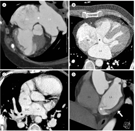
Fig. 11. Four types of ASDs.
A. Secundum ASD in a 74-year-old woman. CT angiography reveals a large interatrial septal defect (asterisk) measuring approximately 22 mm with free mixing of blood between the two atria.
B. Primum ASD in a 54-year-old man. Axial CT angiography shows an interatrial septal defect (asterisk) mea- suring approximately 12 mm, adjacent to the atrioventricular canal.
C. Sinus venosus ASD with partial anomalous pulmonary venous return in a 61-year-old man. Axial contrast- enhanced CT shows pathological interatrial communication beyond the confines of the atrial septum with overriding of the SVC. Notice the abnormal return of the right upper pulmonary veins (asterisk) draining di- rectly into the SVC.
D. Coronary sinus ASD with atresia of the coronary sinus ostium in a 19-year-old man. Contrast-enhanced CT shows no visible communication between the coronary sinus and the RA. However, there is an abnormal venous drainage from the coronary sinus (arrow) to the LA via a direct communication (asterisk) between the two structures.
ASD = atrial septal defect, LA = left atrium, RA = right atrium, SVC = superior vena cava A
C
B
D
LA LA LA
LA RA
SVC RA
RA RA
1 mm/div
1 mm/div
TERARECON
Spin: -45 Tilt: -16
좌심방 띠(left atrial band)는 드문 선천성 변이로 심방 내막의 두 지점을 잇는 섬유근 구조물이 다. 가장 흔한 기시점은 좌측 난원와이고 보고된 바에 의하면 키아리 네트워크(Chiari’s network) 와 난원공 개존증(PFO)과 같은 변이를 잘 동반한다(44). 대부분 무증상이지만 승모판 역류(mitral regurgitation) 및 심방세동과의 연관성도 보고되고 있다. 영상에서 좌심방띠는 삼심방증(cor tri- atriatum)으로 오인될 수 있어 판독에 주의를 요하며, 심도자술 시 카테터 진행을 방해할 수 있어 시술 전 영상 검사에서 해당 변이의 위치를 파악하여 판독문에 기재해주어야 한다(45).
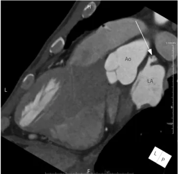
Fig. 12. Accessory left atrial append- age in a 48-year-old man.
The CT angiography multiplanar re- formatted image shows a cauliflower- like lesion (arrow) with a narrow base, protruding from the superoanterior portion of the left atrial wall (height: 12 mm; width: 9 mm; neck: 3 mm).
Ao = aorta, LA = left atrium
Fig. 13. Left atrial diverticulum in a 46-year-old man.
The CT angiography multiplanar re- formatted image shows a smooth sac-like lesion (arrow) with a broad base, protruding from the superoan- terior portion of the left atrial wall (height: 5 mm; width: 3 mm; neck: 7 mm).
LA = left atrium
L P LA Ao H
L
F
1 mm/div
1 mm/div
TERARECON
LA
L P H
A
F
1 mm/div TERARECON
1 mm/div
결론
CT는 가장 널리 보급된 영상기법들 중 하나로서 최적의 이미지 획득을 위한 다양한 기술들이 개발됨에 따라 심장의 평가에도 유용한 도구로 부상하였다. 좌심방의 재형성, 혈전, 종괴 및 선천 성 기형과 같은 좌심방 본체 및 좌심방이의 다양한 병리적 상태들은 CT 소견만으로도 잠정 진단 을 내리기에 충분한 경우가 많다. 하지만 CT는 기능 분석과 조직 특성화에 약점을 보이기 때문에 MR과 심초음파와 같은 영상기법들을 보완적으로 활용해야 한다. 또한 CT 영상은 좌심방에 대해 뛰어난 해부학적 묘사가 가능하기 때문에 성공적인 수술과 카테터 기반 시술의 밑거름이 된다. 이 러한 목적으로 촬영한 심장 CT라면 심장의 기하학적 특징들(모양, 크기 그리고 주변 구조물과의 공간 관계)과 정상 변이(좌심방이 부속물, 게실, 좌심방 띠 및 쿠마딘 능선)를 체계적으로 평가해 주어야 한다.
Author Contributions
Conceptualization, Y.J.Y.; data curation, Y.J.Y., S.M.; formal analysis, Y.J.Y., S.M.; investigation, Y.J.Y., S.M.; methodology, Y.J.Y.; project administration, K.S.J., Y.J.Y.; resources, K.S.J., Y.J.Y., K.H.J., K.M.Y.;
supervision, Y.J.Y.; visualization, Y.J.Y., S.M., K.H.J., K.M.Y.; writing—original draft, Y.J.Y., S.M.; and writing—review & editing, K.S.J., Y.J.Y., S.M.
Conflicts of Interest
The authors have no potential conflicts of interest to disclose.
REFERENCES
1. Balli O, Aytemir K, Karcaaltincaba M. Multidetector CT of left atrium. Eur J Radiol 2012;81:e37-46
2. Lacomis JM, Ocak I, Knollmann F, Voigt A, Bazaaz R. Cardiac CT: electrophysiological applications. In Scho- epf UJ, ed. CT of the heart. Contemporary medical imaging. Totowa: Humana Press 2019:531-552
3. Ho SY, Cabrera JA, Sanchez-Quintana D. Left atrial anatomy revisited. Circ Arrhythm Electrophysiol 2012;
Fig. 14. Coumadin ridge mistaken for a cardiac mass in a 68-year-old woman.
A, B. CT angiographic images show an approximately 7 mm-sized hypoattenuated nodular lesion (arrows in A and B) between the LSPV and the orifice of the LAA.
C. Transesophageal echocardiography shows a 6 × 7 mm-sized echogenic mobile mass at the Q-tip.
LA = left atrium, LAA = left atrial appendage, LSPV = left superior pulmonary vein
A B
C
LA LAA
LAA LSPV
LSPV
5:220-228
4. Barbero U, Ho SY. Anatomy of the atria. A road map to the left atrial appendage. Herzschrittmacherther Ele- ktrophysiol 2017;28:347-354
5. Di Biase L, Natale A, Romero J. Thrombogenic and arrhythmogenic roles of the left atrial appendage in atri- al fibrillation. Circulation 2018;138:2036-2050
6. Korsholm K, Jensen JM, Nielsen-Kudsk JE. Cardiac computed tomography for left atrial appendage occlu- sion: acquisition, analysis, advantages, and limitations. Interv Cardiol Clin 2018;7:229-242
7. Xu B, Betancor J, Sato K, Harb S, Abdur Rehman K, Patel K, et al. Computed tomography measurement of the left atrial appendage for optimal sizing of the Watchman device. J Cardiovasc Comput Tomogr 2018;
12:50-55
8. Rajwani A, Nelson AJ, Shirazi MG, Disney PJS, Teo KSL, Wong DTL, et al. CT sizing for left atrial appendage closure is associated with favourable outcomes for procedural safety. Eur Heart J Cardiovasc Imaging 2017;18:1361-1368
9. Ismail TF, Panikker S, Markides V, Foran JP, Padley S, Rubens MB, et al. CT imaging for left atrial appendage closure: a review and pictorial essay. J Cardiovasc Comput Tomogr 2015;9:89-102
10. January CT, Wann LS, Calkins H, Chen LY, Cigarroa JE, Cleveland JC Jr, et al. 2019 AHA/ACC/HRS focused up- date of the 2014 AHA/ACC/HRS guideline for the management of patients with atrial fibrillation: a report of the American College of Cardiology/American Heart Association Task Force on Clinical Practice Guidelines and the Heart Rhythm Society in collaboration with the Society of Thoracic Surgeons. Circulation 2019;
140:e125-e151
11. Yaghi S, Chang AD, Akiki R, Collins S, Novack T, Hemendinger M, et al. The left atrial appendage morphology is associated with embolic stroke subtypes using a simple classification system: a proof of concept study. J Cardiovasc Comput Tomogr 2020;14:27-33
12. Thomas L, Abhayaratna WP. Left atrial reverse remodeling: mechanisms, evaluation, and clinical signifi- cance. JACC Cardiovasc Imaging 2017;10:65-77
13. Negishi K. Incremental predictive value of left atrial parameters over clinical risk scores for subsequent atrial fibrillation: function beyond size. JACC Cardiovasc Imaging 2019;12:990-992
14. Oliver W, Matthews G, Ayers CR, Garg S, Gupta S, Neeland IJ, et al. Factors associated with left atrial remod- eling in the general population. Circ Cardiovasc Imaging 2017;10:e005047
15. Kuchynka P, Podzimkova J, Masek M, Lambert L, Cerny V, Danek B, et al. The role of magnetic resonance im- aging and cardiac computed tomography in the assessment of left atrial anatomy, size, and function.
Biomed Res Int 2015;2015:247865
16. Olsen FJ, Bertelsen L, De Knegt MC, Christensen TE, Vejlstrup N, Svendsen JH, et al. Multimodality cardiac imaging for the assessment of left atrial function and the association with atrial arrhythmias. Circ Cardio- vasc Imaging 2016;9:e004947
17. Whitaker J, Rajani R, Chubb H, Gabrawi M, Varela M, Wright M, et al. The role of myocardial wall thickness in atrial arrhythmogenesis. Europace 2016;18:1758-1772
18. Shirani J, Alaeddini J. Structural remodeling of the left atrial appendage in patients with chronic non-valvu- lar atrial fibrillation: implications for thrombus formation, systemic embolism, and assessment by trans- esophageal echocardiography. Cardiovasc Pathol 2000;9:95-101
19. Yu HT, Lee JS, Kim TH, Uhm JS, Joung B, Hong GR, et al. Advanced left atrial remodeling and appendage contractile dysfunction in women than in men among the patients with atrial fibrillation: potential mecha- nism for stroke. J Am Heart Assoc 2016;5:e003361
20. Hozawa M, Morino Y, Matsumoto Y, Tanaka R, Nagata K, Kumagai A, et al. 3D-computed tomography to com- pare the dimensions of the left atrial appendage in patients with normal sinus rhythm and those with par- oxysmal atrial fibrillation. Heart Vessels 2018;33:777-785
21. Kosiuk J, Uhe T, Stegmann C, Ueberham L, Bertagnolli L, Dagres N, et al. Morphological determinators of platelet activation status in patients with atrial fibrillation. Int J Cardiol 2019;279:90-95
22. Kim IC, Chang HJ, Cho IJ, Shim CY, Hong GR, Heo JH, et al. Benefit of four-dimensional computed tomogra- phy derived ejection fraction of the left atrial appendage to predict thromboembolic risk in the patients with valvular heart disease. Korean Circ J 2019;49:173-180
23. Lazoura O, Ismail TF, Pavitt C, Lindsay A, Sriharan M, Rubens M, et al. A low-dose, dual-phase cardiovascular CT protocol to assess left atrial appendage anatomy and exclude thrombus prior to left atrial intervention.
Int J Cardiovasc Imaging 2016;32:347-354
24. Pathan F, Hecht H, Narula J, Marwick TH. Roles of transesophageal echocardiography and cardiac comput- ed tomography for evaluation of left atrial thrombus and associated pathology: a review and critical analy- sis. JACC Cardiovasc Imaging 2018;11:616-627
25. Yasuoka R, Kurita T, Kotake Y, Akaiwa Y, Hashiguchi N, Motoki K, et al. A novel method to estimate blood flow velocity in the left atrial appendage using enhanced computed tomography: role of Hounsfield unit density ratio at two distinct points within the left atrial appendage. Heart Vessels 2017;32:893-901
26. Kassop D, Donovan MS, Cheezum MK, Nguyen BT, Gambill NB, Blankstein R, et al. Cardiac masses on cardi- ac CT: a review. Curr Cardiovasc Imaging Rep 2014;7:9281
27. Malik SB, Kwan D, Shah AB, Hsu JY. The right atrium: gateway to the heart--anatomic and pathologic imag- ing findings. Radiographics 2015;35:14-31
28. Araoz PA, Mulvagh SL, Tazelaar HD, Julsrud PR, Breen JF. CT and MR imaging of benign primary cardiac neoplasms with echocardiographic correlation. Radiographics 2000;20:1303-1319
29. Kim AY, Kim JS, Yoon Y, Kim EJ. Multidetector computed tomography findings of a papillary fibroelastoma of the aortic valve: a case report. J Korean Med Sci 2010;25:809-812
30. Laura DM, Donnino R, Kim EE, Benenstein R, Freedberg RS, Saric M. Lipomatous atrial septal hypertrophy: a review of its anatomy, pathophysiology, multimodality imaging, and relevance to percutaneous interven- tions. J Am Soc Echocardiogr 2016;29:717-723
31. Chiles C, Woodard PK, Gutierrez FR, Link KM. Metastatic involvement of the heart and pericardium: CT and MR imaging. Radiographics 2001;21:439-449
32. Lichtenberger JP, Reynolds DA, Keung J, Keung E, Carter BW. Metastasis to the heart: a radiologic approach to diagnosis with pathologic correlation. AJR Am J Roentgenol 2016;207:764-772
33. Is¸Ik O, Akyüz M, AyIk MF, Levent E, Atay Y. Cor triatriatum sinister: a case series. Turk Kardiyol Dern Ars 2016;44:20-23
34. Nassar PN, Hamdan RH. Cor triatriatum sinistrum: classification and imaging modalities. Eur J Cardiovasc Med 2011;1:84-87
35. Malik A, Fram D, Mohani A, Fischerkeller M, Yekta A, Mohyuddin Y, et al. Cor triatriatum: a multimodality im- aging approach. Can J Cardiol 2008;24:e19-20
36. Pizzino F, Khandheria B, Carerj S, Oreto G, Cusmà-Piccione M, Todaro MC, et al. PFO: button me up, but wait
… Comprehensive evaluation of the patient. J Cardiol 2016;67:485-492
37. Yasunaga D, Hamon M. MDCT of interatrial septum. Diagn Interv Imaging 2015;96:891-899
38. Holˊda MK, Krawczyk-Oz.óg A, Koziej M, Pawlik W, Sorysz D, Wszolˊek K, et al. Cardiac computed tomography compared with two-dimensional transesophageal echocardiography for the detection and assessment of atrial septal pouches. Int J Cardiovasc Imaging 2018;34:1305-1313
39. Mazur M, Jasinska KA, Walocha JA. The morphology, clinical significance and imaging methods of the atrial septal pouch: a critical review. Transl Res Anat 2018;13:7-11
40. Nicolay S, Salgado RA, Shivalkar B, Van Herck PL, Vrints C, Parizel PM. CT imaging features of atrioventricular shunts: what the radiologist must know. Insights Imaging 2016;7:119-129
41. Holˊda MK, Koziej M, Wszolˊek K, Pawlik W, Krawczyk-Oz.óg A, Sorysz D, et al. Left atrial accessory appendag- es, diverticula, and left-sided septal pouch in multi-slice computed tomography. Association with atrial fi- brillation and cerebrovascular accidents. Int J Cardiol 2017;244:163-168
42. Terpenning S, White CS. Imaging pitfalls, normal anatomy, and anatomical variants that can simulate dis- ease on cardiac imaging as demonstrated on multidetector computed tomography. Acta Radiol Short Rep 2015;4:2047981614562443
43. Lodhi AM, Nguyen T, Bianco C, Movahed A. Coumadin ridge: an incidental finding of a left atrial pseudotu- mor on transthoracic echocardiography. World J Clin Cases 2015;3:831-834
44. Okajima K, Imamura K, Kanda G, Shimane A. Left atrial anomalous muscular band detected by computed tomography before catheter ablation in a patient with atrial fibrillation. J Arrhythm 2015;31:64-65
45. Nnaoma C, Sandhu G, Sossou C, Vavilin I, Bustillo J, Garg A. A band that causes leaky valves: severe mitral regurgitation due to left atrial fibrous band-a case report and literature review. Case Rep Cardiol 2019;2019:
2458569
좌심방과 좌심방이의 전산화단층촬영 소견:
해부학, 정상변이 및 질환에 관한 임상화보
송민지
1· 김성진
1,2· 구현정
3· 김문영
4· 유진영
1*
전산화단층촬영(이하 CT) 영상 기술의 발전으로 박동하는 심장에 대한 섬세한 영상의학적 평가가 가능해졌다. 심초음파 및 MRI에 비해 심장 CT의 강점은 대부분의 의료기관에 보급 되어 있고 고품질 이미지의 빠른 생산이 가능하며 해부학적 묘사가 뛰어나다는 것이다. 좌심 방과 좌심방이에서 생기는 대부분의 변이 혹은 병리적 상태들은 CT 상의 소견만으로도 추정 진단을 내릴 수 있다. 또한 CT 영상은 성공적인 카테터 기반 시술 또는 수술에 중요한 해부학 적 정보들을 제공한다. 특히 좌심방과 좌심방이는 심방세동 환자들의 치료 및 관리에 중요한 데, 이는 다양한 카테터 기반 시술들이 두 구조물의 기계적 혹은 전기적 차단을 목표로 하기 때문이다. 따라서 임상적으로 의미 있는 판독을 위해서는 병리적 상태의 CT 소견 기술과 함 께 좌심방 및 좌심방이의 모양, 크기 및 주변 구조물과의 상대적 위치 관계 등에 대한 포괄적 인 검토를 해야 한다.
1충북대학교병원 영상의학과,
2충북대학교 의과대학 충북대학교병원 영상의학과,
3울산대학교 의과대학 서울아산병원 영상의학과,
4서울특별시보라매병원 영상의학과