ORIGINAL ARTICLE
화상센터에서 치료한 괴사성근막염의 임상적 고찰
김의명ㆍ전진우
1ㆍ김영민
1ㆍ윤재철
1ㆍ임해준
1ㆍ조용석
1ㆍ김도헌
1ㆍ허 준
1ㆍ전 욱
1한림대학교동탄성심병원 외과, 1한림대학교한강성심병원 화상외과
The Clinical Investigation of Necrotizing Fasciitis in Burn Center
Euimyung Kim, M.D., Jin Woo Chun, M.D.
1, Young Min Kim, M.D.
1, Jae Chul Yoon, M.D.
1, Hae Jun Lim, M.D.
1, Yong Suk Cho, M.D.
1, Dohern Kim, M.D.
1, Jun Hur, M.D.
1and Wook Chun, M.D.
1Department of Surgery, Hallym University Dongtan Sacred Heart Hospital, Hwaseong, 1Department of Burn Surgery, Hangang Sacred Heart Hospital, College of Medicine, Hallym University, Seoul, Korea
Purpose: The necrotizing fasciitis is a terrifying infectious disease that can rapidly spreads to surrounding tissues when fascia
is infected and it can cause sepsis to death if not properly diagnosed and treated. The purpose of this study is to investigate the characteristics, causes, and treatment methods of necrotizing fasciitis in Korea through reviewing patients admitted to our burn center.Methods: 21 patients with necrotizing fasciitis were selected for this study among those inpatients with electronic medical re-
cords (EMR) admitted to Hallym University Hangang Sacred Heart Medical Center from Jan 1, 2008 to June 30, 2019. The medical records and wound photos of those 21 selected subjects were reviewed.Results: There were 13 male and 8 female patients and mean age was 58.76 years old. 13 of 21 subjects were survived and
8 died (38% mortality rate). The surgical treatments performed were I&D, fasciotomy, debridement, allograft, burring, STSG, flap, and amputation. The most common causes were burns in 9 subjects (6 contact burns) and cellulitis occurred on skins in 5 subjects. And other various causes were observed as fournier’s gangrene, stab wound, intramuscular injection, tumor and bleu toe syndrome (toe necrosis). The infected areas were 11 feet and legs, 7 hips, 3 abdomen and trunk in 21 subjects. Of the 8 deaths, 3 were infected in feet and legs, 2 were infected in hips, and 2 were infected in abdomen and trunk. As for under- lying diseases, 12 patients with hypertension or diabetes were the highest and others such as cancer and stroke were found.Conclusion: The only method to increase the survival rate is to ‘suspect’ the disease as much as possible and perform early
extensive excision. It is advisable to treat the disease by the burn center to properly provide adequate and optimal wound management, infection control, medical care and nutritional supports. (J Korean Burn Soc 2019;22:66-70)Key Words: Necrotizing fasciitis, Burn center, High mortality
접수일: 2019. 10. 30, 수정일: 2019. 11. 27, 승인일: 2019. 11. 28 책임저자:전욱, 서울시 영등포구 버드나루로 7길 12
07247, 한림대학교한강성심병원 화상외과 Tel: 02-2639-5001, Fax: 02-2633-7571 E-mail: [email protected]
서 론
괴사성근막염은 흔치 않지만 빠르게 진행하는 질환으로 근막에 염증이 발생하며 초기에는 근막 위 피부와 근막 아 래 근육은 제외되어 발생하게 된다
1,2). 근막 감염을 일으키 는 균은 그룹 A 베타-용혈성 연쇄구균(Group A B-hemo-
lytic streptococcus) 뿐만 아니라 포도알균(Staphylococcus),
비브리오균(Vibrio) 심지어 혐기성세균(anaerobic bacteria)
도 원인이 된다. 여러 균이 동시에 원인이 되는 경우도 보고
되고 있다
3,4). 근막 감염을 일으키는 원인으로는 제왕절개
술, 회음절개, 복부수술, 복강경수술, 지방흡입술 등 다양하
다. 초기 진단이 어려운 이유는, 초기에 질환이 피부를 포함
하는 정도가 제한적이기 때문이라 생각된다. 주된 치료방
법으로 광범위한 외과적 절제술이 우선 필요하며, 이후 부
분층식피술이 시행되기도 한다. 이밖에 쇼크에 대한 수액
치료, 항생제 사용, 영양지원 등이 필요할 수 있다. 저자는
괴사성근막염을 치료하면서 화상외과의가 가장 적절한 치
료의사란 것을 깨달았으며, 여러 증례를 검토함으로서 우
Table 1. Characteristics of Patients
NO Sex Age Hospitalstay (days) Op. name Cause Suvive/
death Study Site Underlying,
medical history
1 F 55 106 Debri (2), STSG (3) CoB S (-) Rt.foot & leg RA
2 M 71 123 Debri, STSG (3) Slip down S (-) Rt.hip & thigh DM, HTN
3 F 1 38 Debri, allo,
STSG (1)
Cellulitis S (-) Lt.hip, thigh, flank, Rt.arm
IM injection
4 M 42 72 Debri, STSG (4),
burring
Cellulitis S (-) Both foot & leg DM, HTN
5 F 85 43 STSG Contaminated
syringe
D (-) Rt.leg FB 13%,HTN
6 F 79 169 Debri,allo, rotation flap, STSG
Cellulitis D MRI Cellulitis, perineum DM, HTN
7 M 41 177 Debri (2), local flap (3)
Slip down, cellulitis
S (-) Rt.buttock paraplegia
8 F 72 74 I&D, local flap (2) SB S (-) Lt.perinum & buttock DM
9 F 76 20 Debri (2),
Lt. BK amp
ChB (medicated patch)
D (-) Lt.foot, ankle, lower leg
DM
10 M 55 19 Debri & STSG Cellulitis S (-) Rt.abdomen & flank HTN, brain tumor op (3)
11 F 72 181 I&D (3), STSG (2) CoB D CT Sacral area HTN, DM, cervix
cancer
12 M 51 66 Debri, STSG, Local
flap
ChB (medicated patch)
S MRI Rt.foot & leg pre STSG site (1yr ago, TA)
13 M 47 96 Debri (3), STSG (2) CoB S MRI Lt.foot DM, HTN
14 M 46 43 Debri, STSG CoB S (-) Rt.leg spodylitis, local
clinic I&D (3)
15 M 81 10 Debri CoB D (-) Lower abdomen,
Lt.flank
local clinic manage (6 monthes) 16 M 40 154 STSG (3), I&D (5),
anal fistla op (2)
Stab wound by iron bar
S CT Both inguinal, buttock
17 M 90 6 Previous
fasciotomy
D (-) Rt.upper inner thigh HTN, previous care (40 days) 18 F 72 35 I&D(3), Debri (3) Fournier
gangrene
D (-) Buttock, inguinal HTN
19 M 58 172 I&D,Lt.BKamp, STSG (2)
Blue toe syndrome
S (-) Lt.foot & lower leg schizo, PTCA
20 M 55 66 Debri, burring,
Curettage (4)
CoB S (-) Rt.foot DM
21 M 45 21 I&D, Debride &
CPA
Fournier gangrene
D CT Abdomen gastric cancer
CoB = contact burn, ChB = Chemical burn, RA = rheumatoid arthritis, BK = below knee, Debri = Debridement, CPA = Cryo-preserved allograft skin coverage, PTCA = percutaneous transluminal coronary angioplasty.
리나라에 발생하는 괴사성근막염의 특징과 원인 그리고 치 료방법에 대해 자세히 알아보고자 하였다.
대상 및 방법
한림대학교한강성심병원에서 전자차트(EMR)가 사용된 2008년부터 2019년6월30일까지 입원 환자 중 화상센터에서 치료한 괴사성근막염 환자 21명을 대상으로 하였다. 21명
의 의무기록과 상처사진을 검토하였다.
통계는 연속 변수들이 정규 분포가 있는 경우 독립 t-test
을 시행했고, 정규분포를 따르지 않을 경우 Mann-Whitney
U-test로 분석했다. 범주형 변수들은 카이제곱검정(Chi-square
test)과 Fisher 정확검정(Fisher’s exact test)를 이용하여 평
가했다. P-value가 0.05이하일 경우 통계학적으로 유의성이
있는 것으로 보았으며, 모든 통계적 분석은 R-project 프로
그램 버전 3.6.0에서 이루어졌다.
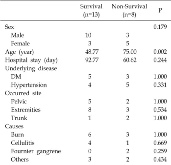
Table 2. Comparison Survivals and Non-Survivals
Survival(n=13)
Non-Survival
(n=8) P
Sex 0.179
Male 10 3
Female 3 5
Age (year) 48.77 75.00 0.002
Hospital stay (day) 92.77 60.62 0.244
Underlying disease
DM 5 3 1.000
Hypertension 4 5 0.331
Occurred site
Pelvic 5 2 1.000
Extremities 8 3 0.534
Trunk 1 2 1.000
Causes
Burn 6 3 1.000
Cellulitis 4 1 0.669
Fournier gangrene 0 2 0.259
Others 3 2 0.434
결 과
21명의 입원환자에게 괴사성근막염의 진단이 적용되었 다. 남자가 13명, 여자가 8명 이었다. 평균나이는 58.76세, 평균입원기간은 80.5일이었다. 본원에서는 20명에서 수술 적 치료가 시행되었으며 화상외과 전문의에 의해 시행되었 다. 21명 중 13명이 생존하였고 8명이 사망하였다(사망률 38%). 생존자들의 평균나이는 48.77세, 사망자들의 평균나 이는 75.00세였다. 평균 재원일수는 생존자의 경우 92.77일, 사망자의 경우 60.62일 이었다. 진단은 6예에서 MRI와 CT 가 사용되었고, 15예에서는 임상적 소견만으로 진단하였다.
수술적 치료방법은 변연절제술(debridement), 절개 및 배 농술(I&D), 피판술(flap), 버링술식(burring), 동종피부이식 술(allograft), 부분층식피술(STSG), 절단술(amputation)등 이 사용되었다. 적은 크기의 괴사성근막염은 여러 번의 절 개 및 배농술과 국소피판술(local flap)로 치료하였고, 큰 크 기의 괴사성근막염은 광범위절제술(wide excision), 동종피 부이식술(allograft), 부분층식피술(STSG)을 사용하여 치료 하였다. 발생원인으로는 화상이 9명(접촉화상이 6명)으로 제일 많았고 피부에 발생한 봉와직염(cellulitis)가 5명, 회음 부 괴사성근막염(fournier’s gangrene), 미끄러져 넘어짐 (slip down)시 충격 등 다양하였다. 발병 부위는 발 다리가 11명, 엉덩이(hip), 회음부(perineum), 둔부(buttock), 천골 부(sacral area)를 포함하는 골반부위가 7명, 복부 및 몸통이 3명이었다. 기저질환(underlying disease)에는 고혈압이 9 명, 당뇨가 8명에서 동반되어 제일 많았다(Table 1).
고 찰
본원의 경우 사망률이 38%이며, 괴사성근막염은 치사률 이 매우 높은 질환임을 알 수 있다. 전세계적으로 괴사성근 막염의 사망률은 6%에서 74%까지 보고되었으며, 평균은 38%정도이다. 만일 당뇨병이 동반되었다면 사망률은 63∼
71%까지 증가하게 된다
5).
근막에 외상이나 창상, 화상 등 기타이유로 감염이 발생 하고, 감염을 일으킨 균이 근막 층을 통해 인근 부위로 퍼져 나가게 되어 결국 광범위한 감염을 일으키게 된다. 이러한 과정에는 2가지의 핵심적 요인이 관여하게 되는데 첫 번째 요인은 근막 층은 감염의 수평적 전파를 막을 수 있는 물리 적 방어벽이 존재하지 않는다는 것이고, 두 번째 요인은 근 막은 피부 밑에 존재하기 때문에 초기 근막의 염증이 피부 에 의해 차폐(masking)가 되어 진단이 지연될 수 있으며, 염증이 진행하는 경우에는 피부 밑에서 압력하 농양
(pus-under pressure) 상태가 되어, 쉽게 패혈증으로 진행 될 수 있다는 점이다.
광범위화상환자의 가피절제술 방법 중 근막위절제술 (fascial level excision)이 있는데, 쉽게 근막과 피하지방층 이 분리되는 것을 보면 근막이 감염의 전파가 매우 용이하 다는 것을 알 수 있겠다. 또한 질병 초기에는 피부의 침범이 제한되어있기 때문에 더더욱 질병의 진단이 어렵게 된다.
21명의 증례환자 중에서 13명이 생존하고 8명이 사망하 였다. 어느 정도 병이 진행된 후 본원으로 전원 된 경우가 대부분인 점을 감안하더라도 사망률이 매우 높은 질환이 다. 사망자의 경우 평균나이가 75세로 생존자의 평균나이 인 48.77세보다 높았으며 통계적 의의가 있었다. 전체 발생 연령대는 40∼70대가 대부분이었고 대부분 기저질환이 동 반되어있었다. 하지만 생후 2개월된 아이도 이환 되었으며, 아마도 근육주사기가 원인이 아니었나 유추해본다. 주사기 가 원인인 다른 경우가 85세 환자도 있었기에, 영아나 유아, 혹은 노인에서는 오염된 주사기도 충분히 원인이 될 수 있 음을 알 수 있다.
남자 환자 13명중 3명이 사망하였으나 여자 환자 8명 중
5명이 사망하여 여자에서 예후가 더욱 좋지 않았으나 통계
적 의의는 없었다. 발생부위별 비교에서는 몸통부위는 발
다리(12례)나 골반부위(7례)보다 발생빈도는 적었으나 사
망률은 상대적으로 높았다(3명 중 2명 사망). 증례 수가 적
어 통계적 의의는 없었지만, 아마도 감염 부위가 심장과 폐
와 가까워지고 몸통의 경우 감염의 전파가 빠르며 광범위
한 괴사를 일으키기 때문이라 생각된다(Table 2).
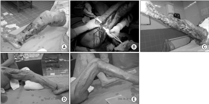
Fig. 1. Clinical case (occurred on Rt. leg). (A) Initially small contact burn wound on Rt. foot→aggravated, suspect necrotizing fasciitis
(transferred to our burn center). (B) Wound exploration, conform necrotizing fasciitis, wound extension. (C) Wide wound excision including muscle fascia. (D) 1st skin graft. (E) 2nd skin graft.발생원인으로는 화상이 9례, 봉와직염(cellulitis)가 5례, 회음부 괴사성근막염(Fournier gangrene)이 2례, 기타 이유 가 5례였다. 회음부 괴사성근막염(Fournier gangrene) 2례 모두 사망하였다. 피부에 발생한 화상이나 봉와직염 (cellulitis)은 예전에는 흔하지 않은 원인이었지만, 본원이 화상센터가 특화되어 이러한 증상을 가지는 환자들을 많이 치료하기 때문에 원인들 중 차지하는 빈도가 높았을 수도 있겠다.
괴사성근막염에 대한 외국 논문은 대부분 1900년도 후반 까지 있었으며, 2000년 이후에는 보고가 거의 없었다. 아마 도 예전에는 오염된 수술 기구로 복부 수술 혹은 산과수술 등을 하다가 발생하는 경우가 있었지만 멸균법의 발전으 로, 최근에는 그러한 원인이 감소하면서 질환의 유병률도 크게 줄었기 때문이라 생각된다.
치료에서 제일 중요한 핵심 포인트는 ‘질환의 의심’이다.
괴사성근막염의 가능성을 염두하고 항상 상처를 확인해야 한다. 경우에 따라 피부를 절개해야 확인 가능함으로 부담 감이 있지만, 질환의 진행이 워낙 빠르기 때문에 고민하다 가 환자를 놓칠 수 있다. 만일 임상적으로는 의심되지만 육 안적으로 불명확하다면 MRI를 촬영하여 진단이 가능하다.
MRI 이용이 어렵다면 CT나 초음파 촬영도 도움을 받을 수 있겠다. 괴사성근막염이 저명하게 의심된다면 전신마취 하 에 절개 및 배농술(I&D)을 시행하는 것이 좋으며, 이후 육 안으로 괴사성근막염이 확인된다면 절개(incision)를 충분
히 연장하여야 하고 감염된 근막이 있는 부위 상층의 피하 지방 및 피부는 절제(excision) 해야 한다(un-roofing tech- nique). 물론 감염된 근육도 동시에 제거가 필요하다(Fig.
1). 절제범위가 넓다면 사체피부이식을 동시에 시행하는 것 이 좋다. 수술 시 주의할 점은 피부가 괴사된 경계선보다 그 밑의 근막 감염은 적어도 5∼10 cm 이상 넘어간 부위까 지 진행되어있는 수가 있기 때문에, 반드시 감염된 근막을 먼저 확인하고 그 위의 정상으로 보이는 피부도 동시에 제 거해야 한다(Fig. 2). 이러한 술식을 통해 압력하 농양 (pus-under pressure) 상태의 위험으로부터 최대한 피해갈 수 있다. 처음 수술 시에 제거되지 못한 감염된 근육과 근막 은 여러 번의 이어진 변연절제술(serial wide excision)을 통 해 제거될 수 있다. 변연절제술 간에는 마페니드 아세테이 트(mafenide acetate) 연고나 용액을 이용하여 드레싱을 한 다. 상처가 크다면 영양지원이 필요하며, 상처표면에 육아 조직이 어느 정도 발생하였을 때 피부이식술을 시행한다.
괴사조직이 모두 제거되었다면 통상적으로 14일정도 경과 하면 육아조직이 형성되기 때문에, 최대한 서둘러 부분층 식피술을 시행한다. 관절부위에는 무세포동종진피를 동시 에 적용할 수 있겠다.
결 론
괴사성근막염은 적절한 치료에 불구하고 사망률이 매우
Fig. 2. Clinical case (occurred on trunk). (A) Rapidly aggravated necrotizing fasciitis (transferred to our burn center). (B) Wide excision
(C) Un-roofing technique. (D, E) Covered with cadaveric allograft skin.높은 질환이다. 감염된 주사기, 강아지 등 반려 동물의 교 상, 넘어지면서 엉덩이의 충격, 조그마한 봉와직염이나 화 상 등 미약한 상처나 충격도, 환자가 당뇨와 같은 동반질환 을 가지며 면역력이 감소되어있다면, 이 치명적인 질환의 원인이 될 수 있다. 더욱이 피부에 의해 근막 염증이 차폐 (masking)될 수 있으며, 이로 인해 진단이 더욱 지연될 수 있다. 생존률을 높일 수 있는 유일한 방법은 최대한 이 질환 을 ‘의심’하고, 광범위한 절제를 조기에 시행하는 것이다.
광범위한 피부 및 근막의 절제 후 상처 관리, 감염관리, 내 과적 관리 및 영양지원을 위해서 되도록 화상센터에서 치 료하는 것이 좋겠다
6).
REFERENCES
1) Barillo DJ, Cancio LC, Kim SH, Shirani KZ, Goodwin CW.
Fatal and near-fatal complications of liposuction. South Med J 1998;91:487–92.
2) Green RJ, Dafoe DC, Raffin TA. Necrotizing fasciitis. Chest 1996;110:219–29.
3) Brook I, Frazier EH. Clinical and microbiological features of necrotizing fasciitis. J Clin Microbiol 1995; 33: 2382–7.
4) Lille ST, Sato TT, Engrav LH, Foy H, Jurkovich GJ. Necrotizing soft tissue infections: obstacles in diagnosis. J Am Coll Surg 1996;182:7–11.
5) Golshani S, Simons AJ, Der R, et al. Necrotizing fasciitis following laparoscopic surgery. Case report and review of the literature. Surg Endosc 1996;10:751–4.
6) Barillo DJ, McManus AT, Cancio LC, Sofer A, Goodwin CW.
Burn center management of necrotizing fasciitis. J Burn Care Rehabil. 2003 May-Jun;24:127-32.