2013, Vol. 57, No. 6
Printed in the Republic of Korea
http://dx.doi.org/10.5012/jkcs.2013.57.6.859 단신
(Notes)
Synthesis and Crystal Structures of Ni(II)/(III) and Zn(II) Complexes with Schiff Base Ligands
Bon Kweon Koo*
Department of Chemistry, Catholic University of Daegu, Gyeongbuk 712-702, Korea.
*E-mail: [email protected]
(Received September 27, 2013; Accepted October 26, 2013)
Key words: Ni(II)/(III), Zn(II), Schiff base, Crystal structure
Coordination polymers are of great interest due to their intriguing structural motifs and potential applications in optical, electronic, magnetic, and porous materials.1−3 The most commonly used strategy for designing such materials relies on the utilization of multidentate N- or O- donor ligands which have the capacity to bridge between metal centers to form polymeric structures.4 The Schiff bases with N,O,S donor atoms are an useful source as they are readily available and easily form stable complexes with most transition metal ions.5,6 Schiff bases are also important intermediates in synthesis of some bioactive compounds7 and are potent anti-bacterial, anti-fungal, anticancer and antiviral compounds.8
In this work, the Schiff bases, Hapb and Hbpb, derived from 2-acetylpyridene or 2-benzoylpyridine and benzhy- drazide were taken as trifunctional (N,N,O) monobasic ligand (Scheme 1). This ligand is of important because the π-delocalization of charge and the configurational flexi- bility of their molecular chain can give rise to a great vari- ety of coordination modes. Although many metal–Schiff base complexes have been reported, the 1D, 2D, and 3D networks of coordination polymers linked through the bridging of ligands such as dicyanamide, N(CN)2− as coli- gand have been little published.9,10 In the process of work- ing to extend the dimensionality of the metal-Schiff base complexes using benzilic acid as a bridging ligand, we obtained three simple metal (II)/(III) complexes of acetylpy-
ridine/2-benzoyl pyridine based benzhydrazide ligand. There- fore, we report here the synthesis and crystal structures of the complexes.
EXPERIMENTAL Chemicals and Measurements
All chemicals are comercially available and were used as received without further purification. Elemental analyses (CHN) were performed on a Vario EL EA-Elementar Ana- lyzer.
Preparation of [Zn(bpb)2] (1)
To a methanolic solution (30 mL) of Hbpb ligand (0.302 g, 1.0 mmol) was added Zn (ClO4)2·6H2O (0.372 g, 1.0 mmol).
To the resulting pale yellow solution was added a meth- anolic solution (3 mL) of benzylic acid (0.228 g, 1.0 mmol) and triethylamine (0.152 g, 1.5 mmol). The solution turned to yellow and was refluxed for 3 h to yield yellow solid. The yellow solid was isolated by filtration and air-dried. The yellow filtrate was kept at room temperature to give yel- low block crystals in good quality for X-ray crystallogra- phy. Yield: 63% (0.419 g) based on Zn. Elemental Anal.
Calcd. For C38H28N6O2Zn: C, 68.52; H, 4.24; N, 12.62.
Found: C, 68.30; H, 4.57; N, 12.30%.
Preparation of [Ni(bpb)2] (2)
The compound was prepared similarly by the method described above for the preparation of 1, with use of Ni(ClO4)2·6H2O instead of Zn(Cl−O4)2·6H2O. Yield 58%
(0.382 g) based on Ni. Elemental Anal. Calcd. for C38H28N6O2Ni: C, 69.22; H, 4.28; N, 12.75. Found: C, 68.72; H, 4.52; N, 12.28%.
Preparation of [Ni(apb)2]·ClO4·CH3OH (3)
The compound was prepared similarly by the method Scheme 1. Chemical structures of Schiff bases and their abbre-
viations.
860 Bon Kweon Koo
described above for the preparation of 1, with use of Ni(ClO4)2·6H2O and Hapb instead of Zn(ClO4)2·6H2O and Hbpb ligand, respectively. Yield 65% (0.432 g) based on Ni. Elemental Anal. Calcd. for C29H28N6O7ClNi: C, 52.24;
H, 4.23; N, 12.61. Found: C, 51.85; H, 4.48; N, 12.31%.
X-ray Structure Determination
Single crystals of 1−3 were obtained by the method described in the above procedures. Structural measure- ment for the complexes were performed on a Bruker SMART APEX CCD diffractometer using graphite monochroma- tized Mo-Kα radiation (λ = 0.71073 Å) at the Korea Basic Science Institute. The structures were solved by direct method and refined on F2 by full-matrix least-squares pro- cedures using the SHELXTL programs.11 All non-hydrogen atoms were refined using anisotropic thermal parameters.
The hydrogen atoms were included in the structure factor calculation at idealized positions by using a riding model, but not refined. Images were created with the ORTEP12 or
DIAMOND program.13 The crystallographic data for com- plexes 1−3 are listed in Table 1.
RESULTS AND DISCUSSION
The complexes of 1−3 were prepared from the methanolic solution of M(ClO4)2·xH2O (M = Ni, and Zn), Schiff base ligand, and bnzilic acid. Our first aim in this work was to obtain the coordination polymers which metal centers are bridged by the benzilic acid as mixed ligand. However, the benzilic acid present in the initial reaction mixture was not found in the crystalline product. Unfortunately, attempts to obtain the product containing the benzilic acid and to improve yields by varying stoichiometry, temperature, and other reaction parameters proved to be generally unsuc- cessful.
Description of the Structures
The molecular structure of complex 1 consists of one Table 1. Crystal data and structure refinement for complexes 1−3
Complex 1 2 3
Empirical formula C38H28N6O2Zn C38H28N6O2Ni C29H28N6O7ClNi
Formula weight 666.03 659.37 666.73
T(K) 200(2) 200(2) 296(2)
Crystal system Triclinic Triclinic Monoclinic
Space group P−1 P−1 P2(1)/n
a (Å) 10.6428(6) 10.7854(5) 8.648(2)
b (Å) 12.5122(7) 12.3971(5) 10.625(2)
c (Å) 12.9696(7) 12.6910(6) 33.376(6)
α (°) 66.234(1) 66.726(1)
β (°) 83.802(1) 83.472(1) 96.438(4)
γ (°) 83.602(1) 82.652(1)
V (Å3) 1567.0(2) 1542.3(1) 3047.3(1)
Z 2 2 4
µ (mm−1) 0.829 0.675 0.780
F(000) 688 684 1380
θ (°) 1.72 to 28.29 1.80 to 28.30 1.23 to 28.31
Absorption correction none none multi-scan
Tmin = 0.814, Tmax = 0.901 Limiting indices −14≤h≤14, −16≤k≤16, −10≤k≤17 −14≤h≤14, −16≤k≤16, −16≤l≤10 −10≤h≤11, −14≤k≤13, −44≤l≤39
Reflections collected 11790 11603 22130
Independent reflections 7684 [R(int) = 0.0464] 7545 [R(int) = 0.0459] 7523 [R(int) = 0.0642]
Observed reflections [I≥2σ(I)] 3588 4017 4094
Goodness-of-fit on F2 0.890 1.092 1.103
R1 [I≥2σ(I)] 0.0523 0.0691 0.0520
wR2 [I≥2σ(I)] 0.0690 0.1055 0.1154
R1 0.1332 0.1399 0.1208
wR2 0.1013 0.1706 0.1617
Largest peak and hole (eÅ−3) 0.844 and −1.384 1.273 and −2.108 0.645 and −0.624
Zn(II) ion and two coordinated bpb− ligands (Fig. 1(a)).
Zn(II) ion center is six-coordinated by four nitrogen atoms (N1, N2, N4, N5) and two oxygen atoms (O1, O2) from two bpb− ligands. The Zn−N bond lengths are slightly longer than the Zn−O distances. The basal plane (N2N4N5O2) is nearly planar (mean deviation 0.053(3) Å) and the Zn1 is displaced by 0.041(1) from the plane toward O1. The structural data are listed in Table 2 and are in agreement with those of the Zn(II) complexes which exhibit the sim- ilar geometry.14,15 Three planes [N1C1−C5, C7−C12 and C14−C19] in bpb− are nearly planar with the largest devi- ations of atoms from the mean planes: C1; 0.005(4), C9;
−0.003(5), and C17; 0.003(4) Å, respectively. The dihe- dral angles between two phenyl rings (C7−C12 and C14−
C19) and between two planes N1C1−C5 and C14−C19 are 71.97 (13) and 18.39(11)o, respectively.
The principal feature of the crystal packing is the forma- tion of a three-dimensional network by C−H...N and π...π contacts. Two monomeric molecules in the unit cell are linked by π...π stacking between pyridine and phenyl rings of neighboring molecules with inter-ring distance of 4.146 (1) Å. The unit cell is further extended by intermolecular H-bonds (C12−H12...N3) to give 1D chain network along a-axis (Fig. 1(b)). This chains also constructs 2D plane (ab-plane) by the π...π stacking between adjacent pyri- dine rings (centroid-to-centroid distance of 3.771(1) Å) (Fig. 1(c)). The 3D supramolecular structure is formed by inter-layer face-to-face π...π stacking (4.352(1) Å) between phenyl rings of adjacent 2D layers along c-axis. The pack- ing diagram is shown in Fig. 1(d).
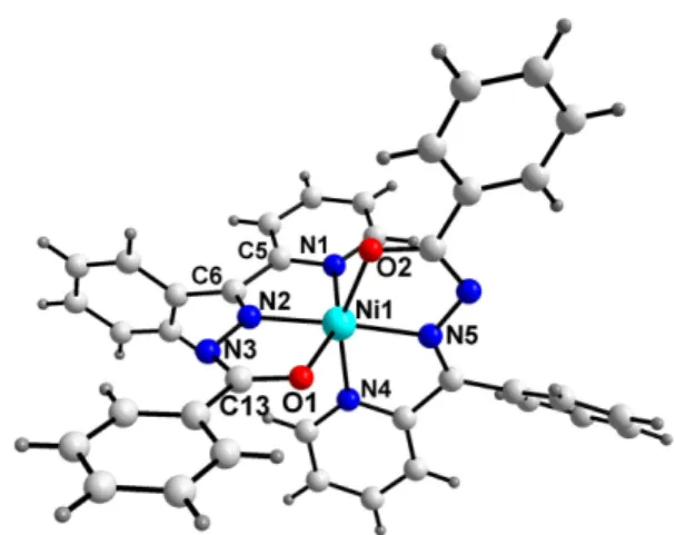
The complex 2 has basically the same structure of 1 (Fig. 2). Nickel(III) ion exhibits a distorted octahedral Figure 1. (a) Molecular structure of complex 1 with atomic labeling. (b) The 1D chain formed by H-bond and π−π interactions in 1 along the a-axis. Geometric details describing the H-bond operating in the crystal structure: C12i−H12...N3 = 2.46Å with angle at H12 = 139.4o for symmetry operation i: 2−x, 1−y, 1−z. (c) The 2D layer framework of complex 1 formed by H-bond and π−π inter- actions. (d) Packing diagram of complex 1. The hydrogen bonds and π−π interactions have been shown as dashed and dotted lines, respectively. All H-atoms in (b)−(d) have been omitted for clarity.
862 Bon Kweon Koo
environments with the cis- and trans-L-Ni-L angles in the range of 76.8(2)−109.6(2)o and 154.8(2)−172.7(2)o, respec- tively (Table 2), compared to the complex 1 (cis- and trans- L−Zn−L angles in the range of 74.3(1)−116.1(1)o and 148.9(1)−166.9 (1)o, respectively). The deviation from the ideal values for the octahedral structure is relatively small and more close to the octahedral compared to the complex 1. The average bond distances of Ni−N and Ni−O are 2.056(4) and 2.073(3) Å, respectively. The distances are slightly shorter than the distances (Zn−N = 2.145(3) and Zn−O = 2.080(3) Å) of complex 1. Most notably, the distance of Ni−N is smaller than Ni−O bond distances, contrast to the complex 1.
Complex 2 is also consolidated into a three-dimen- sional network by a C−H...N (C8i−H8...N3ii = 2.52 Å and C8i...N3ii =3.28(1) Å with angle at H8 = 141o for symmetry operation i: x, 1+y, −1+z; ii: 2−x, 2−y, −z) and π...π inter- actions. Two monomeric molecules in the unit cell are linked by π...π stacking between pyridine and phenyl rings of neighboring molecules with inter-ring distance of 4.157(1)
Å and intermolecular H-bonds (C8i−H8...N3ii) to give 1D chain network along a-axis as shown in complex 1. The chains constructs 2D framework by the π...π stacking between adjacent pyridine rings (centroid-to-centroid dis- tance of 3.815(1) Å) and finally, 3D network is accom- plished by inter-layer face-to-face π...π stacking (4.400(1) Å) between phenyl rings of adjacent 2D layers along c- axis.
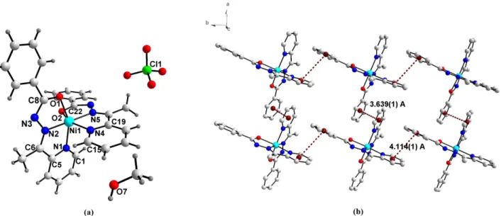
While, complex 3 consists of one Ni(III) ion, two coor- dinated apb ligands, one perchlorate ion, and one solvent methanol molecule, respectively (Fig. 3(a)). Here, the oxidation state of nickel is +3 in contrast to Ni(II) of start- ing material. This oxidation to Ni(III) can be assumed that the starting Ni(II) salt undergoes aerial oxidation in the presence of the Schiff base ligand in methanolic solution during preparation of the complex.16−18
The structure of complex 3 is similar to those of the complexes 1 and 2 with bpb− ligand. Two planes of apb− ligand, N4C15−C19 and C23−C28, are nearly plane with rms = 0.003(4), and 0.006(5) Å, respectively. The dihedral angle between them is 15.1(1)o. While, the dihedral angle between two pyridine planes (N1C1−C5 and N4C15−C19) of two apb− ligands is 89.9(1)o. The distances and bond angles are listed in Table 2. The structural data are similar to those of the Mn (II) and Zn(II) complexes with apb ligand, respectively.14
As shown in Fig. 3(b), the monomeric units construct 2D network by π...π stacking between pyridine and phe- nyl rings with inter-ring distance of 3.639(1) (a-axis) and 4.114(1) Å (b-axis), respectively. In contrast to complexes 1 and 2, there is not intermolecular H-bonding which con- struct 3D net work. For all complexes, the relatively short C13−N3 (for 1 and 2) and C8−N3 (for 3) bond distances (normal single bond is 1.52 Å),19 coupled with the length- Table 2. Selected bond lengths (Å) and angles (o) for complexes
1−3 Complex 1
Zn1–O2 2.056(2) Zn1–O1 2.104(3)
Zn1–N5 2.076(3) Zn1–N2 2.082(3)
Zn1–N4 2.210(3) Zn1–N1 2.213(3)
O1–C13 1.271(4) N3–C13 1.367(4)
O2–Zn1–N5 75.8(1) O2–Zn1–N2 116.1(1) N5–Zn1–N2 166.9(1) O2–Zn1–N4 150.3(1) N5–Zn1–N4 74.5(1) N2–Zn1–N4 93.7(1) N2–Zn1–N1 74.3(1) O1–Zn1–N1 148.9(1) Complex 2
Ni1–O2 2.063(3) Ni1–O1 2.082(3)
Ni1–N5 2.003(4) Ni1-N2 2.002(4)
Ni1–N4 2.098(4) Ni1-N1 2.122(4)
O1–C13 1.277(6) N3–C13 1.348(6)
N2–Ni1–N5 172.7(2) N2–Ni1–O2 109.6(2) N5–Ni1–O2 77.1(2) N2–Ni1–O1 76.8(2) N2–Ni1–N4 95.1(2) N5–Ni1–N4 78.3(2) O2–Ni1–N4 155.2(2) O1–Ni1–N1 154.8(2) Complex 3
Ni1–N2 1.983(3) Ni1–N5 2.016(3)
Ni1–O1 2.072(2) Ni1–N1 2.090(3)
Ni1–N4 2.109(3) Ni1-O2 2.153(2)
N3–C8 1.331(4) C8–O1 1.287(4)
N2–Ni1–N5 174.7(1) O1–Ni1–N1 154.6(1) N2–Ni1–N4 99.2(1) N5–Ni1–N4 76.6(1) N2–Ni1–O2 108.4(1) N5–Ni1–O2 76.0(1) N4–Ni1–O2 152.3(1)
Figure 2. Molecular structure of complex 2 with atomic labeling.
ened C13−O1 (for 1 and 2) and C8−O1 (for 3) distances which is typical of ketonic linkage (1.23 Å)20,21 indicate that the ligands act as enol form.
In conclusion, by the reactions of Ni(II)/Zn(II) ions and Schiff base ligands, three new metal(II)/(III) complexes, [Zn(bpb)2] (1), [Ni(bpb)2] (2), and [Ni(apb)2]·ClO4·CH3OH (3), have been synthesized. In each complex, metal ion is six-coordinated with N4O2 donors. The Schiff base is coordinated to metal(II)/(III) ions as a trifunctional ligand.
The structures are further extended into supramolecular framework by hydrogen bonds and π...π stacking inter- actions. Unfortunately, it has failed to obtain the poly- meric complexes of Schiff base through benzilic acid in this work. The development of synthetic routes to the sys- tems containing Schiff base is still required for the ratio- nal design and synthesis.
Acknowledgments. This work was supported by the 2012 Research Fund of Catholic University of Daegu. The author also acknowledges the Korea Basic Science Insti- tute for providing the crystal structure results.
Supporting Information. Crystallographic data in CIF format have been deposited with the Cambridge Struc- tural Database CCDC 927522 (for 1), 927523 (for 2), and 927521 (for 3), respectively. These data can be obtained free of charge at www.ccdc.cam.ac.uk/conts/retrieving.html (or from the Cambridge Crystallographic Data Centre, 12, Union Road, Cambridge CB2 1EZ, UK; Fax: +44-1223/336-033;
E-mail: [email protected]).
REFERENCES
1. Yaghi, O. M.; O’Keeffe, M.; Ockwig, N. W.; Chae, H. K.;
Eddaoudi, M.; Kim, J. Nature 2003, 423, 705.
2. Hagrman, P. J.; Hagrman, D.; Zubieta, J. Angew. Chem.
Int. Ed. 1999, 38, 2638.
3. Erxleben, A. Coord. Chem. Rev. 2003, 246, 203.
4. Suna,Y.-X.; Youb Z.-L. Z. Anorg. Allg. Chem. 2006, 632, 1566.
5. Shaabani, B.; Khandar, A. A.; Dusek, M. M.; Mahmoudi, P. F. Inorg. Chim. Acta 2013, 394, 563.
6. Nayak, S.; Gamez, P.; Kozlevcar, B.; Pevec, A.; Roubeau, O.; Dehnen, S.; Reedijik, J. Polyhedron 2010, 29, 2291.
7. Mohamad, G. G.; Omar, M. M.; Ibrahim, A. A. Spectrochim.
Acta, Part A 2010, 75, 678.
8. Rapheal, P. F.; Manoj, E.; Kurup, M. R. P.; Suresh, E.
Polyhedron 2007, 26, 607.
9. Yue, Y.-F.; Gao, E.-Q.; Fang, C.-J.; Xu, S.; Yan, C.-H.; J.
Mol. Struct. 2007, 841, 67.
10. Karmakar, R.; Choudhury, C. R.; Hughes, D. L.; Yap, G..
P. A.; Fallah, M. S. El.; Desplanches, C.; Sutter, J.-P.; Mitra, S. Inorg. Chim. Acta 2006, 359, 1184.
11. Sheldrick, G. M. SHELXTL: version 6; Bruker AXS Inc.;
Madison, Wisconsin, USA, 2001.
12. Farrugia, L. J. J. Appl. Cryst. 1997, 30, 565.
13. Brandenburg, K. DIAMOND: version 2.1; Crystal Impact GbR: Bonn, Germany, 1998.
14. Jang,Y. J.; Lee, U.; Koo, B. K. Bull. Korean Chem. Soc.
2005, 26, 925.
15. Anitha, C.; Sheela, C. D.; Tharmaraj, P. S.; Raja, J. Spec- trochim. Acta, Part A 2012, 98, 35.
16. Pinho, D.; Gomes, P.; Freire, C.; Castro, B. Eur. J. Inorg.
Chem. 2001, 1483.
Figure 3. (a) Molecular structure of complex 3 with atomic labeling. (b) 2D layer in complex 3. All H atoms, CH3OH, and ClO4− have been omitted for clarity.
864 Bon Kweon Koo
17. Franks, M.; et al. Inorg. Chem. 2013, 52, 660.
18. Freire, C.; Castro, B. Polyhedron 1998, 17, 4227.
19. Dutta, S.; Basy, P.; Chakravorty, A. Inorg. Chem. 1991, 30, 4031.
20. Mangalam, N. A.; Sivakumar, S.; Sheeja, S. R.; Kurup, M.
R. P.; Tiekink, E. R. T. Inorg. Chim. Acta 2009, 362, 4191.
21. Patole, J.; Sandbhor, U.; Padhye, S.; Deobagkar, D. N.;
Anson, C. E.; Powell, A. Bioorg. Med. Chem. Lett. 2003, 13, 51.